This study was submitted and approved by the Research Ethics Committee of the São Leopoldo Mandic Research Institute (SP, Brazil) under registration number #1,963,827. Before inclusion, all patients signed the informed consent, permitting the inclusion of the tooth. The recruitment period and study duration were from April 2022 to July 2023.
Sample Characteristics and Eligibility Criteria
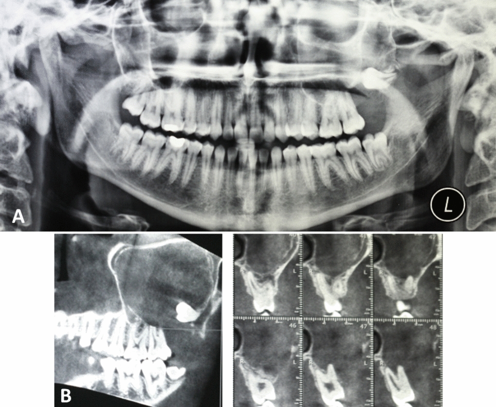
For the inclusion of the patients, it was followed: (1) 12 freshly extracted human third molars (one upper or one lower per patient) with fully formed roots were selected from six healthy individuals (without any systemic condition); (2) tooth referred for extraction due to orthodontic reasons or prevention of adjacent tooth resorption; (3) patients ≥ 18 years old; and (4) with completely included and impacted tooth. It was excluded: (1) pregnant patients; (2) smokers; (3) patients with any systemic condition other than healthy; or (4) extraction of not impacted/totally included tooth.
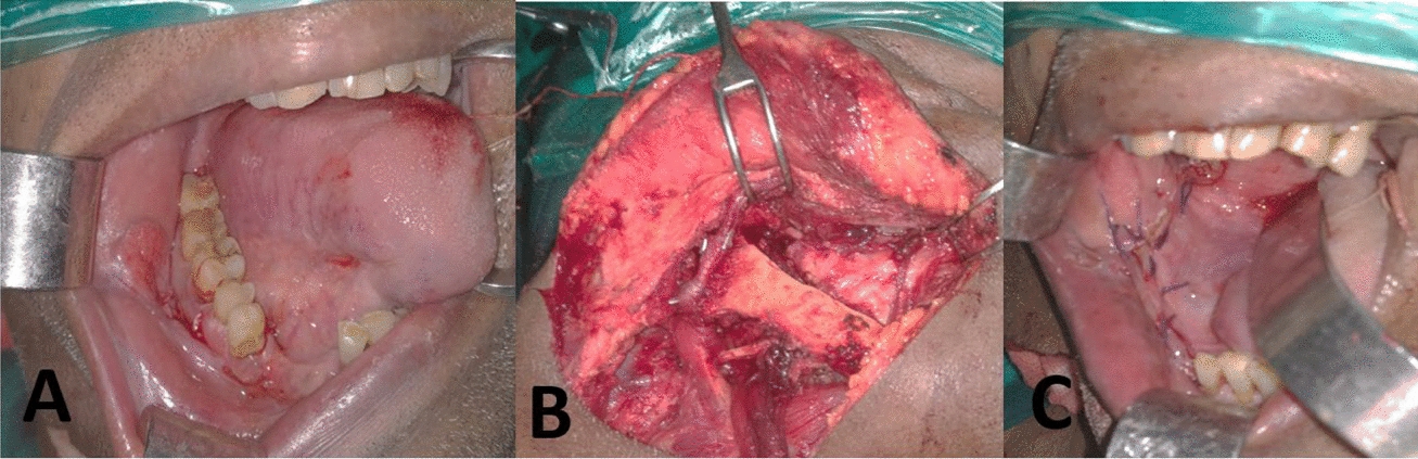
The extractions were performed by the same professional under local anesthesia (2% lidocaine and epinephrine at 1:100,000). The soft tissues surrounding the tooth were incised and removed to facilitate luxation and subsequent extraction. There was no section in the tooth, which was a motive for exclusion. The extracted tooth was abundantly rinsed with saline solution to remove blood and debris.
Groups
The teeth were divided into four test groups (n = 3) and a control group: Group 1. Control Group—no tooth-derived material was used—the cell culture was only supplemented with culture medium to keep the pre-osteoblastic cells viable; Group 2. FT (full tooth): the tooth was wholly preserved: enamel, dentin, periodontal ligament, cementum, and pulp; Group 3. WE (without enamel): enamel was removed using new diamond burs at high speed and water cooling; Group 4. WPL (without periodontal ligament): only the periodontal ligament was removed using periodontal curettes; and Group 5. WEPL (without enamel and periodontal ligament): enamel and periodontal ligament were removed.
After extraction, the teeth were randomly distributed (head or tail) by the groups until three teeth were achieved. They were immediately ground (according to the group distribution) using a manual bone-crushing pestle until the granules were visually (macroscopically) homogeneous.
Protein Extraction and Quantification
After grinding, the teeth were deposited in 15-ml polystyrene test tubes containing 1.5 mL of RIPA lysis buffer solution (Thermo Scientific, USA), containing 50 mM Tris HCl (pH 7.4), 150 mM NaCl, 1 mM EDTA, 1% Triton X-100, 1% sodium deoxycholate and 0.1% sodium dodecyl sulfate (SDS), plus a protease inhibitor cocktail (Sigma, St. Louis, MO, USA) at the final concentration of 1%, for each sample. Each tube was then shaken on a shaker (Phoenix AP 56) for 30 s to homogenize the elements contained in the samples.
The tubes were centrifuged (Eppendorf 5804 R) at 4 °C and 156G for 15 min, resulting in a three-phase solution. The supernatant corresponding to the protein extracts was pipetted out and transferred to new polystyrene tubes (Eppendorf, USA), duly identified.
The samples' total protein quantification was performed using the BCA Protein Assay kit (Thermo Scientific, USA), following the manufacturer's guidelines. The different concentrations of protein extracts (10 and 100 ng/nL) of the groups were added to the cell culture, and the results were compared to the control group.
Cell Cultures
The mouse’s pre-osteoblastic cell line (MC3T3-E1) used in this study was obtained from the American Type Culture Collection (ATCC, VC, USA). Pre-osteoblastic cells were cultured in alpha modification of minimum essential medium (α-MEM) supplemented with 10% fetal bovine serum (Cultilab®, Campinas, SP, Brazil) and 100 U/mL penicillin and 100 μg/mL streptomycin (Sigma, St. Louis, Missouri, U.S.A.).
All procedures were performed in a laminar flow hood to maintain the sterility of the materials and substances used for cell culture. The cells were kept in an incubator at 37 °C in a humid atmosphere containing 95% air and 5% carbon dioxide. The culture medium was changed every 2–3 days, and the culture progression was evaluated using phase microscopy in cultures grown on polystyrene. The concentrations tested in the present study were obtained from a concentration-effect curve.
Cell Proliferation Assay
For the cell proliferation assay, the Trypan blue vital exclusion method was used. The cells were cultured to a density of 110 cells/mm2 in 24-well plates and exposed to different protein extracts. After 1, 3, and 7 days, the cultures were enzymatically removed from the plates, and the cell pellet resulting from the centrifugation was suspended in 1 ml of medium. 10 μL of the cell suspension was removed, and 10 μL of Trypan blue was added to it. 1 μL of this suspension was then placed onto a hemocytometer (Neubauer-Fisher Scientific chamber, Pittsburgh, PA, USA) for manual counting under an inverted phase microscope (Nikon, Eclipse TS100). The data obtained were expressed in the number of cells × 104.
Cytotoxicity Assay (MTT)
Cell cultures were tested for cell viability using the MTT assay. In the cytotoxicity assay, cells were cultured at a density of 110 cells/mm2. After 1, 3, and 7 days of exposure to protein extracts from the different groups, the culture medium was removed and a new medium containing MTT (5 mg/mL, Sigma-Aldrich, U.S.A.) was added; cell cultures were incubated for 3 h at 37 °C. After this period, 100 μl of DMSO (Dimethylsulfoxide, LGC, São Paulo, Brazil) was added for 15 min at room temperature. The solubilized crystals were quantified in an ELX800 microplate reader (Biotek Instruments, Inc.) at 590 nm. The data were expressed as absorbance.
mRNA Expression of Genes Involved in Osteogenic Differentiation
The expression of genes encoding type I collagen (COL1), runt-related transcription factor 2 (RUNX-2), and bone morphogenetic protein 2 (BMP-2) was assessed by the real-time polymerase chain reaction (RT-PCR). The MC3T3-E1 cells were cultured in 24-well plates and treated with protein extracts for 1 and 3 days. Total RNA was extracted to make the cDNA using the Platus Transcriber Rnase H-cDNA First Strand kit (Sinapse inc).
Real-time PCR reactions were performed on a 7500 Fast Real-Time PCR System (Applied Biosystems) using the SYBR Green/ROX qPCR reagent (Thermo Fisher) as the detection system. The sequence of the primers used is listed in Table 1. Expression of the COL1, RUNX-2, and BMP-2 genes was normalized in relation to the endogenous gene GAPDH and calculated using the 2−ΔΔCt method. The values were expressed in arbitrary units (AU).
Table 1 Primer sequences used in this studyVon Kossa Staining
The pre-osteoblastic cells were cultured with the protein extract for 14 days, and the culture medium/protein extract was changed every 2–3 days. After 14 days, the culture medium was removed, and 2 ml of silver nitrate (AgNO3) was placed in each well for 20 min. The wells were then rinsed with distilled water, and 2 ml of hydroquinone (C6H4 (OH) 2) was added for 2 min. 2 ml of thiosulfate (S2O3−2) was then added for 2 min. The samples were rinsed with distilled water and prepared for analysis under light microscopy (Nikon Eclipse E800, Japan). Phosphate deposits were identified by the presence of black-colored deposits by means of macroscopic photos converted into binary data. Image analysis was performed using the ImageJ software (NIH).
Statistical Analysis
All experiments were performed in triplicate. The mean values and standard deviations were compared using a two-way analysis of variance (ANOVA) and Tukey/Dunnett-Sidak tests at a significance level of 5% (p < 0.05). The analyses were performed on GraphPad Prism 9.5.1 (GraphPad Software, LLC).


留言 (0)