We reported a case where prolonged prone positioning led to VP shunt obstruction, worsening hydrocephalus, and delayed emergence from anesthesia. There have been reports of increased intra-abdominal pressure during laparoscopic surgery causing VP shunt malfunction [9]. However, to our knowledge, no reports have identified intraoperative positioning as a factor in VP shunt obstruction.
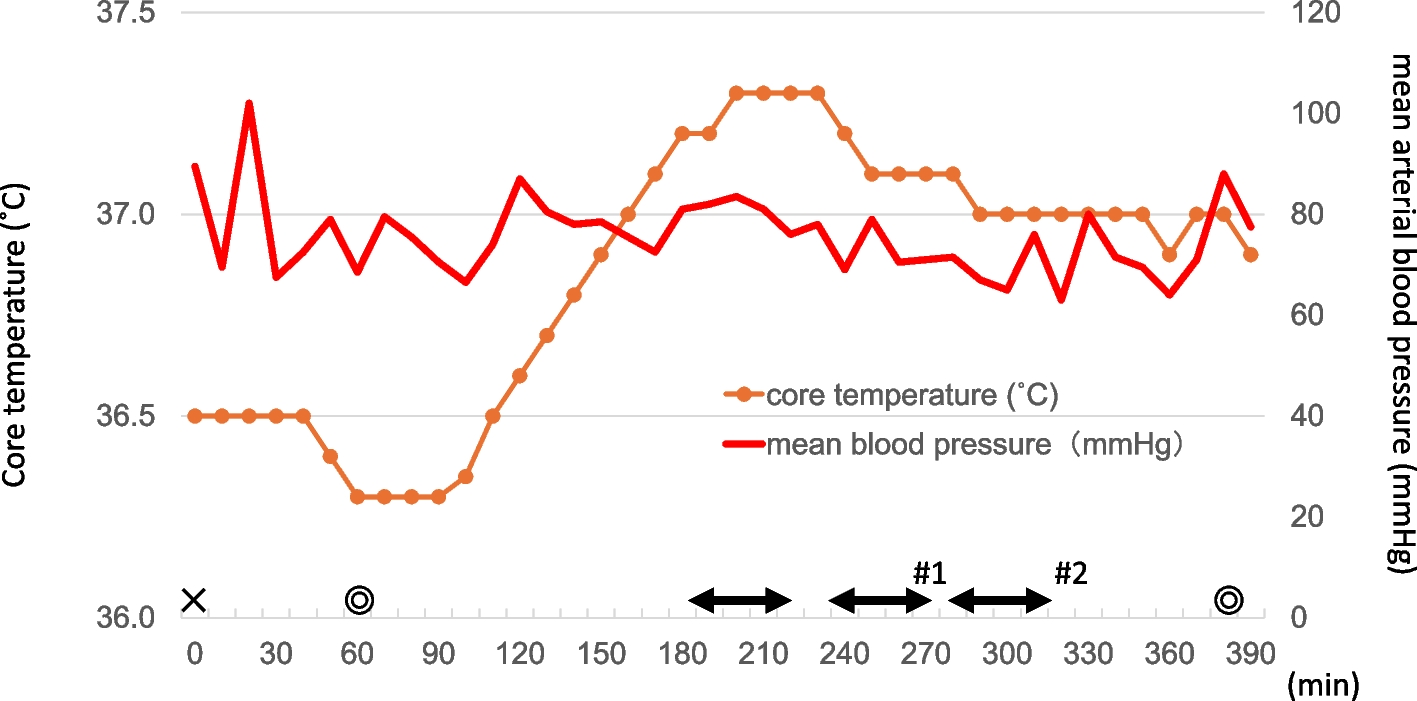
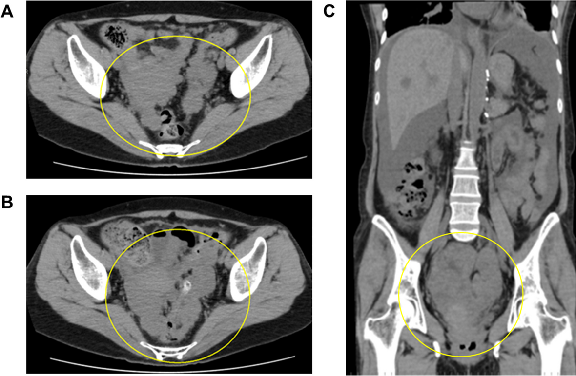
Several factors can induce delayed emergence from anesthesia [10, 11]. Residual effects of sedatives are unlikely to be the cause in this case because of intraoperative BIS values between 40 and 60, normal liver and renal function, and normal preoperative hypoalbuminemia or hypothermia. Blood gas data, glucose, and electrolytes were also within normal ranges. Based on differential diagnosis, we considered that a complication in the central nervous system might have occurred. Postoperative CT revealed hydrocephalus despite the presence of a VP shunt. To promote the flow of CSF, the unidirectional reservoir of the shunt was pumped. A few minutes after pumping the reservoir, the patient’s eyes opened. This suggests that the VP shunt might not have functioned intraoperatively, leading to hydrocephalus and delayed recovery of consciousness after the cessation of anesthetics.
The detailed mechanisms of VP shunt occlusion remain unclear. The VP shunt may have kinked at the cervical level due to the forward flexion of the head during occipital surgery. Another possibility is that the prone position itself may have occluded the VP shunt tube. The VP shunt tube was placed subcutaneously in the anterior thoracic region (Fig. 1) and may have been compressed by the body. Other possibilities include increased intra-abdominal pressure due to the prone position, which may have obstructed the CSF flow.
The preoperative CSF outflow resistance was set at 14 cmH2O. This value may have been appropriate because the patient’s preoperative level of consciousness was normal [12]. Therefore, it is unlikely that the CSF outflow resistance contributed to the delayed recovery of consciousness after anesthesia.
While we had recognized that the patient had a VP shunt, the possibility of intraoperative obstruction or malfunction of the VP shunt due to the surgical position had not been considered. Previously, increased intra-abdominal pressure during laparoscopic surgery has been shown to induce malfunction of a VP shunt [9]. To avoid this complication, several reports have demonstrated the usefulness of transcranial Doppler for CSF flow monitoring [7, 8]. In the perioperative management of patients with VP shunts, it is important to consider the risk of shunt malfunction perioperatively, protect the shunt route, and monitor CSF flow with transcranial Doppler intraoperatively, depending on the situation.
In conclusion, we reported a case of VP shunt malfunction and exacerbation of hydrocephalus during neurosurgery under prolonged prone positioning, which resulted in a delayed recovery of consciousness after anesthesia. It is important to manage anesthesia with the possibility of intraoperative shunt malfunctions in mind, depending on the position of the patient with a VP shunt implantation.


留言 (0)