
記住我


The growing demand for better esthetics during orthodontic treatment has led to the development of appliances that combine both acceptable esthetics for the patient and adequate technical performance for the clinician.[1,2] Esthetic of fixed labial appliances have evolved through the inclusion of ceramic brackets, esthetic ligatures, and tooth-colored archwires.[3] However, coated archwires are still not used widely because their coating is not durable. Previous studies have reported that there is a 25% coating loss and deterioration of surface quality within 33 days in vivo.[4] Characteristics such as optical, biological, and mechanical properties, coating stability, force transfer values, color stability, and plaque accumulation of esthetic archwires have been evaluated previously and have been reported to be not ideal. Despite these disadvantages, esthetic wires are still commercialized and used in clinical practice.[5]
Insufficient data are available comparing properties of as received coated wires and orally exposed wires since most of the tests have been conducted in laboratories and do not simulate real clinical conditions.[6] Moreover, only the segments of wire were used instead of the entire archwire, making clinical simulation incomplete and representing a limitation.[7]
Aims and objectivesThe aim and objectives of the study were to evaluate and compare the coating stability and surface characteristics of different types of esthetic archwires as received from the manufacturer and after 21 days of oral exposure.
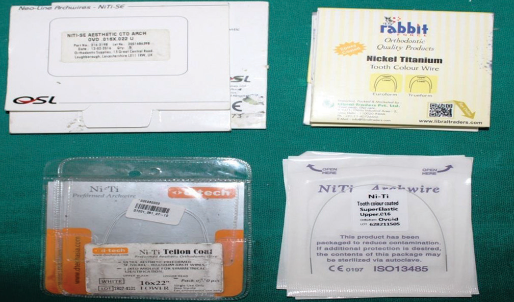
MaterialsEsthetic wires: [Figure 1]
Epoxy-coated wire (0.016 × 0.022”), manufactured by Rabbit force, Libral Traders Pvt Ltd., Okhla industrial area, 2- New Delhi
Polymer-coated wire (0.016”), manufactured by Nexus Medodent, NS Road Mulund, Mumbai)
Teflon-coated (0.016 × 0.022”), manufactured by D tech Orthodontic Private Ltd, Pune
Rhodium-coated (0.016 × 0.022”), manufactured by OSL Neo- line arch wires, GD- 17 lane no 4, Hari Nagar, New Delhi.
Oral hygiene products
Toothbrush: Colgate ortho
Toothpaste: Colgate.
Ultrasonic cleaner
UDS-J wood pecker, Med Net Bork Strasse 10.48167 Muenster Germany.
Glass slide
Stereomicroscope
Scanning electron microscope (SEM; Model JSM 6100 (Jeol)
3DProfilometer (zeta) by Pace Analytical Systems, Bengaluru.
Export to PPT
MethodologyFour types of nickel-titanium (NiTi) esthetic orthodontic archwires (epoxy, polymer, Teflon, and rhodium) were evaluated for the study [Table 1].
Table 1: Characteristics of archwires used in the study
Group Manufacturer Cross-section size Types of coating I Rabbit Force, Libral Traders PTV.LTD Okhala industrial area 2-New Delhi 110020 india 0.016 X 0.022” Epoxy Coated II Nexus Medodent, NS Road Muland Mumbai 400080 India 0.016” Polymer Coated III D Tech orthodontic Pvt Ltd , Pune Nagar road Wagholi Pune 412207 0.016 X 0.022” Teflon Coated IV OSL Neo-Line arch wires, GD-17, lane no-4, Hari Nagar, New delhi 110064 0.016 X 0.022” Rhodium Coated Evaluation and comparison of as received esthetic archwiresSpecimens with a length of 10 mm were prepared from straight sections of arch wire as received from the manufacturer from each group and were evaluated for coating thickness and dimension under stereomicroscope at ×30 magnification (n = 1). The coating thickness of each group was measured using Image Pro Plus software. Graph paper with a width of 10 mm was fixed on a glass slide and served as a template. Each wire was positioned with the midpoint on the center of the graph paper, and the ends were fixed with wax. Two 5-mm-long images were obtained of the same sample, one on its right side and one on its left side. Thus, the 10-mm segment of each coated wire located in the inter bracket space was fully evaluated. For each coated surface, three coating thickness readings were measured randomly. Micro-morphological characteristics of the labial surface were evaluated under SEM (n = 1), whereas surface roughness was assessed under 3D profilometer (n = 1).
Oral exposureA sample size calculation and parameters were based on formula described by Pandis et al.[8] α = 0.05, β = 0.2, and a difference to be detected of 30% in coating loss percentage (On average, 28.71% coating loss was observed in coated NiTi archwires). This calculation indicated the need for 11 segments of each archwire and, hence, a total of 44 segments of esthetic archwires. Subjects undergoing fixed orthodontic treatment in the leveling and alignment stage with 0.022 MBT brackets in the Department of Orthodontics and Dentofacial Orthopedics, Swami Devi Dyal Dental College and Hospital, Barwala, Panchkula, were selected. The selection criteria considered healthy patients with good oral hygiene, no caries, and complete permanent dentition. The ethical approval for this study was obtained from the Institutional Ethical Committee.
The archwires were ligated into the 0.022 MBT brackets using elastomeric modules for patients under fixed orthodontic treatment during the leveling and alignment phase. Oral hygiene instructions were given, and the patient used the same toothbrush and toothpaste (Colgate) throughout the study. The subjects were instructed not to use any other oral subjects, including oral irrigators or anti-microbial mouthwash. The archwires were placed and removed by the same operator. After 21 days, the archwires were removed and placed individually in an ultrasonic cleaner (Digital ultrasonic cleaner) immersed in (Ambersil cleaning liquid) for 30 min to remove organic debris. The wires were then subjected to the following laboratory analysis.
All the retrieved archwires (n = 44) were evaluated for coating loss under stereomicroscope at ×30 magnification. Two archwires from each group were evaluated for micromorphological characteristics under SEM and for surface roughness under 3D profilometer.
The data obtained were analyzed with conventional and descriptive statistics. All the analyses were performed with commercial statistical software Statistical Package for the social Sciences version 17.0.
RESULTS Evaluation of coating thickness and dimension Coating thickness of as – received esthetic NiTi wiresMaximum coating thickness was observed in Group I (Epoxy coated 0.00344 ± 0.00009”) and minimum in Group II (Polymer coated 0.00196 ± 0.00013”). The order of coating thickness was seen in the following order: Group I: Epoxy coated (0.00344 ± 0.00009”) >Group IV: Rhodium coated (0.00287 ± 0.00014”) >Group III: Teflon coated (0.00244 ± 0.00014”) >Group II: Polymer coated (0.00196 ± 0.00013”) [Table 2 and Figure 2].
Table 2: Coating thickness of as received wires (inch).
Group Mean Standard deviation Minimum Maximum I 0.00344 0.00009 0.00337 0.00356 II 0.00196 0.00013 0.00158 0.00202 III 0.00244 0.00014 0.00224 0.00256 IV 0.00287 0.00014 0.00249 0.00303 P value 0.550 Statistically insignificant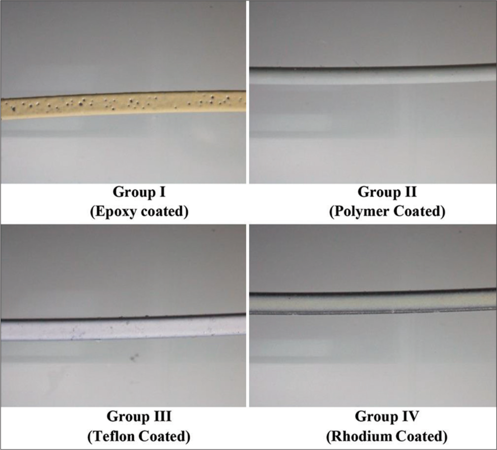
Export to PPT
Coating thickness after oral exposure of 21 daysAfter oral exposure, maximum coating loss was observed in Group I (Epoxy coated: 0.00197 ± 0.00036”) and the minimum in Group II (Polymer coated, 0.00083 ± 0.00016”). The coating loss was observed in the following order: Group I (Epoxy coated: 0.00197 ± 0.00036”) >Group III (Teflon coated: 0.00148 ± 0.00035”) >Group IV (Rhodium coated: 0.00135 ± 0.00025”) >Group II (Polymer coated: 0.00083 ± 0.00016”) [Table 3 and Figure 3].
Table 3: Coating thickness of wires after oral exposure, inch
Group Mean Standard deviation Minimum Maximum P I 0.00197 0.00036 0.00137 0.00241 0.001 II 0.00083 0.00016 0.00063 0.00105 0.001 III 0.00148 0.00035 0.00104 0.00187 0.001 IV 0.00135 0.00025 0.00104 0.00168 0.001 P value P- 0.001 Statistically significant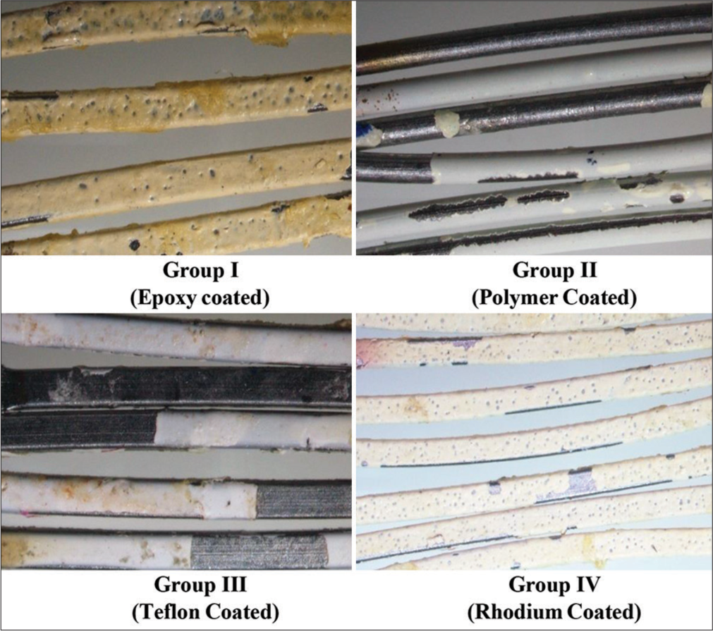
Export to PPT
Measurement of coating lossThe maximum percentage of coating loss was observed in Group II (Polymer coated 57.3983%), and the minimum percentage of coating loss belongs to Group III (Teflon coated 39.4018%). The coating loss was observed in the following order: Group III Teflon coated (39.4018%) >Group I Epoxy coated (42.5134%) >Group IV Rhodium coated (52.6969%) >Group II Polymer coated (57.3983%) [Table 4].
Table 4: Lost Coating Percentage
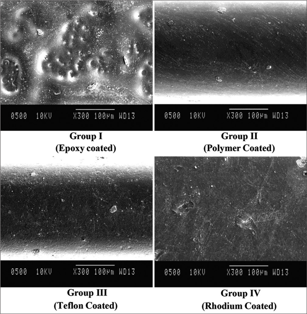
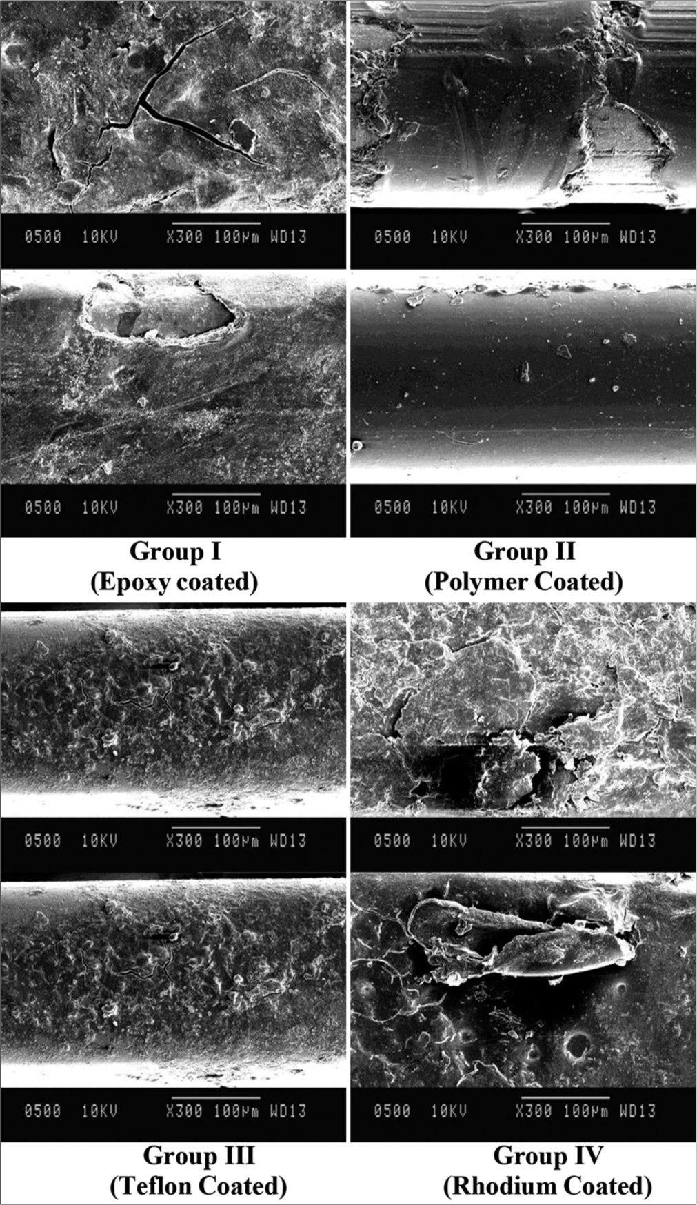
Group Mean Standard Deviation Minimum Maximum I 42.5134 10.61748 28.53 59.26 II 57.3983 10.77231 33.30 70.40 III 39.4018 15.71010 16.51 59.11 IV 52.6960 9.47400 37.74 63.70 Evaluation of Surface characterization Surface topographyOn the visual evaluation of SEM images of received archwires [Figure 4], it was seen that Group I wire (Epoxy coated) had larger surface defects with surface elevations dispersed as fields. Group II (Polymer coated) showed a small number of grooves and very fine striations that were not parallel to the long axis of the wire. Group III (Teflon coated) has minor surface irregular defects with fine striations, whereas Group IV (Rhodium coated) shows minor surface defects throughout its surface.
Export to PPT
After oral exposure [Figure 5], Group I (Epoxy coated) showed microcracks, and Group II (Polymer coated) wires showed areas of entire coating loss, exposing the underlying wire; they also showed large-sized striations. Group III (Teflon-coated) wires showed finer cracks and much surface irregularity, whereas Group IV (Rhodium-coated) wire showed large-sized grooves with overall destruction of coating regularity.
Export to PPT
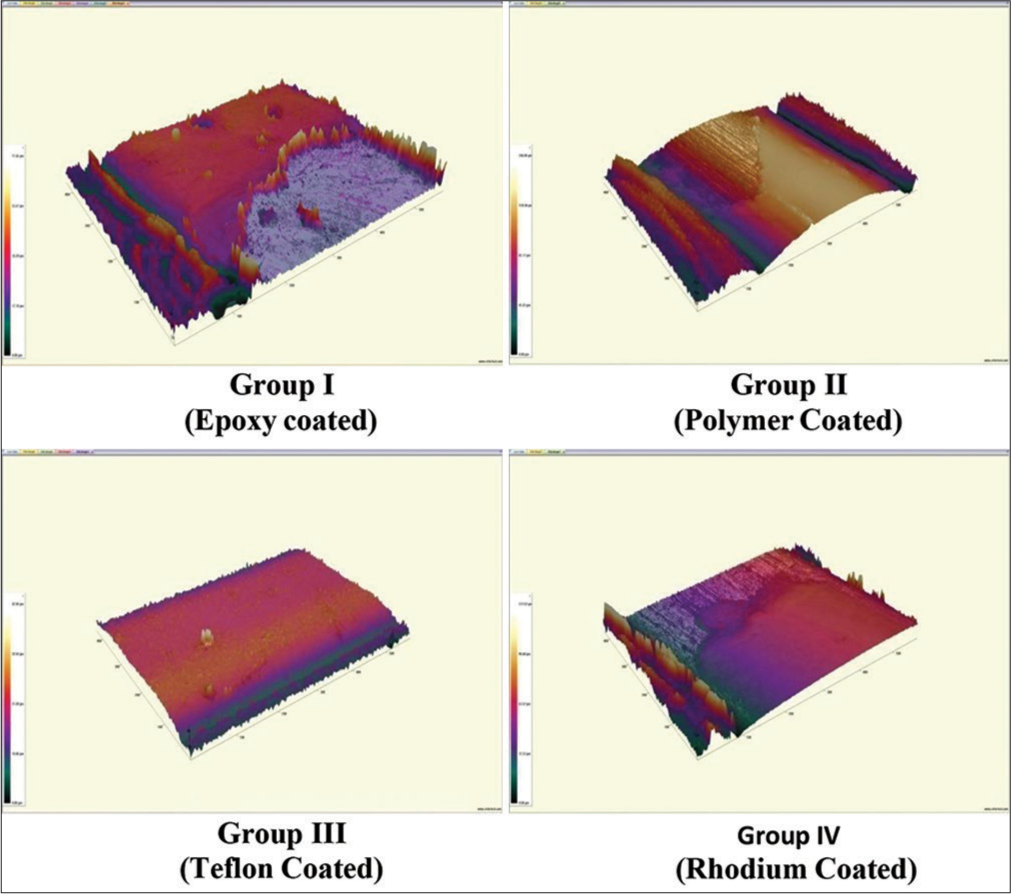
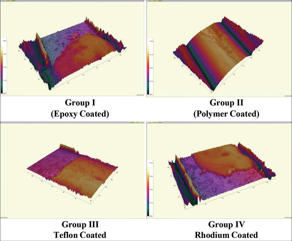
Surface roughnessOn interpreting the statistical details of the 3D profilometer [Figure 6], it was observed that the wire with minimum surface roughness (minor surface irregular defects with fine striations) belonged to Group III (Teflon coated, 0.8060 µm) and the wire with maximum surface roughness belonged to Group II (Polymer coated: 2.194 µm) [Table 5]. After oral exposure for 21 days [Figure 7], the maximum surface roughness (cracks and surface irregularity) belonged to Group III (Teflon coated 6.674 µm), and the wire with the minimum surface roughness belonged to Group IV (Rhodium coated 1.372 µm) [Table 6]. Hence, the smoothest coating was observed to be rhodium coated, and the roughest wire was Teflon coated.
Export to PPT
Export to PPT
Table 5: Surface roughness of as received wires, micrometers
Group Arithematic mean Root mean square I (Epoxy Coated) 1.320 2.609 II (Polymer Coated) 2.194 2.711 III (Teflon Coated) 0.8060 1.053 IV (Rhodium Coated) 1.372 1.712Table 6: Surface roughness of coated wires after oral exposure
Group Arithematic mean Root mean square I (Epoxy Coated) 2.080 2.518 II (Polymer Coated) 4.269 5.110 III ( Teflon Coated) 6.674 9.323 IV (Rhodium Coated) 1.372 1.712 DISCUSSIONOrthodontic treatment usually extends over months or years. Therefore, the appearance of orthodontic appliances has become a significant factor in orthodontic treatment decisions, particularly due to increasing demands from adult patients. These demands mean that esthetic considerations now extend beyond ceramic or composite brackets and ligatures and are now a concern for archwires as well, which has led to the advent of esthetic orthodontic wires. The esthetic coating used can be epoxy-resin, Teflon or polytetrafluoroethylene (PTFE), parylene or silver-polymer, rhodium, or, less commonly, palladium. These archwires vary on the basis of the thickness of the coating, area of coating (full coverage or labial surface only), manufacturing process, and mechanical properties.[1] However, the coating is likely to peel off in daily tooth brushing and under orthodontic force.[4]
In our study, maximum coating thickness was observed in epoxy-coated archwires, whereas minimum was observed in polymer-coated archwires. Epoxy-coated arch wires also had large surface defects with surface elevations under SEM, whereas other esthetic archwires showed minor surface defects under SEM examination. It has been mentioned earlier that epoxy coating is achieved by a method called electrostatic coating or E – coating in which a high voltage charge is applied to the archwire, and atomized liquid epoxy particles are air sprayed over the wire surface. This gives a 0.002-inch thick epoxy covering around the wire. Whereas the polymer coating (0.000127 mm thick) is reported as having a double-layered coating structure, the inner layer is silver and platinum, and the outer layer is polymeric. The outer layer imparts durability and wear resistance, and the inner layer gives a tooth-colored appearance to the archwires.[9,10] Maximum coating loss was observed in polymer-coated archwire, and minimum coating loss was observed in Teflon-coated archwire after oral exposure. On comparing as received coated archwires, maximum surface roughness belonged to polymer coated and minimum surface roughness was observed in Teflon-coated archwire. Coating loss depends on the manufacturing process of the archwire; hence, the amoun-t of coating does not correlate with coating loss.
A similar study was conducted in 2013 by Da Silva et al.[11] wherein labial surface topography of as received coated wires had delamination and irregularities, and they did not show a uniform coating thickness pattern. The coating layer tended to be thicker in the center and thinner on the edges of labial surfaces or all surfaces. The coating layer peeled off in many areas during oral exposure, leaving surface defects similar to our study.
Maximum coating loss was observed in polymer-coated archwires and minimum in Teflon-coated archwires. Teflon-coated archwires also showed minimum surface roughness under a 3D profilometer, and maximum surface roughness was observed in polymer-coated archwires. This significant difference in the surface roughness of the coated wires could be related to the effects of different surface treatments, manufacturing techniques, thickness of the coating, and chemical composition of the coatings. Previous study Elayyan et al.[5] has shown that the coating suffers from high and variable amounts of deterioration. Many of the specimens were characterized by delamination of the coating over large areas. This clearly affected the esthetic value of coated archwires. On average, 25% of the coating was lost within 33 days in vivo, and surface morphology showed severe deterioration, which led to a significant reduction of the esthetic qualities. Teflon, or PTFE, is a material characterized by a completely fluoridated chain. This chain is responsible for its physical and chemical characteristics. Since Teflon has a low coefficient of friction, archwires with a Teflon coating could reduce resistance to sliding, which could be the reason for minimum coating loss.[11] An earlier study on Teflon-coated wires reported that Teflon-coated archwires produced lower friction than the corresponding uncoated archwires (P < 0.01). The best frictional results were registered with a combination of Teflon-coated archwires and Quick brackets. The findings suggested that coating orthodontic archwires with Teflon has the potential to decrease resistance to sliding. Coating with Teflon has excellent esthetic properties: the tooth-like color of Teflon-coated archwires, together with their improved frictional performance, may lead to widespread use of this type of archwire in future orthodontic practice.[12]
However, after oral exposure of 21 days, the smoothest surface was observed in rhodium-coated archwires, and the roughest was observed in Teflon-coated archwires. Earlier studies revealed that epoxy-coated wires had the highest values for surface roughness, followed by PTFE wires. The rhodium-coated wire was the best-coated wire in terms of surface roughness values comparable with that of uncoated wires, which are similar to our results[13]. Rhodium is a hard, silvery-white transition metal that is a member of the platinum group. It is usually alloyed with platinum and palladium and applied in highly resistant and corrosion-resistant coatings. It is applied using a plasma-immersion ion implantation technique.[9]
The oral exposure of 21 days had an important role in coating loss and in the surface quality of the coated wires. The color stability of esthetic archwires during orthodontic treatment is clinically important. Ideally, the color of esthetic wires should match that of teeth and esthetic brackets. Da Silva et al.[11] stated that all esthetic archwires assessed in their study showed noticeable color changes after 21 days in staining. Some studies conclude that coffee was the most chromogenic agent when it was compared. Surface characteristics, friction coefficient, and coating stability may have correlation but are not statistically significant due to multifactorial conditions in the oral environment.[14]
CONCLUSIONThe average coating thickness of as received wires was observed to be 0.002” in dimension
Maximum percentage of coating loss was observed in Group II (polymer coated- 57.3983 ± 10.77231%) and the minimum in Group III (Teflon coated-39.4018 ± 15.71010%)
In terms of surface roughness, maximum surface roughness was observed in Group II (polymer-coated – RMS-2.711) and the minimum surface roughness was observed in Group III (Teflon coated- RMS- 1.053)
All the wires had low esthetic values, as they presented a non-durable coating after oral exposure
On visual examination of SEM image, all four group esthetic wires showed variations ranging from microcracks and large sized striations to overall destruction of coating regularity
Coinciding the orthodontic treatment duration, none of the orthodontic wires studied presented ideal characteristics for clinical use.
Limitations of the studyThe limitations include a short duration of oral exposure. There are few studies in the literature to compare our results which make the conclusion based mainly on our finding. It was interpreted that an ideal esthetic archwire should have uniform coating thickness pattern, great esthetic aspects with surface ultra-structures, and mechanical properties comparable to conventional NiTi archwires.
留言 (0)