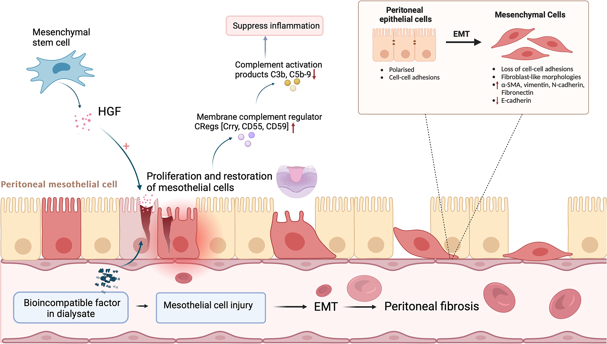
Isolation, culture and identification of umbilical cord-derived mesenchymal stem cells
UC-MSCs were obtained from Xuzhou Regional Cell Preparation Center Co., Ltd. Fresh umbilical cord tissue was collected and washed with phosphate-buffered saline (PBS), after which the Wharton's jelly was separated from the umbilical cord tissue using sterile scissors and forceps. The Wharton's jelly was then cut into small pieces approximately 1 mm3 in size. These pieces were cultured in 15 mL of serum-free medium in T75 flasks. Upon reaching approximately 80% confluence, the cells were passaged. We used fifth passage cells for subsequent experiments. Cells expressed CD73, CD90, and CD105 but did not express CD19, CD34, CD45, CD79a, CD11b, or HLA-DR. These cells met the current international standards for defining mesenchymal stem cells derived from bone marrow [27]. Flow cytometry results were provided by Nanjing Kingmed for Clinical Laboratory.
PET imaging of 89Zr labeled MSCs in DN model
To prepare the 89Zr-oxine complex, we obtained a certain volume of oxalic acid 89Zr solution, added 9 times the volume of HEPES buffer solution (0.1 mol/L) and added 0.375 times the volume of Na2CO3 solution (1 mol/L) to adjust the pH to 7. Then, we labeled mesenchymal stem cells with 89Zr-oxine. The cells were centrifuged and distributed into 15 mL sterile centrifuge tubes. Then, 10 μCi of labeled 89Zr-oxine was added to each 1 × 10⁶ stem cell sample and incubated for 15 min at room temperature. The cells were intermittently agitated to ensure that the cells were suspended. Then, the mixture was centrifuged at 300×g for 5 min at room temperature and washed 3 times with DPBS to remove unbound 89Zr-oxine and free 89Zr ions to obtain 89Zr-stem cells (89Zr-MSCs). Finally, we intravenously injected 89Zr-labeled mesenchymal stem cells into the rats. To eliminate free Zirconium from the body, 200 μL of 50 mg/mL DFO was injected into the muscle of each rat 30 min before the injection of 89Zr-MSCs. At 1 h, 3 h, 6 h, 24 h, 48 h, 72 h, 5 d, 10 d, or 12 d after 89Zr-MSCs administration, an Inveon MicroPET scanner (Siemens Medical Solutions) was used for static PET scanning. PET images were quantitatively analyzed using a previously reported method [28].
Antibody arrays
Rat Cytokine arrays (GSR-CYT-3-1, RayBiotech, Norcross, GA, USA) were used according to the manufacturer’s instructions to measure the expression levels of 27 cytokines in in the renal cortex of rats. The slide scanning was performed using InnoSan 300 Microarray Scanner (Innopsys, Parc d'Activités Activestre; 31 390 Carbonne-France). Differentially expressed proteins were arranged using hierarchical clustering and represented as a heat map. Data analysis was conducted using the GSR-CYT-3 data analysis software.
Drugs and chemicals
Irbesartan (Sanofi-Aventis, France, 30 mg/kg/day, IG) and streptozotocin (STZ) (Sigma Chemical Co., St. Louis, MO, USA, 35 mg/kg, IP) were used. A Rat Cytokine Array GS3 was used (RayBiotech, USA). Nephrin (Santa Cruz, sc-376522), WT-1 (Abcam, ab267377), and NPHS2 (Proteintech, 20384-1-AP) were used. Serum creatinine (Scr) and blood urea nitrogen (BUN) levels were measured by a Hitachi automatic analyzer (Hitachi Co. Ltd., Tokyo, Japan). Urinary albumin was measured by standard methods using commercial ELISA kits (Wuhan Newqidi Biological Technology Co., Ltd.).
Animal
Animal experiments were approved by the Experimental Animal Ethics Committee of Xuzhou Medical University (202207s017). Forty male Sprague-Dawley (SD) rats weighing 160–180 g were purchased from Beijing Vital River Laboratory Animal Technology Co., Ltd., and raised in the Animal Center of Xuzhou Medical University. The sample size of the experimental and control groups was determined based on previous studies regarding the treatment of DN using MSCs transplantation combined with other drugs [27, 29] and our pilot trial. The laboratory temperature was 22 °C, and an alternative 12-h light/dark cycle was used. There were 4–5 rats per cage. Ear tags were used for labeling. These rats had free access to food and water and could freely move in their cages.
Animal experiments
After the animals were acclimatized and fed for one week, they were randomly divided into the following five groups using a random number generator: (1) the control group (CON), (2) the diabetic nephropathy group (DN), (3) the mesenchymal stem cells treatment group (MSCs), (4) the irbesartan treatment group (Irb), and (5) the combined administration group (MSC + Irb). In order to control experimental bias, we requested a researcher unfamiliar with the animal modeling situation to perform blind grouping, with the number of rats that contributed data for analysis in each experiment indicated in figure legends. Rats in the control group were fed normal chow, and the remaining rats were fed high-fat chow (Research Diets, Shanghai Synergy; XTHF45 rodent diet with 45 kcal% fat) for 6 weeks. Except for control rats the rats were intraperitoneally injected with streptozotocin (35 mg/kg; Sigma-Aldrich, St. Louis, MO, USA) dissolved in 0.1 M sodium citrate (pH 4.5), and control rats were injected with 0.1 M sodium citrate [6]. Three days after STZ injection, blood glucose levels were measured by tail vein blood sampling for three consecutive days, and blood glucose levels above 16.7 mmol/L were considered to indicate diabetes [30]. Body weight was monitored weekly, and blood glucose levels were tested every two weeks. After 4 weeks of streptozotocin injection, the diabetic rats exhibited significant increases in urine output, and urine protein levels greater than 20 mg/24 h indicated successful modeling [17]. During the experiment, we tried to minimize the pain of the animals and did not experience any adverse events related to the experimental drug. After animal modeling, we closely observe the state of the experimental animals. Due to the serious progression of some animal diseases that meet humanitarian priorities and end of experiment indicators, such as anorexia and weakness, they should be removed from the experiment and euthanasia should be implemented in a timely manner. The rats in the MSCs group and the MSC + Irb group were injected with 5th generation UC-MSCs (2 × 10⁶ cells, 1 mL) via the tail vein 3 times every 10 days [31]. The remaining groups were injected with an equal amount of PBS via the tail vein. Rats in the Irb group and MSC + Irb group were administered irbesartan (30 mg/kg/d) by gavage. The remaining groups received the same amount of saline by gavage. Ensure that each group has at least 6 rats.
For the pharmacokinetic experiment, animal breeding and modeling were similar to the pharmacological experiments, and the animals were divided into two groups. (1) the 89Zr-MSCs-CON (C + M) group (n = 5); (2) the 89Zr-MSCs-DN (D + M) group (n = 5). The meaning is respectively the injection of 89Zr-labeled mesenchymal stem cells in control group and diabetic nephropathy group. 10 SD rats in total.
Before dissection, rats were euthanized with an overdose of pentobarbital sodium. Our animal studies were reported in accordance with the ARRIVE guidelines 2.0. (Additional file 1).
Assessment of glucose tolerance
After 3 weeks of drug treatment, the rats were fasted for 12 h, and glucose solution (1 g/kg) was injected intraperitoneally. Blood glucose was measured by collecting blood from the tail vein at 0, 25, 30, 60, and 120 min after injection. Insulin resistance was assessed by the steady-state test (HOMA-IR) and was calculated using the following formula: HOMA-IR = glucose concentration (mmol/L) × insulin (μU/L)/22.5.
Renal histopathological evaluation
Kidney tissues were fixed in 10% formalin for 24 h, embedded in paraffin, sectioned at a thickness of 3 μm, stained with hematoxylin-eosin (HE) and periodic acid-Schiff (PAS), and examined under a microscope. To prepare ultrathin sections, 1 mm3 of kidney tissue was fixed with 2.5% glutaraldehyde (pH 7.4) for 24 h at 4 °C. The tissue was then fixed in 1% osmium tetroxide for 2–3 h, dehydrated in acetone and ethanol, and embedded in epoxy resin. Sections were cut to a thickness of 50 nm with an ultramicrotome and then stained with 3% uranyl acetate and lead citrate. The sections were examined by transmission electron microscopy.
Urine and blood samples and tissue specimens
Urine samples were collected from the rats before the end of the experiment, and to prevent the effects of feces and food on urinary proteins, the rats were placed in metabolic cages (Shanghai, China) and were fasted but watered. Urine was collected and centrifuged at 1500 rpm for 5 min, and the supernatant was collected and stored at −80 °C. These samples were used to measure urinary microalbumin and creatinine. The urine microalbumin/creatinine ratio (UACR) was subsequently calculated. Before the end of the experiment, the rats were fasted for 12 h and anesthetized with sodium pentobarbital for blood sampling. The collected blood was centrifuged at 3000×g for 15 min, and the supernatant was stored at −80 °C. These samples were used to measure blood creatinine, blood urea nitrogen, and inflammatory factor levels. Left kidney tissue was fixed in 10% formalin for histological examination. The other part of the renal cortex was cut into 1 mm × 1 mm × 1 mm cubes and placed in electron microscopy fluid for storage (stored at 4 ℃ in the dark). The right cortex was quickly frozen in liquid nitrogen and stored at −80 °C for biochemical and immunoblot analyses.
Western blotting
Kidney tissue was added to lysis buffer (Bi Biotechnology, Shanghai, China), cut into pieces with scissors and processed with a grinder. Total protein was separated by SDS-PAGE and transferred to an NC membrane (Millipore, MA, USA). The membranes were blocked with protein-free rapid blocking solution for 15 min and rapidly washed with 1× PBS for 2 min. The NC membranes were incubated overnight at 4 ℃ with the following antibodies: anti-nephrin (1:1000; Santa Cruz; sc-376522), anti-NPHS2 (1:1000; Proteintech; 20384-1-AP), anti-WT-1 (1:1000; Abcam; ab267377), and β-actin (1:10000; Bioworld; AP0060). Then, the membranes were incubated with secondary antibodies at room temperature. After 1 h, the protein bands were examined using near-infrared fluorescence imaging technology. The band intensity was analyzed using ImageJ software (NIH, Bethesda, MD, USA).
Quantitative reverse transcription PCR (real-time PCR)
Total RNA was extracted from approximately 30 mg of kidney tissue using TRIzol® reagent. The RNA concentration was measured by a super-trace ultraviolet spectrophotometer (Thermo). Total RNA (1 μg) was reverse transcribed. The following primer pairs were used: GAPDH forward 5′-CTGGAGAAACCTGCCAAGTATG-3′, reverse 5′-GGTGGAAGAATGGGAGTTGCT-3′; β-NGF forward 5′-GAGACTCTGTCCCTGAAGCCCA-3′, reverse 5′-CCACAGTGATGTTGCGGGTCT-3′; GM-CSF forward 5′-TACAGTTTCTCAGCACCCACC-3′, reverse 5′-TCTTCGTTCTTTTCGTTCTCCAG-3′; TNF-α forward 5′-CCAGGTTCTCTTCAAGGGACAA-3′, reverse 5′-GGTATGAAATGGCAAATCGGCT-3′; and IL-1β forward 5′-TGTGACTCGTGGGATGATGAC-3′, reverse 5′-CCACTTGTTGGCTTATGTTCTGTC-3′. Relative mRNA expression was normalized to the GAPDH level and calculated by the 2−ΔΔCT method.
Statistical analysis
Statistical analysis was performed using SPSS statistics 20.0 software and GraphPad Prism 9.0 (GraphPad Software, Inc., San Diego, CA, USA). All data are presented as the mean ± SEM. Comparisons among groups were performed using one-way analysis of variance (ANOVA). Differences between two groups were assessed with Student’s t test. P < 0.05 indicated statistical significance.


留言 (0)