Chemicals and reagents
HPLC grade acetonitrile, ethanol absolute, methanol p.a., acetone p.a., and HPLC grade water were purchased from Fisher Scientific (Loughborough, United Kingdom). Di-potassium hydrogen phosphate, acetic acid (100%), formic acid (98–100%), aqueous sodium hydroxide solution (1 M) and β-glucuronidase/aryl sulfatase from Helix pomatia were obtained from Merck (Darmstadt, Germany). 5F-MDMB-P7AICA DBA (1 mg in 100 µl acetonitrile) and AB-FUBINACA-d4 (1 mg/mL in methanol) were purchased from LGC Standards (Wesel, Germany). 5F-MDMB-P7AICA (1 mg) was obtained from Cayman Chemical (Ann Arbor, USA). Furthermore, a larger amount of 5F-MDMB-P7AICA (~ 1 g, 80% purity, 20% non-toxic degradation products) was purchased as ‘research chemical’ from an internet provider (www.buyresearchchemicals.de) falsely labelled by the vendor as 4′N-5F-ADB (Richter et al. 2019). Molecular formula, CAS number, SMILES ID and InChi code of 5F-MDMB-P7AICA, DBA metabolite and AB-FUBINACA-d4 each are listed in Supplementary Table 1.
The buffers were prepared as described in a previous study (Schaefer et al. 2015). Briefly, for the phosphate buffer (pH 9, 0.1 M) 22.82 g di-potassium hydrogen phosphate was dissolved in 1 L of deionized water. The acetate buffer (pH 4, 0.1 M) was prepared by diluting 5.7 mL anhydrous acetic acid and 16 mL aqueous sodium hydroxide solution (1 M) in 1 L deionized water.
Calibrators used for the standard addition approach
For preparation of standard stock solutions of 5F-MDMB-P7AICA (1 mg/mL), 5 mg solid substance were dissolved with 5 mL ethanol. To generate working solutions the standard stocks (5F-MDMB-P7AICA) or the liquid standard references (DBA) were diluted with ethanol. The concentrations of the calibrators used for standard addition are listed in Table 1.
Table 1 Calibrator concentrations of 5F-MDMB-P7AICA and its dimethyl butanoic acid (DBA) metabolite used for the standard addition approach divided between the different approaches as well as the various specimens in ng/g tissue/ body fluid specimenAll solutions were stored at − 20 °C.
Animals
The experiments were conducted in compliance with the German legislation on protection of animals and the National Institutes of Health Guide for the Care and Use of Laboratory Animals (permission number 32/2018). Six domestic male pigs (Swabian Hall strain; body weight [BW] 40–51.2 kg, 3 months old) were kept with free access to water and standard daily food (OlymPig fattening feed, Raiffeisen, Münster, Germany). One night prior to the experiments, the animals were kept fasting. The animals had a dark/light cycle of 12 h. The room temperature was 22 ± 1 °C with a humidity of 55 ± 10%.
Surgical procedures
Surgical procedures were performed as described elsewhere (Doerr et al. 2020; Schaefer et al. 2017, 2019, 2020a) for anesthesia, ventilation, intravital collection of specimens and surveillance of vital parameters. Details are listed in the Supplementary Material. Vital parameters at the time of death: blood pressure, pulse, rectal temperature and O2-saturation are depicted in Supplementary Table 3.
Study design
As previously described (Doerr et al. 2020; Schaefer et al. 2019, 2020a), an ethanolic solution of 5 mg/mL 5F-MDMB-P7AICA was prepared. An aliquot of 1.600–2.048 mL was filled up with ethanol to a total volume of 2 mL, if needed to achieve a concentration of 200 µg per kg BW. The SC was administered inhalatively over 6.5–8 min, using a M-Neb flow + ventilation ultrasonic nebulizer MN-300/7 (Nebutec, Elsenfeld, Germany) in the inspiration-triggered mode.
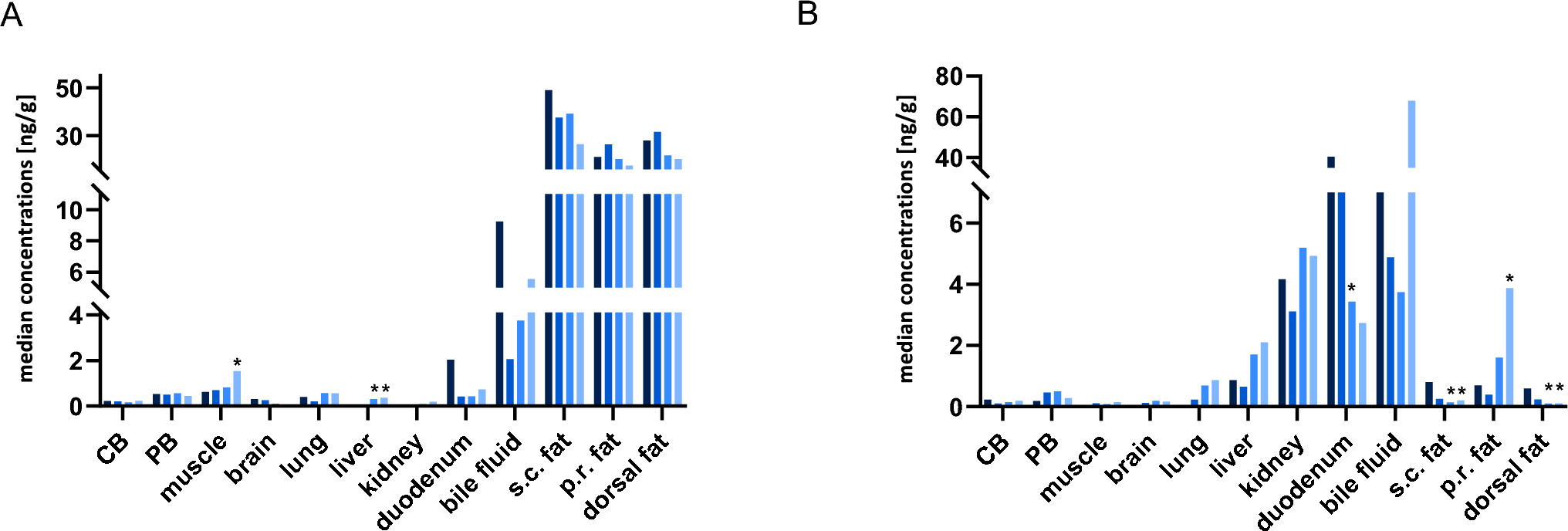
Animal euthanasia was conducted eight hours after the drug administration using T 61 (embutramide, 0.12 mL/kg BW, Intervet Deutschland GmbH, Unterschleißheim, Germany). Afterwards, the abdominal cavity was opened. Samples of the following organs, tissues and body fluids were collected (PMI 0) and stored at − 20 °C until further analysis: Brain (cerebrum), lungs and liver with no differentiation between the lobes, kidneys, muscle tissue (from the hindleg), adipose tissue (subcutaneous (sc), dorsal, perirenal), bile fluid, duodenum content, urine (only at PMI 0) and PB (V. jugularis) as well as CB.
The abdominal cavity was sutured leaving the organs in situ and the animal bodies were kept at room temperature in a supine position. Analogously, samples were taken again after 24, 48 and 72 h (PMI 1–3), respectively. Yet, PM PB specimens were obtained by sampling the coagulated blood from the V. femoralis or V. brachialis. For this purpose, the whole vessel was sampled and the blood was drawn therefrom using a pipette with a wide lumen.
Sample preparationTissue specimens and body fluids
Specimens were prepared according to a previous published method (Schaefer et al. 2017, 2019, 2020a) with changes regarding the applied buffers and the amount of acetonitrile. An amount of 2 g of solid tissue (brain, lung, liver, kidney and muscle tissue) was homogenized (1:5 w/w with water), respectively and 1 g of body fluids (bile fluid, duodenum content, and urine) was diluted (1:10 w/w for bile and duodenum content, 1:5 w/w for urine, respectively, with water). The samples were stored at − 20 °C.
To determine the standard addition calibration curves, four 0.5 g aliquots were added to 20 µL of an ethanolic stable-isotope-labeled internal standard solution (SIL-IS, 1 ng/20 µL AB-FUBINACA-d4) and 25 µL of ethanol or an ethanolic solution of the analytes.
Subsequently, the solution was mixed with 500 µL of acetate buffer and 50 µL of β-glucuronidase/arylsulfatase and incubated for 2 h at 60 °C to induce enzymatic hydrolysis of the glucuronidated DBA (phase-II metabolite).
For the following protein precipitation, the samples were mixed with 500 µL of acetonitrile and centrifuged at 3500g for 8 min. The supernatants were transferred to 1 mL phosphate buffer (pH 9) vortexed and centrifuged at 3500g for 8 min again.
Solid phase extraction (SPE) was carried out using Strata C18 end capped cartridges (Phenomenex, Aschaffenburg, Germany), previously conditioned with 2 × 3 mL methanol and 3 mL phosphate buffer. After loading the samples, the columns were washed with 3 mL phosphate buffer, 3 mL acetic acid (0.25 M) and 3 mL deionized water, respectively. 60 μL acetone was added and columns were dried for 5 min using negative pressure (about 33 kPa). Thereafter, the analytes were eluted with a mixture of 1.5 mL methanol-acetone (1:1, v/v) and the eluate was evaporated under a gentle stream of nitrogen at 60 °C. The dry residues were resuspended in 100 μL of a 1:1 (v/v) mixture of mobile phases A (0.1% aqueous formic acid) and B (0.1% formic acid in acetonitrile). 20 µL were injected for the analysis into the liquid-chromatography tandem-mass-spectrometry (LC–MS/MS) system.
Blood specimens
As the small amount of matrix did not allow for a standard addition method in PB, a previously validated method was applied for these samples (recovery ~ 75% and more, no relevant matrix effects, linear calibration with a weighting factor of 1/x2 for parent and 1/x for metabolite, calibration range 0.5 ng/mL-50 ng/mL (both analytes), limit of detection 0.05 ng/mL (both analytes), lower limit of quantification 0.5 ng/mL) (Walle et al. 2021). Briefly, 20 µL of a SIL-IS solution was mixed with 25 µL ethanol, 50 µL water and 50 µL blood. Precipitation was performed by adding 500 µL of acetonitrile and shaking for about 5 min. After centrifugation for 5 min at 12,000g, the supernatants were transferred to a new vial and gently evaporated under a nitrogen flow at 60 °C. The residues were reconstituted in 50 µL of a mixture of mobile phases A and B (1:1, v/v) and 20 µL were injected onto the LC–MS/MS system. The concentrations were quantified by a calibration.
Standard addition method
5F-MDMB-P7AICA and DBA were quantified in tissue and body fluid samples using the standard addition method. To determine the calibration curves, four 0.5 g aliquots were prepared: one native and three with addition of differently concentrated standard mixtures consisting of the two analytes (see Table 1). The analyte/SIL-IS area ratio was plotted against the calibrator concentration. The regression equations could be determined from the curves by the term y = a x + b. Calculation was performed using Microsoft Office Excel 2016 (Redmond, WA, USA). The unknown concentration corresponds to the intersection point of the axis of abscissa and results from the slope (a) and the point of intersection with the axis of ordinate (b) (Schaefer et al. 2020a).
Apparatus
LC–MS/MS conditions including the chromatographic, instrumentation, and mass spectrometric conditions were identical to a recently published study (Walle et al. 2021) and are listed in detail in the Supplementary material and Supplementary Table 2.
Statistical tests
For the evaluation of concentration changes over the time of observation, a non-parametric Friedman-test (p < 0.05) followed by a Dunn’s multiple comparison post hoc test was applied for each matrix. Calculations were performed using GraphPad Prism 9.0.1 (GraphPad Software, San Diego, CA, USA).

留言 (0)