
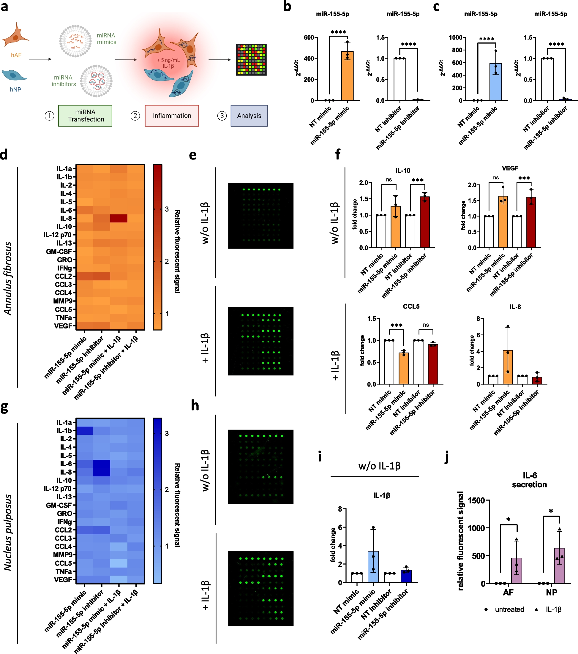
記住我


In a previous study we showed that knockdown of DGCR8, an indispensable component of miRNA biogenesis, results in abnormal cytokine secretion and the premature onset of cellular senescence in hMSCs [18, 24]. Considering the impact of miRNAs in hMSCs, we hypothesized that additional dysregulated functions are linked to global miRNA knockdown. This was explored by performing a GSEA for functional enrichment using our previously generated microarray profiling dataset with siGFP and siDGCR8-transfected hMSCs (GSE149171). Interestingly, the top 10 Gene Ontology (GO)_cellular components (GO_CCs) associated with DGCR8 knockdown were related to the actin filament bundle, actomyosin, contractile fiber, and cortical actin cytoskeleton (Fig. 1A). Specifically, a GO_biological process (GO_BP) related to the regulation of wound healing and including 127 genes was downregulated upon DGCR8 knockdown (Fig. 1B). These enrichment analyses suggested that DGCR8 knockdown correlates with cellular migration defects. This relationship was further examined in a Transwell migration assay, after confirming a significant reduction (0.47-fold) in the level of DGCR8 mRNA in siDGCR8-transfected hMSCs by quantitative real-time PCR (qRT-PCR) (Fig. S2A). Consistent with the enrichment results using GSEA, the migration of siDGCR8-transfected hMSCs was markedly reduced compared to siNC-transfected cells (0.69-fold, P < 0.01) (Fig. 1C).
Fig. 1
Overexpression of miR-29a-3p rescues the migration impairment caused by DGCR8 knockdown in hMSCs. (A) Bubble plot of the top 10 Gene Ontology (GO)_cellular components (CCs) significantly enriched in siDGCR8 vs. siGFP-transfected hMSCs by gene set enrichment analysis (GSEA). Each dot represents a GO_CC term; dot size and color indicate the number and false discovery rate (FDR), respectively. (B) A gene set of regulation of wound healing in siDGCR8 vs. siGFP-transfected hMSCs. Normalized enrichment scores (NES) and p-values are indicated. (C) Representative images of a Transwell migration assay. Scale bars, 100 μm. (D) Migrated cells were enumerated and statistically analyzed (error bars indicate the standard errors of mean of four experiments, **P < 0.01, ***P < 0.001 by Student’s two-tailed t-test). “siD8” indicates siRNA targeting DGCR8.
Next, we set out to identify the miRNAs that could ameliorate the migration defect in siDGCR8-transfected cells. Candidate miRNAs were identified after investigating the upstream regulators linked to DEGs (siGFP vs. siDGCR8, P < 0.05, 2,383 transcripts from GSE149171) in DGCR8-knockdown versus control hMSCs using IPA software. The top five diseases and cellular functions associated with DGCR8 knockdown included cell death and disease, gene expression, cellular assembly and organization, cellular function and maintenance, and the cell cycle (Table S3). Because cellular migration is closely related to cellular assembly and organization and to cellular function and maintenance, the upstream regulators of those functions were considered. In total, 30 miRNAs were identified based on statistical parameters (P < 0.0001). Among these candidates, miR-29a-3p was of particular interest, due to conflicting reports concerning its role in migration and invasion in cancer cells [25,26,27] and because its effects on hMSC migration are largely unknown. The ability of miR-29a-3p to ameliorate the migration defect in siDGCR8-transfected cells was therefore examined by overexpressing miR-29a-3p in DGCR8-knockdown hMSCs. Transfection of a miR-29a-3p mimic led to a 31-fold increase in the miR-29a-3p level in siDGCR8-transfected cells according to qRT-PCR (Fig. S2B). Notably, the overexpression of miR-29a-3p significantly enhanced migration by 2.2-fold compared to siDGCR8-transfected hMSCs (Fig. 1C, D, P < 0.001). Compared to siNC-transfected cells, which have normal migration, the overexpression of miR-29a-3p resulted in a 1.5-fold improvement in migration (Fig. 1D, P < 0.01). To confirm the specific effect of miR-29a-3p on the impaired migration of siDGCR8-transfected hMSCs, we introduced an LNA miRNA inhibitor to wild-type hMSCs to suppress miR-29a-3p. The miR-29a-3p level decreased 0.09-fold according to qRT-PCR, resulting in a 25% decrease in the migration of LNA-miR-29a-3p–transfected hMSCs (Fig. S3). Taken together, these results indicated that the global loss of miRNAs in hMSCs leads to defects in cellular migration and miR-29a-3p can rescue the impaired migration of DGCR8-knockdown hMSCs, thus identifying miR-29a-3p as a key regulator of hMSC migration.
Potential mRNA targets of miR-29a-3p in hMSCs exhibit enrichment in processes related to cellular assembly and organizationThe molecular mechanism by which miR-29a-3p enhances hMSC migration was investigated by performing target prediction analyses followed by experimental validation. Two miRNA target prediction tools and our previously published microarray gene profiling data of control and DGCR8-depleted hMSCs (GSE149171) were employed [18]. First, we compiled a list of putative miR-29a-3p targets using TargetScan v. 8.0 and the miRNA target filter in IPA software. This yielded 1,702 (miRNA target filter) and 1,265 (TargetScan v. 8.0) potential targets with a total of 2,146 non-redundant genes (Fig. 2A). In the context of mRNA expression profiles, we hypothesized that the levels of potential direct target mRNAs of miR-29a-3p would be upregulated in DGCR8-knockdown hMSCs compared to siGFP-transfected cells. Of those assessed, 1,117 genes were upregulated in siDGCR8-transfected hMSCs. We therefore focused on the 62 genes overlapping across all platforms to identify putative mRNA targets (Table S4). Pathway analysis using IPA revealed the top five molecular and cellular functions associated with these 62 genes (Table S5). Consistent with the impaired migration in DGCR8-knockdown cells, we found enrichment of functions related to cellular assembly and organization, including the orientation of the Golgi apparatus, formation of FAs, and formation of actin. Interestingly, the leading-edge subsets, which represent the core group of genes contributing the most to the enrichment signal in the GSEA, exhibited significant enrichment in several phosphatases (Fig. 2B). Because PTPRK and PTEN were predicted targets of miR-29a-3p and both phosphatases are negative regulators of cell migration, they were chosen for further investigation after confirming their upregulation in DGCR8-knockdown hMSCs at the mRNA (Fig. 2C, D) and protein (Fig. 2E, F) levels. To investigate whether miR-29a-3p influences the expression levels of PTPRK and PTEN in hMSCs, we overexpressed miR-29a-3p in a DGCR8-knockdown background. The PTPRK and PTEN mRNA (Fig. 2C, D) and protein levels (Fig. 2E, F) were downregulated upon miR-29a-3p overexpression. Next, we explored the effect of silencing PTEN and PTPRK on hMSC migration. Using siRNAs, we transiently silenced PTPRK and PTEN expression, which was confirmed by qRT-PCR and Western blotting (Fig. S4). Interestingly, knocking down PTPRK or PTEN enhanced the migration of hMSCs 1.7- and 1.3-fold, respectively (Fig. 2G, H), mimicking the effects of miR-29a-3p overexpression. In summary, we identified a subset of genes, including PTPRK and PTEN, as potential targets of miR-29a-3p; these genes were downregulated upon miR-29a-3p overexpression. Moreover, their knockdown enhanced cell migration.
Fig. 2
Putative mRNA targets of miR-29a-3p in hMSCs are enriched in processes associated with cellular assembly and organization. (A) Venn diagrams showing the numbers of putative targets according to miRNA target filter and TargetScan v. 8.0 and upregulated genes in DGCR8-knockdown cells. Sixty-two genes were identified by both target prediction algorithms and expression profiling (overlap in the Venn diagram). (B) Overlapping leading-edge genes in DGCR8-knockdown hMSCs. (C, D) mRNA levels of PTPRK (C) and PTEN (D) as determined by quantitative real-time PCR in DGCR8-knockdown hMSCs with or without miR-29a-3p overexpression. Data were normalized to the GAPDH mRNA level. (Error bars indicate standard errors of the mean of three and six experiments, *P < 0.05, **P < 0.01 by Student’s two-tailed t-test). (E, F) Immunoblot analysis of PTPRK (E) or PTEN (F) in DGCR8-knockdown cells with or without miR-29a-3p forced expression. GAPDH was used as the loading control. (G, H) Representative images of a Transwell migration assay in siPTPRK (G) and siPTEN (H) hMSCs. Scale bars, 100 μm. Migrated cells were quantified and statically analyzed (error bars indicate standard errors of the mean of four experiments, *P < 0.05, **P < 0.01 by Student’s two-tailed t-test)
miR-29a-3p enhances hMSC migration by directly suppressing PTPRK and PTENTo ascertain whether PTPRK and PTEN are direct targets of miR-29a-3p, we cloned the full 3’-UTR of PTPRK (1–1448) and the partial 3’-UTR of PTEN (504–855 or 1564–1913) downstream of the Renilla luciferase reporter gene. Subsequently, we cotransfected the reporter constructs with miR-29a-3p mimics into 293T cells (Fig. 3A, C). Compared to mock- and miR-control–transfected cells, luciferase activity significantly decreased 0.76-fold for PTPRK (Fig. 3B). For PTEN, the site 1 binding site (PTEN WT1) and site 2 binding site (PTEN WT2) were reduced 0.41- and 0.53-fold, respectively (Fig. 3D). Mutagenesis of the putative miRNA binding sites led to increased luciferase activity (Fig. 3B and D). In summary, our findings indicate that miR-29a-3p enhances hMSC migration in part by directly repressing PTPRK and/or PTEN.
Fig. 3
miR-29a-3p directly represses PTPRK and PTEN. (A, C) Schematic of miR-29a-3p binding sites in the 3’-UTR of PTPRK (A) and PTEN (C). Mutated nucleotides are underlined and highlighted in red. (B, D) Luciferase reporter assay. 293T cells were co-transfected with luciferase reporters carrying the wild-type or mutated 3′-UTR, as well as 50 nM negative control or miR-29a-3p mimics. Data were normalized to firefly luciferase expression. Values for mock-transfections set to 1 as denoted by the dashed line (error bars indicate standard errors of the mean of three experiments, *P < 0.05 and **P < 0.01 by Student’s two-tailed t-test)
miR-29a-3p regulates cell polarization and migration during wound healingTo determine how miR-29a-3p influences the stepwise process of hMSC migration at the cellular level, we first examined its role in cell polarization and cytoskeletal dynamics. In a wound healing assay, DGCR8 gene knockdown led to reduced wound closure. The decrease in migration was significantly restored upon miR-29-3p overexpression (Fig. 4A, B). The initial establishment of polarization and repositioning of the Golgi apparatus towards the leading edge are essential for cell migration during wound healing [28, 29]. Polarization of the Golgi apparatus was examined by GM130 staining, within a 120° sector facing the wound. Unlike control cells, most DGCR8-knockdown cells in the foremost row were unpolarized (Fig. 4C, depicted as “–”). However, the reintroduction of miR-29a-3p reorganized the Golgi apparatus. Specifically, 56.94% of siNC control cells exhibited a polarized Golgi, compared to only 31.77% of DGCR8-knockdown cells, while 64.98% of miR-29a-3p–overexpressing cells displayed polarization (Fig. 4D). These results demonstrated the crucial role of miR-29a-3p in restoring Golgi polarization in DGCR8-knockdwon cells. Cell polarization in response to wound healing also involves a reorganization of the actin cytoskeleton at the leading edge [30]. At 4 h post-wounding, actin fibers were organized perpendicular to the wound edge in both control and miR-29a-3p–overexpressing hMSCs (Fig. 4E, arrowhead indicated). By contrast, actin fibers in DGCR8-knockdown cells were predominantly oriented parallel to the wound edge. The percentage of cells at the wound edge with a polarized distribution of F-actin was 53.54% in control cells, 18.51% in DGCR8-knockdown cells, and 46.18% in miR-29a-3p overexpressing cells (Fig. 4F). These results showed that miR-29a-3p restores not only Golgi polarization but also the proper orientation of actin fibers, both of which are required for effective cell migration during wound healing.
Fig. 4
miR-29a-3p modulates hMSC polarization during wound healing. (A) Representative images of a wound healing assay of siNC, siDGCR8, and siDGCR8 overexpressing miR-29a-3p (scale bars, 500 μm). (B) The percentage of wound closure was calculated as follows: (A0 − A8)/A0. (A0 is the initial (0 h) wound area, and A8 is the wound area after 8 h). (siNC; n = 26 wells, siDGCR8; n = 26 wells, siDGCR8 + miR-29a-3p; n = 19 wells, ***P < 0.001 by Student’s two-tailed t-test). (C) Representative immunofluorescence images of Golgi (GM130, green) and nucleus (DAPI, blue) in hMSCs 4 h after wounding. (+) polarized; (–) non-polarized. Scale bars, 100 μm. (D) Percentage of polarized hMSCs at the wound edge (siNC; n = 53 cells, siDGCR8; n = 87 cells, siDGCR8 + miR-29a-3p; n = 84 cells, *P < 0.05 and **P < 0.01 by Student’s two-tailed t-test). (E) Representative immunofluorescence images of F-actin in hMSCs 4 h after wounding. White arrowheads indicate the orientation of actin fibers. Scale bars, 100 μm. (F) Percentage of hMSCs with actin fibers perpendicular to the wound (siNC; n = 49 cells, siDGCR8; n = 76 cells, siDGCR8 + miR-29a-3p; n = 98 cells, *P < 0.05 by Student’s two-tailed t-test). “siD8” indicates siRNA targeting DGCR8.
miR-29a-3p modulates actomyosin contractility and cellular traction forces in hMSCsHaving established the role of miR-29a-3p in cell polarization, we next investigated the impact of miR-29a-3p on actomyosin contractility and cellular traction, which are required for cell migration and reflect a balance between adhesion and contraction [28, 31, 32]. To address the impact of miR-29a-3p on contractility during cell migration, we monitored active pMLC (Thr18/Ser19). The level of pMLC2 was decreased in DGCR8-knockdown cells but was restored upon miR-29a-3p overexpression (Fig. 5A). Notably, F-actin and pMLC2 staining showed a significant non-overlapping pattern in DGCR8-knockdown cells (Fig. 5B). These abnormal distributions were reversed by miR-29a-3p, as indicated by the co-immunolocalization of pMLC2 with actin fibers and Pearson’s correlation coefficient (Fig. 5C). Because actomyosin-mediated contractility generates the mechanical stress necessary for cell migration [33] and pMLC2 immunofluorescence does not represent the magnitude and distribution of cellular traction forces, actomyosin contractility was quantified by measuring traction forces via TFM (Fig. 5D). Compared to control siNC-transfected cells, DGCR8-knockdown hMSCs exhibited a lower total traction force, consistent with the pMLC2 results (P < 0.05, Fig. 5D, E). Overexpression of miR-29a-3p in siDGCR8-transfected cells increased the contractile force to normal levels (P < 0.05, Fig. 5D, E). Therefore, miR-29a-3p influences actomyosin contractility and the generation of traction forces in hMSCs.
Fig. 5
miR-29a-3p regulates actomyosin contractility and cellular traction force in hMSCs. (A) Representative fluorescence images of F-actin (red) and pMLC2 (green). A higher-magnification image shows actin and pMLC2. Scale bars, 50 μm. (B) Intensity profiles of F-actin (red) and pMLC2 (green) in siNC or siDGCR8 hMSCs with or without miR-29a-3p overexpression. (C) Pearson’s correlation coefficients of F-actin and pMLC2 (siNC; n = 19 cells, siDGCR8; n = 22 cells, siDGCR8 + miR-29a-3p; n = 27 cells, **P < 0.01 and ***P < 0.001 by Student’s two-tailed t-test). “siD8” indicates siRNA targeting DGCR8. (D) Representative images and stress maps by traction force microscopy of DGCR8-knockdown cells with or without miR-29a-3p forced expression. Scale bars, 50 μm. Force scale bar is in Pascals (Pa). (E) Total traction force of control and DGCR8-knockdown cells with or without miR-29a-3p overexpression. Central box, first to third quartile; middle line, median. (siNC; n = 20 cells, siDGCR8; n = 17 cells, siDGCR8 + miR-29a-3p; n = 17 cells, *P < 0.05 by Student’s two-tailed t-test). (F) Representative images show a Transwell migration assay in siDGCR8 hMSCs overexpressing miR-29a-3p and treated with the non-muscle myosin II inhibitor blebbistatin (blebb) (10 µM), the actin polymerization inhibitor latrunculin A (Lat-A) (0.1 µM), the Rho-associated protein kinase (ROCK) inhibitor Y-27632 (10 µM), or the myosin light chain kinase inhibitor ML-7 (10 µM). Scale bars represent 100 μm. Migrated cells were quantified and statistically analyzed (error bars indicate standard errors of the mean from four experiments, **P < 0.01, ***P < 0.001 by Student’s two-tailed t-test)
The mechanisms underlying the activity of miR-29a-3p during cellular migration were investigated using the pharmacological inhibitors blebbistatin (blebb, a non-muscle myosin II inhibitor), latrunculin A (Lat-A, an actin polymerization inhibitor), Y-27632 (a Rho-associated, coiled-coil containing protein kinase (ROCK) inhibitor), and ML-7 (a myosin light chain kinase inhibitor). Despite miR-29a-3p overexpression in a DGCR8 knockdown background, cell migration was significantly inhibited by blebb, Lat-A, and ML-7, but not by Y-27632 (Fig. 5F). This result suggests that the promotion of cellular migration by miR-29a-3p is dependent on the actin cytoskeleton and pMLC2.
miR-29a-3p modulates focal adhesion and actin cytoskeleton in hMSCsBuilding on our findings regarding actomyosin contractility, we focused on the role of miR-29a-3p in regulating FAs and the actin cytoskeleton, both of which are also critical for cellular migration [32]. Compared to siNC-transfected hMSCs, the expression of rhodamine phalloidin, an actin cytoskeleton marker, and of paxillin, a FA marker, were reduced in siDGCR8-transfected cells. In siNC-transfected control cells, paxillin showed typical elongated staining aligned with F-actin at the peripheral region (Fig. 6A). However, in DGCR8-knockdown hMSCs, paxillin was smaller, more dispersed, and randomly distributed throughout the cell. Overexpression of miR-29a-3p in siDGCR8-transfected cells fully restored the intensity and distribution of paxillin and F-actin (Fig. 6A, see enlarged images). The absolute number of FAs per cell and their size were significantly reduced in DGCR8-knockdown hMSCs compared to the control. This reduction was reversed by miR-29a-3p overexpression (Fig. 6B, C). FA morphology was then characterized by quantifying the proportion of enlarged FAs per cell and their aspect ratios (the ratio of the major and minor axes). Consistent with the abnormal FAs in DGCR8-knockdown cells, the percentages of enlarged FAs and their aspect ratios were significantly decreased. These aberrations were reversed by miR-29a-3p (Fig. 6D, E). Paxillin is an FA-associated adaptor protein whose function and localization are regulated by phosphorylation at multiple Tyr and Ser residues [34]. We therefore examined the phosphorylation of paxillin (pY118-paxillin), an essential modification for cell migration, using a phospho-specific pY118-paxillin antibody. miR-29a-3p-overexpression siDGCR8 hMSCs exhibited a significant increase in pY118-paxillin (Fig. 6F). Because miR-29a-3p directly represses the expression of PTPRK and PTEN phosphatases, we explored the effect of PTPRK and PTEN knockdown on paxillin and pY118-paxillin levels. Interestingly, PTPRK knockdown in hMSCs increased the level of pY118-paxillin, whereas PTEN knockdown by siRNA reduced the levels of paxillin and pY118-paxillin (Fig. 6G). Next, we examined the interactions between endogenous paxillin and PTPRK by co-immunoprecipitation; paxillin co-precipitated with PTPRK (Fig. 6H). These results indicate that miR-29a-3p regulates FAs during cell migration, at least in part by modulating PTPRK and paxillin.
Fig. 6
miR-29a-3p modulates focal adhesion formation and maturation by regulating PTPRK and paxillin in hMSCs. (A) Representative immunofluorescence images of actin cytoskeleton (F-actin, red) and focal adhesions (paxillin, green) in siNC or siDGCR8 hMSCs with or without miR-29a-3p overexpression. A higher-magnification image shows actin and paxillin. Scale bars, 50 μm. (B) Number of focal adhesions per cell, (C) average size of focal adhesions per cell, (D) percentage of cells with enlarged focal adhesions (greater than the median focal adhesion size), and (E) aspect ratio of focal adhesions (siNC; n = 21 cells, siDGCR8; n = 35 cells, siDGCR8 + miR-29a-3p; n = 24 cells; lines, medians, **P < 0.01 and #P < 0.0001 by Student’s two-tailed t-test). (F) Western blotting of the levels of paxillin and pY118-paxillin (p-Paxillin) in siNC or siDGCR8 hMSCs with or without miR-29a-3p overexpression. GAPDH is the loading control. (G) Western blotting of the levels of paxillin and p-Paxillin in siNC, siPTPRK, and siPTEN hMSCs. (H) Co-immunoprecipitation of paxillin and PTPRK in immortalized hMSCs. Immunoprecipitation was performed using an anti-paxillin antibody, and precipitated proteins were examined by western blotting using anti-PTPRK and anti-paxillin antibodies
miR-29a-3p enhances hMSC migration in vivoGiven that miR-29a-3p influences nearly every key stage of cellular migration in vitro, its function in vivo was investigated after a Transwell migration assay confirmed that its overexpression in wild-type immortalized hMSCs (Fig. S5A) enhances cell migration towards SDF-1α, a chemokine released during inflammation or injury [23, 35, 36] (Fig. 7A). This finding was further supported by a collagen gel contraction assay, which provides insights into interactions between cells and the extracellular matrix (ECM) in 3D environments [37]. Replenishment of miR-29a-3p in hMSCs significantly increased their ability to contract collagen-rich gels (Fig. 7B), consistent with the observed increase in pMLC2 in hMSCs due to miR-29a-3p overexpression in vitro (Fig. 5). The enhanced migration of hMSCs caused by miR-29a-3p overexpression in vivo was demonstrated in an SDF-1α-releasing Matrigel plug assay in immunocompromised mice, creating a chemoattractant gradient to attract hMSC. Matrigel plugs, with or without SDF-1α, were subcutaneously implanted into the right and left sides of NOD/SCID mice, respectively [22]. Qtracker™ 800-labeled miR-29a-3p overexpressing cells and miR-NC-transfected cells were subcutaneously injected at the center and tracked using the IVIS Spectrum Imaging System (Fig. 7C). Before injection, the similar viabilities of control mimic- and miR-29a-3p–transfected hMSCs was confirmed (Fig. S5B). The mice were imaged by IVIS at 0 h to identify labeled hMSCs and at 24 and 46 h to detect MSC migration towards the Matrigel plugs. Compared to control miR-NC-transfected cells, which exhibited marginal migration towards the Matrigel plug with SDF-1α (Fig. 7D, top), miR-29a-3p-overexpression hMSCs showed significantly increased migration (Fig. 7E, top). Fluorescence intensity profiles indicated that miR-29a-3p enhanced the in vivo migration of hMSCs (Fig. 7D, E, bottom). Cell migration was enhanced to the greatest degree in miR-29a-3p-overexpression hMSCs at 46 h post-injection (Fig. 7F). Therefore, miR-29a-3p enhances cell migration in vitro and in vivo.
Fig. 7
miR-29a-3p enhances hMSC migration in vivo. (A) Representative images of a Transwell migration assay of hMSCs with control or miR-29a-3p overexpression. SDF-1α was used as the chemoattractant. Scale bars, 100 μm. Migrated cells were enumerated and statistically analyzed (error bars indicate standard errors of the mean of four experiments, *P < 0.05 by Student’s two-tailed t-test). (B) Gel contraction by miR-NC and miR-29a-3p overexpression hMSCs (error bars indicate standard errors of the mean of three experiments, *P < 0.05 by Student’s two-tailed t-test). (C) Schematic of subcutaneously implanted Matrigel plugs and injected cells in immunocompromised mice. (D, E) (Top) Qtracker™ 800-labeled hMSCs with or without miR-29a-3p overexpression were traced in vivo using the IVIS Spectrum In Vivo Imaging System at 0, 24, and 46 h. (Bottom) Fluorescence intensity profiles, along with injection sites. (F) Labeled cell distribution at the indicated time points (error bars indicate standard errors of the mean of two or three experiments, mice, n = 3/group, miR-29a-3p group at 46 h, n = 2). (G) A working model of the orchestration of cellular migration by miR-29a-3p, encompassing modulation of the polarization, adhesion, and contractility of hMSCs.
留言 (0)