Bioinformatics analysis
We downloaded the dataset GSE48397 from the GEO database, which includes 5 samples of normal lung fibroblasts and 5 samples of CAFs. The data were corrected and normalized using the Limma package, and a heatmap was generated using the ggplot2 package. Additionally, ChIP-seq data for PRRX1 in CAFs were downloaded from the GEO database. MACS2 was employed to analyze PRRX1’s ChIP-seq data, and call4peaking was used to assess the binding of PRRX1 to the CAF genome.
Clinical sample collection
We collected tumor tissue samples from 48 cases of lung cancer from January 2019 to June 2022 in the oncology and respiratory departments of our hospital. These samples were routinely dehydrated and embedded in paraffin for immunohistochemistry (IHC) analysis. Clinical information for all enrolled patients was obtained from our hospital's Health Information System (HIS), including gender, age, staging, pathological features, smoking status, treatment plans, clinical outcomes, and more [24].
Inclusion criteria
1.
Patients aged 18 years and older.
2.
Confirmed diagnosis of lung adenocarcinoma through histopathological examination.
3.
Staged as IV according to the AJCC Eighth Edition Lung Cancer TNM Staging.
4.
Provision of approved histopathological samples.
5.
Met one or more of the following criteria: Eastern Cooperative Oncology Group (ECOG) PS score of 0 or 1.
6.
Sufficient tumor tissue available for PD-L1 expression analysis.
7.
At least one record of PD-1/PD-L1 treatment.
Exclusion criteria
1.
Patients with sensitive gene mutations (EGFR, ALK, ROS1).
2.
Patients who have already received immune checkpoint inhibitor therapy (e.g., PD-1, PD-L1, CTLA-4, etc.).
3.
Patients with known autoimmune diseases, such as systemic lupus erythematosus, rheumatoid arthritis, scleroderma, etc.
4.
Currently undergoing treatment with other experimental drugs.
5.
Patients with severe liver, kidney, or heart diseases.
6.
Other situations that do not meet the study requirements.
Clinical Treatment Response Evaluation: Clinical treatment response was assessed using modified Response Evaluation Criteria in Solid Tumors (mRECIST) criteria, categorized as follows: Complete Remission (CR), Partial Remission (PR),Stable Disease (SD), Progressive Disease (PD). Progression-Free Survival (PFS): PFS is defined as the time from the start of treatment to disease progression or death from any cause. The first treatment response evaluation was conducted after two cycles of treatment, with subsequent evaluations performed every two treatment cycles. Patients who achieved CR, PR, or SD for at least 4 weeks after receiving immunotherapy were classified as responders, while patients with disease progression were classified as non-responders.
Immunohistochemistry
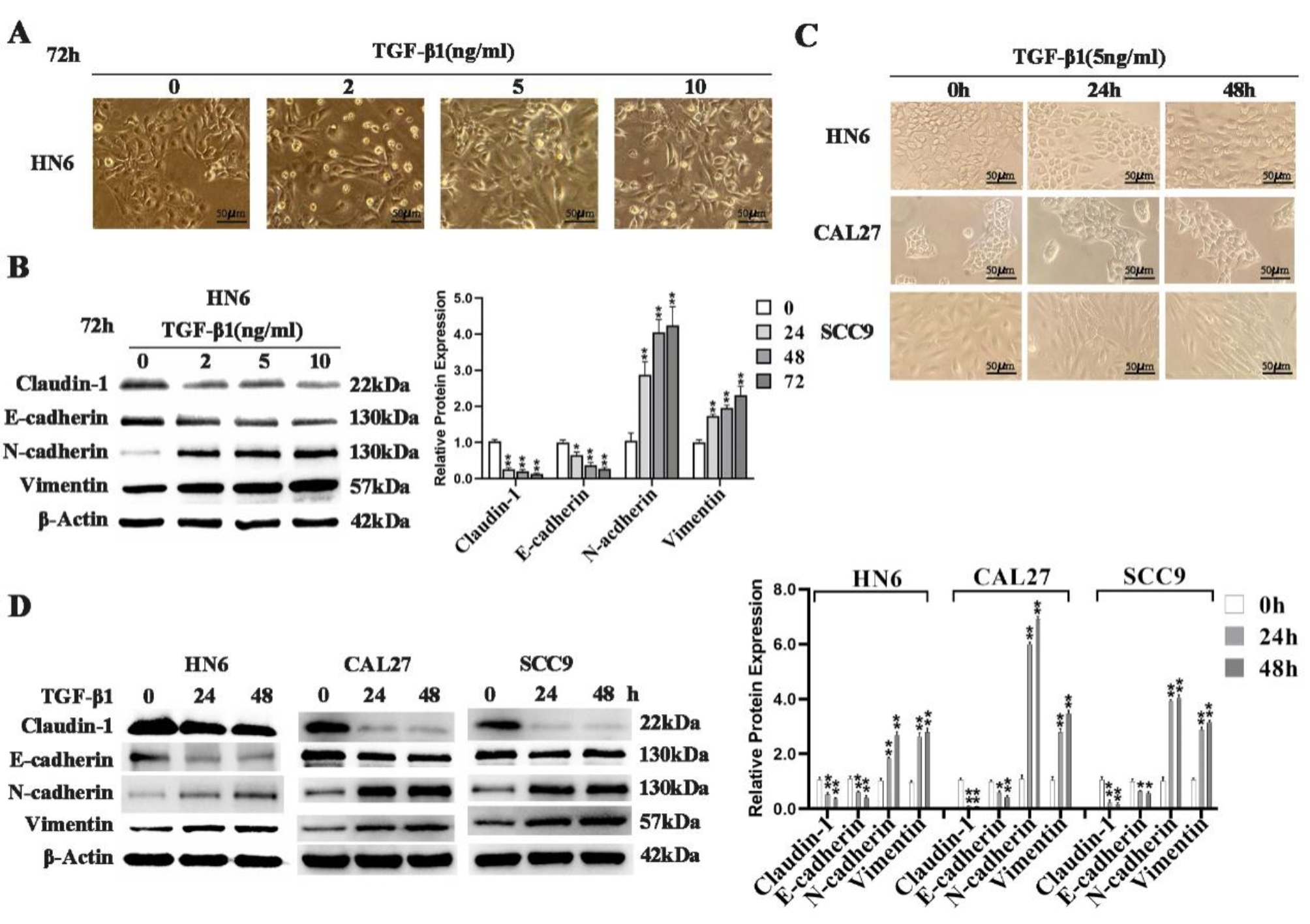
Paraffin-embedded sections of lung adenocarcinoma tissue were used for immunohistochemical staining. The sections underwent deparaffinization in xylene, followed by gradual ethanol dehydration. They were then rinsed three times in PBS for 5 min each time. To block endogenous peroxidase activity, the sections were immersed in a 3% hydrogen peroxide solution. After another rinse with PBS, antigen retrieval was performed in 98 °C citrate solution for 30–60 s, followed by PBS washing. Primary antibodies, including OLR1 (ab232837, 1:200), PRRX1 (ab20034, 1:500), and PD-L1, were applied and incubated overnight at 4 °C. After PBS washing, secondary antibodies were added, and the sections were incubated at room temperature for 40 min. Following another PBS wash, DAB staining was performed. Counterstaining was done with hematoxylin, and sections were dehydrated in a graded ethanol series, cleared in xylene, and mounted with neutral resin for observation under an optical microscope. Data collection was based on the percentage of stained cells and staining intensity. OLR1 was considered positive when localized in the cytoplasm of tumor cells, occasionally with positive membrane expression. Positivity was determined by the presence of brownish-yellow granules in the cytoplasm and/or cell membrane. PRRX1 was considered positive when nuclear staining exhibited yellow or light brown granules. For each slide, five high-power fields with a higher number of stained cells were chosen, and the absolute quantity of PRRX1-positive cells in the tumor stroma was determined by averaging cell counts. Protein expression of OLR1 or PRRX1 was evaluated using the H-score system. “Pi” represents the percentage of cells at a particular intensity level “i,” with values ranging from 0 to 100%. “I” represents the staining intensity, with values of 0 (no staining evidence), 1 (weak staining), 2 (moderate staining), and 3 (strong staining). The final H-score was calculated by summing the products of “I” and “Pi” (H-score = (0P0) + (1P1) + (2P2) + (3P3)), with a range of scores from 0 to 300.
Isolation and extraction of CAFs
CAFs were isolated from fresh LUAD tissues. After surgical resection, samples were collected in tissue storage buffer (Miltenyi) and washed in phosphate-buffered saline (PBS) containing 1% antibiotics-antimycotics (Gibco, Life Technologies). Tissues were then minced into small pieces (1–2 mm) and digested using the Human Tumor Dissociation Kit and gentleMACS Octo Dissociator following the manufacturer's instructions. The digested samples were sequentially filtered through a 70-micron cell strainer. Cells were collected by centrifugation at 250 g for 5 min and cultured in Dulbecco’s Modified Eagle Medium (DMEM) (Gibco, Carlsbad, CA, USA) supplemented with 10% fetal bovine serum (FBS) and 1% antibiotics-antimycotics at 37 °C. The culture medium was changed every 3 days. Primary CAFs were characterized as negative for EpCAM, CD45, and CD31, and positive for FAP and α-SMA. To knock down or overexpress specific genes, primary fibroblasts were transduced with lentiviral vectors [multiplicity of infection (MOI) of 100] at 37 °C with 5 μg mL−1 polybrene (Sigma). The targeting sequences of each shRNA are provided in Table 1.
Immunofluorescence
Cells were plated on sterile glass slides and cultured for 48 h. Subsequently, cells were fixed with 4% paraformaldehyde for 15 min, permeabilized with 0.25% Triton-X 100 for 10 min, and then blocked with 3% BSA for 1 h. Immunofluorescence staining was performed using antibodies against Vimentin, α-SMA, FSP1, F-actin, and Fibronectin (dilution 1:100, Abcam). After washing with phosphate-buffered saline (PBS), cells were incubated with Alexa Fluor 488 or Alexa Fluor 546 secondary antibodies (Life Technologies). Cell nuclei were stained with DAPI, and observations were made using a FV-1000 laser scanning confocal microscope. Quantitative analysis was performed on cells from at least 300 cells in three independent experiments.
qPCR
RNA was extracted from CAFs or LC cells using the RNAeasyTM Plus Animal RNA Isolation Kit with Spin Column (Beyotime, Jiangsu, China). cDNA synthesis was performed using SuperScript III (Takara, Dalian, China) following the manufacturer's protocol. Real-time PCR analysis was carried out using the Applied Biosystems 7500 Real-Time PCR System, as per the manufacturer's instructions. Reactions were performed for three independent experiments, and the results were normalized to β-actin. The primer sequences used can be found in Table 2. The mean ± SD of three independent experiments is presented.
Western blot
Total protein was extracted using cold radioimmunoprecipitation assay (RIPA) buffer containing protease and phosphatase inhibitors. Cell lysates were separated by 8–10% SDS-PAGE and transferred to polyvinylidene fluoride (PVDF) membranes. Rabbit or mouse IgG antibodies conjugated with horseradish peroxidase (Santa Cruz) were used as secondary antibodies. Protein bands labeled on the membranes with antibodies were detected using the G-BOX iChemi XT instrument (Syngene).
Co-culture
Co-culture experiments were established using Transwell membranes with a pore size of 0.4 µm (Merck Millipore, USA) in 12-well plates. Co-culture was conducted for 3–6 days, with LC cells (5 × 10⁴ cells) seeded on the permeable membrane, and CAFs cells (initially 5 × 10⁴ cells) prepared for further cell-based experiments beneath the membrane.
CCK-8 assay
Cell viability was assessed using the Cell Counting Kit-8 (CCK-8) assay kit (Sigma) following the manufacturer’s instructions. LC cells were digested with trypsin, counted, and then seeded in 96-well plates at a density of 2 × 10³ cells per well. Cells were cultured at 37 °C. Adherent cells were incubated with CCK-8 dilution for 1 h, followed by measuring the absorbance at a wavelength of 450 nm for each well.
SA-β-gal assay
Senescence-associated β-Galactosidase (SA-β-Gal) activity was measured using the senescence-associated β-Galactosidase staining kit (Beyotime, Shanghai, China) according to the manufacturer's instructions. Briefly, cells grown in 6-well plates were first washed and fixed for 15 min at room temperature, then incubated overnight at 37 °C with SA-β-gal working solution. For xenograft tumor tissues, frozen tissues were cut into 4 μm sections using a cryostat and mounted on glass slides with a positive charge. The sections were then fixed and stained as described above. SA-β-Gal-positive cells, appearing blue (light or dark blue), were determined as the percentage of stained cells from five random fields relative to the total cell count.
Matrigel invasion assay
The Matrigel invasion assay was performed in 24-well Transwell culture plates. In brief, 40 μL of Matrigel (1 mg/mL, BD) was coated on 8 μm polycarbonate membrane filters. Approximately 3 × 10⁴ H1299 or A549 cells were re-suspended and then seeded in the upper chambers containing FBS-free culture medium and the lower chambers containing complete growth medium with 10% FBS, respectively. Cells were incubated at 37 °C for 24 or 48 h. Non-invasive cells on the upper side of the invasion membrane were removed, and cells on the lower surface were stained with hematoxylin. The average number of cells in each field was determined by counting cells in six random fields per well. Cells were counted in four different fields from three independent experiments.
ChIP-qPCR
ChIP experiments were performed using the EZ-Magna ChIP kit (Millipore, Billerica, MA, USA). Approximately 5 × 10⁶ cells were fixed with 1% formaldehyde (final concentration) at room temperature for 10 min. Fixation was stopped by adding 1/10 volume of 1.25 M glycine and incubating at room temperature for 5 min. The sonication step was carried out in a Covaris sonicator (5 min, 20 s on, 20 s off) with 200 µg of protein-chromatin complexes used for each immunoprecipitation. Antibody-protein complexes were captured using pre-blocked dynabeads protein G (Invitrogen). qPCR analysis was performed using SYBR Green (Takara) on an ABI-7500 instrument (Applied Biosystems), with primers specified in Table 3. The antibodies used included H3K27ac, H3K4me3, PRRX1 (CST), and normal mouse IgG (Millipore).
Table 3 ChIP-qPCR primersDual-luciferase reporter assay
Transcriptional activity was analyzed using the dual-luciferase reporter gene assay according to the manufacturer’s instructions (Promega Corporation, Madison, WI, USA). Reporter gene expression was measured and quantified using the Dual-Luciferase Reporter Assay kit (Promega). Luciferase activity was normalized to Renilla luciferase control activity.
Animal experiments
For in vivo metastasis experiments, each experimental group included 6–8 male C57BL/6 J mice aged 6 weeks. In brief, H1299 cells were mixed with CAFs cells stably expressing low levels of OLR1 in a 1:1 ratio, suspended in 40 μL of serum-free DMEM, and then injected into the tail veins of mice. Starting from the second week, mice were treated with anti-PD-1 therapy (50 mg/kg/week) for 8 weeks, after which the mice were euthanized. Tumor nodules formed on the lung surface were macroscopically identified and counted. Lung tissues were then excised and embedded in paraffin for HE staining to analyze the number of lung nodules, and flow cytometry was used to assess the proportion of immune cell infiltration in the nodules.
ELISA
Analysis was performed using mouse IL-6, IL-8, IFNg, CSF1 (Absion), and TNF-alpha, TGF-β quantikine ELISA kits (Novus, China) according to the manufacturer’s instructions. In brief, 50 μL of samples and standards were added to wells of a microplate. Then, 100 μL of assay reagent was added to each assay well. After incubation for 20 min, absorbance at 450 nm was measured using an ELISA microplate reader.
Statistical analysis
Data analysis was performed using SPSS 21.0 (SPSS, Inc., Chicago, IL, USA) statistical software. The data were assessed for normality using the Kolmogorov–Smirnov test, and as the data followed a normal distribution, results are presented as mean ± standard deviation. Comparisons between two groups were conducted using the t-test, while comparisons among multiple groups were analyzed using One-Way ANOVA (analysis of variance). Post hoc pairwise comparisons following ANOVA were conducted using Tukey's multiple comparisons test. Survival curves were generated using the Kaplan–Meier method, and statistical significance was assessed using the Log-rank test. All tests were two-tailed, and a p-value less than 0.05 was considered statistically significant.


留言 (0)