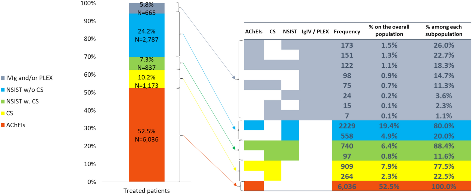
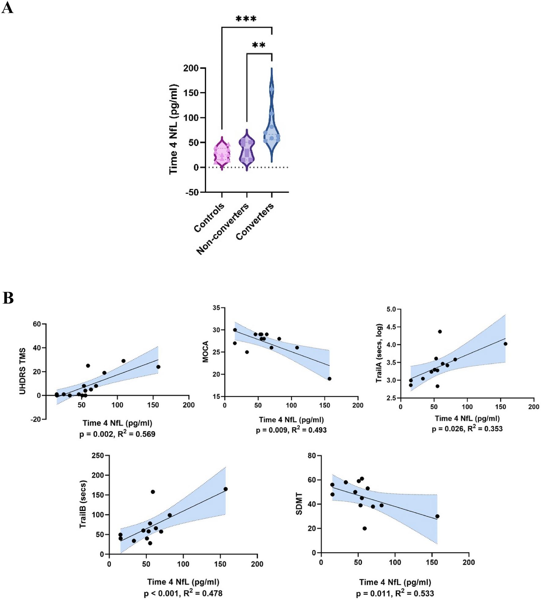
In the present study, we explored in vivo glymphatic dysfunction in a cohort of PD-MCI patients to predict the dementia conversion during the follow-up period. We demonstrated that PD patients with MCI who progressed to dementia exhibited reduced glymphatic activity and enlarged PVS in the basal ganglia in comparison to those nonconverters, and the glymphatic dysfunction was associated with enlarged PVS volume fraction, global cognitive performance, and executive function. Furthermore, the DTI-ALPS index can well identify PDD converters from nonconverters in patients with PD and MCI. Therefore, glymphatic impairment may contribute to the conversion from MCI to dementia in PD, and is likely to be a promising indicator to assist in the dementia prediction and potential neuroprotective strategy for slowing the cognitive deteriorative process in PD.
The assessment of glymphatic function in vivo has been hampered by the lack of direct imaging measures not requiring the injection of contrast agent. To overcome this obstacle, DTI-ALPS index was recently introduced to assess diffusivity along the perivascular space in vivo and hence, as measure of brain glymphatic function. Researchers reported that there is a negative correlation between α-synuclein deposition and AQP4 expression in the brain of PD patients, indicating a link between glymphatic dysfunction and pathologic protein accumulation [25]. This also supports the hypothesis of “prion-like propagation”, where a drainage system like the glymphatic system helps clear the accumulation of toxic proteins or, in the case of glymphatic system failure could contribute to the neurodegeneration and brain pathology [26]. The development of dementia in PD is thought to be a culminating result of the a heavy burden of α-synuclein pathology in limbic and neocortical structures [27], indicating that dementia is related to a more severe glymphatic impairment. Previous studies have also identified the relationship between cognitive impairment and glymphatic dysfunction in schizophrenia [28], AD [29], frontotemporal dementia [30], and PD [14]. Therefore, in accordance with prior studies, our findings also found the reduced glymphatic activity in the patients with MCI who develop dementia and cognitive deterioration. Considering the elevated incidence of dementia in PD patients, a multitude of researchers have been dedicated to identify MRI biomarkers to be indicative of cognitive deterioration in PD in order to facilitate an early identification and prevention. For example, researchers have proposed using cortical thickness on T1WI [31], and white matter structural connectivity on DTI [32] as predictors for PD cognitive progression. Hence, glymphatic dysfunction, assessed by DTI-ALPS, could be used as an additional MRI indicator for predicting cognitive deterioration, and also as a potential supplement for the intervention of cognitive function in patients with PD.
In our study, PDD converters were associated with glymphatic dysfunction in the left hemisphere, but not in the right hemisphere. One possible explanation could be that glymphatic impairments begins in the left hemisphere for right-handed patients as evidenced by motor asymmetry in PD [33, 34]. In line with our study, previous research identified abnormalities in the left-hemispheric DTI-ALPS index in PD [19]. However, Qin et al. investigated the correlation between glymphatic function and motor symptoms, suggesting the right hemispheric DTI-ALPS index was lower than the left side [21]. Therefore, the debate regarding whether PD patients exhibited more pronounced glymphatic impairment in the left hemisphere persists, necessitating further investigation with subgroup analysis. Apart from the DTI-ALPS index in the left hemisphere, PDD converters also exhibited higher diffusivity along the y-axis in the projection neural fibers and diffusivity along the z-axis in the association neural fibers in the left hemisphere. Since diffusivity in the y-axis and z-axis does not align with the perivascular water flow, therefore, the different diffusivity in the projection and association fibers could be attributed to white matter degeneration due to cognitive deterioration in the projection or association fibers as indicated in previous studies [35, 36].
The perivascular space is a region that surrounds arterioles, capillaries, and venules in the brain, and is considered as a part of glymphatic system both structurally and functionally [37]. Animal studies reported diminished glymphatic influx and heightened perivascular α-syn aggregation, as well as blocked glymphatic efflux and increased deposition of α-syn and worsened PD pathology [6].Therefore, glymphatic dysfunction may contribute to the dilation of PVS, which in turn lead to the aggregation and accumulation of misfolded proteins.
Conversely, an elevated PVS burden could further exacerbate the glymphatic dysfunction. Among the three classic locations of PVS burden according to previous reports, only PVS burden in the basal ganglion is significantly enlarged, representing a heavy neurovascular burden in the BG region linked to cognitive progression. Our findings align with recent research showing an elevated presence of PVS in PD patients, with EPVS in the BG region likely more representative [12, 17, 38]. Researchers have proposed that dementia is more strongly associated with visible PVS in the BG than with PVS at other sites [37]. Furthermore, functional and structural alterations in the BG region have been related to the progression of PD pathology and its clinical manifestations [39, 40]. As a result, the heavy vascular burden in BG is linked to cognitive deterioration in patients with PD and MCI.
Besides, both DTI-ALPS index and BG-PVS showed significant correlation with global cognitive performance and the executive function. In line with our findings, DTI-ALPS index and EPVS in BG were reported to be closely related to cognitive function, information processing, as well as executive function [21, 41, 42].Nevertheless, several reports are in contrast to our findings, reporting no statistical correlation between BG-EPVS and cognition [10]. The difference in the definition of the PVS score may be one of the contributing factors. PVS scoring based on the number of EPVS may result in a higher rate of severe vascular burden. Despite the significant correlations between DTI-ALPS, BG-EPVS, and cognitive function, our study did not find statistical mediation effect of BG-EPVS on the relationship between DTI-ALPS and cognitive function. One possible explanation could be that the EPVS in the BG serves as an independent factor influencing cognition [41]. It is closely associated with lacunar infarcts and periventricular white matter hyperintensities, which can directly impact cognitive and executive function by interrupting the prefrontal subcortical circuit [43]. In contrast to our findings, a recent study found that DTI-ALPS acted as a mediator in the relationship between white matter hyperintensities and cognitive performance [44]. Therefore, the potential causal relationship between EPVS, glymphatic dysfunction, and cognition needs further investigation.
This study had several limitations. First, the current study had a small sample size in terms of longitudinal follow-up time. Future studies with a larger sample size and a longer follow-up period are needed to confirm our results. Second, although correlations between glymphatic dysfunction and cognitive deterioration over time were initially established, the causal effect between glymphatic impairment and cognitive conversion during period time was not determined in the current study. Third, the ROIs for the DTI-ALPS calculations were manually delineated, and the PVS was defined by fractional volume in the three specified regions. Despite having perfect inter-observer and intra-observer reliability, utilizing automated or semi-automated delineation methods could reduce potential bias and improve the clinical utility. In addition, future studies are needed to explore the influence of additional factors, such as white matter hyperintensities, on the DTI-ALPS index and cognitive decline in PD patients. Despite the limitations mentioned above, the DTI-ALPS index, as a non-invasive imaging method, still holds potential for investigating glymphatic system function in vivo, and therefore, serves as imaging indicator in forecasting progress to dementia in PD-MCI stage.
Our study found a lower DTI-ALPS in patients with PD and MCI who develop dementia, indicating potential dysfunction in the glymphatic system. Besides, DTI-ALPS index was correlated with enlarged BG-PVS and global cognitive performance, as well as executive function. Therefore, the DTI-ALPS may assist in identifying PD with MCI at a high risk of cognitive deterioration before the onset of dementia, thereby providing potential therapeutic strategies.


留言 (0)