Gerbode and colleagues first described a case series of successful surgical closure for an LV–RA defect [2]. This defect is a rare intracardiac abnormality caused by a deficiency in the membranous ventricular septum that separates the LV from the RA. Gerbode defect is usually congenital, but can also be acquired as a complication of IE, myocardial infarction, blunt chest trauma, or previous cardiac surgery [3]. In cases associated with IE, the infection often extends into the aortic subannular region, involving the high membranous septum. This leads to the rupture of the portion of the septum and results in an LV to RA shunt with an intact tricuspid valve [4].
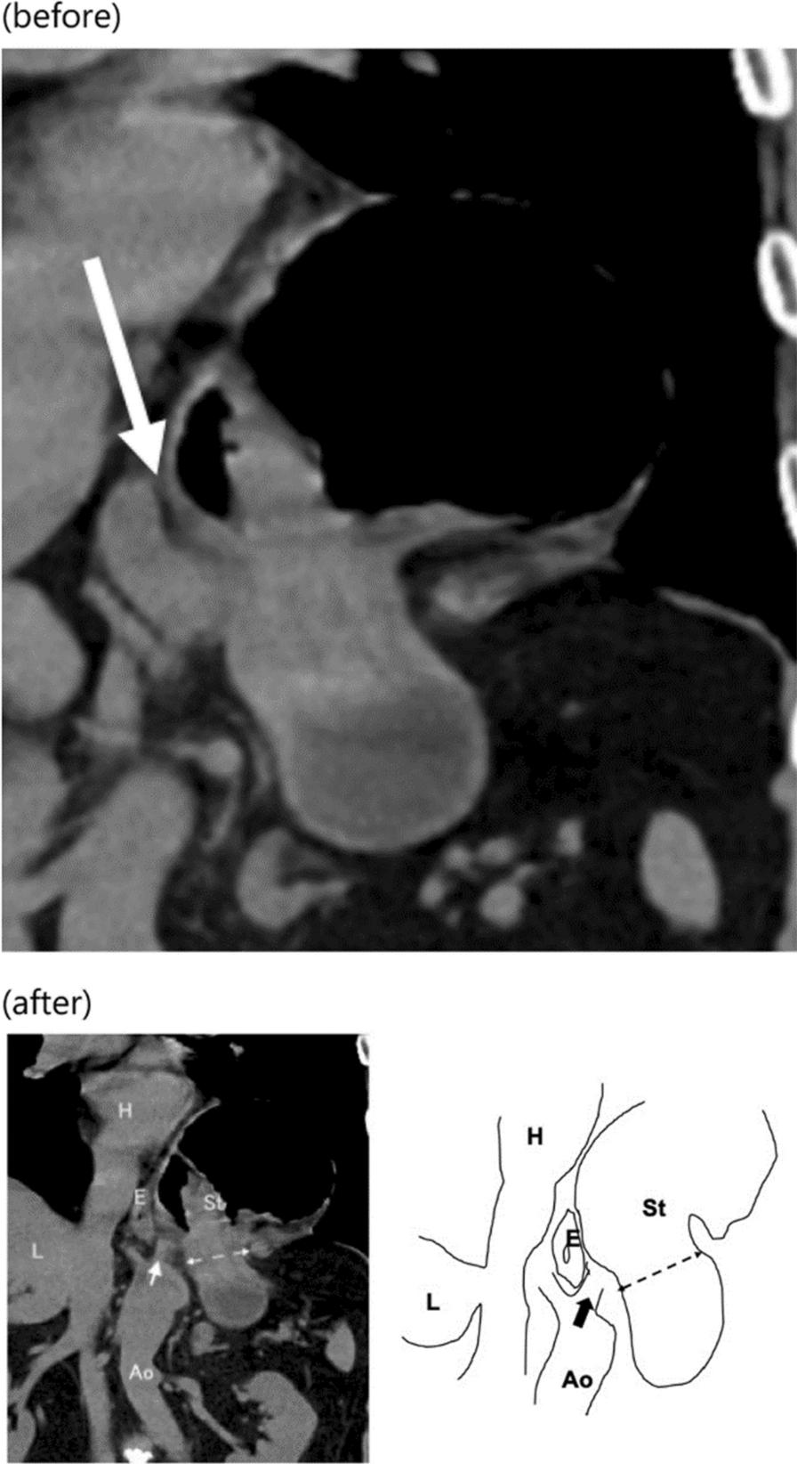
Compared with the more stable congenital Gerbode defects, acute fistulation between the LV and the RA associated with IE is a life-threatening complication that often requires urgent definitive intervention [5]. However, the identification of actual communication is often extremely difficult. Small acquired Gerbode defects are usually asymptomatic and can be easily missed in TTE. Even in our case, the defect could not be detected by TTE. TEE has been demonstrated to be superior to TTE in the detection of vegetations associated with endocarditis and subaortic complications such as fistula formation [6]. In our case, by detecting the presence of the abnormal cavity immediately below the right coronary cusp and in contact with RA using TEE, the possibility of LV–RA communication was suspected. Additionally, the location of the vegetation attachment, not to the leaflet of the tricuspid valve but to the atrioventricular septum, the site of a potential Gerbode defect, was another finding that led to suspicion of a defect. Through careful TEE examination, the Gerbode defect was finally detected. Especially if the defect is small and not visible easily on the TTE, it is important to suspect the presence of a defect from these secondary findings and to perform a careful and meticulous TEE for accurate early diagnosis and successful repair.
In most cases, a direct suture from the RA side is sufficient to close the defect [3, 4]. However, in cases of large defects with extensive tissue destruction, defect patch closure and the reconstruction of the tricuspid valve and/or LVOT besides AVR are mandatory [3, 7]. In our case, a part of the LVOT wall was destroyed, but the infection did not extend extensively, and could be simply repaired by direct suture without using a patch. If the diagnosis had been delayed, the LVOT could have been more extensively destroyed or an LVOT pseudoaneurysm could have formed and required patch repair, which could be much more challenging.
Our case was unusual in that S. agalactiae, a type of GBS was the causative organism of IE. GBS is a beta-hemolytic Gram-positive bacteria that is known to be an important causative organism of sepsis in pregnancy and neonates but has been an uncommon cause of IE. However, GBS IE has recently been increasingly reported [1]. The clinical course of patients with GBS IE tends to progress rapidly and is more likely to be complicated by stroke, systemic embolization, and acute congestive heart failure because it forms large, friable vegetations and has a markedly destructive effect on valvular tissue. Early surgical intervention is necessary to prevent the rapid progression of the disease and reduce mortality. In studies that report data collected after 2000, mortality has ranged from 20 to 33.3% [8]. Although the mortality rate has been lower than in the past due to increased recognition, improved diagnosis, and early surgical intervention, it remains high. In addition, diabetes in particular is a risk factor for the development of invasive GBS disease, consistent with the patient in this case. The presence of GBS bacteremia, especially among people who have a risk factor such as diabetes, should prompt clinicians to have a high suspicion of IE.


留言 (0)