A Peculiar Finding on the Ventrolateral Surface of the Tongue
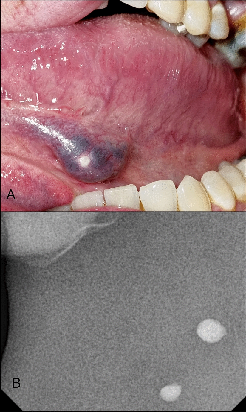
Phleboliths are reported as calcifications that occur in vascular malformations, associated with changes in blood flow dynamics, thrombus formation and subsequent calcifications. Radiological examination, such as cone beam computed tomography (CBCT) could help in demonstrating the presence of a calcifiied mass. A 45-year-old male was referred to our service with an asymptomatic nodular purplish lesion located on the ventrolateral tongue. Within the lesion, a stony mass was also evident on palpation. A digital dental radiograph demonstrated two circumscribed radiopaque structures. Phleboliths associated with vascular malformation was the main diagnostic hypothesis. The patient underwent a sclerotherapy protocol allowing surgical accessibility to the area. Phlebolyts were surgically removed using electrocoagulation. Histopathological examination revealed phleboliths in the context of a vascular malformation with intense fibrosis.


留言 (0)