OSCC has various prognoses according to histological grade, subtype, and clinical prognostic factors [19]. OPSCC is an uncommon subtype of OSCC that was postulated to have a favourable prognosis in comparison to conventional OSCC [5]. TME in OSCC has a significant role in tumor initiation, progression, and metastasis due to the presence of critical components such as CAFs and myofibroblasts [8]. Therefore, recently, CAFs staining by immunohistochemistry has been highly recommended to correlate their density with different histological grades and subtypes of OSCC [20]. Accordingly, for a better understanding of the TME in OPSCC and its prognosis, the current study used immunohistochemical staining with TGF-β and α-SMA to compare their expression in OPSCC with that in conventional OSCC. As well as to correlate their expression with histopathological and clinical prognostic factors.
The current study revealed that OPSCC was more common in middle-aged male patients (from 47 to 64 years old). The most common affected sites were the alveolar mucosa, followed by the buccal mucosa, and finally the lateral border of the tongue. The floor of the mouth and the palate were not involved. Lymph nodes were involved in only a small percentage of cases; however, not all cases were examined. These clinical findings were in agreement with [21], who found that OPSCC was more common in males. However, they contradicted some of our findings, where they found that OPSCC was common in older patients with a peak age of 63 years old, and they also found that the palate was the most common site of involvement. Furthermore, [22] contradicted our findings, as they found that OPSCC was more common in females and in older patients ranging in age from 53 to 83 years old. They also found that OPSCC was common in the buccal mucosa, followed by the gingiva, the lower lip, the palate, the tongue, and finally the floor of the mouth.
On the other hand, the different histological grades of conventional OSCC in this study occurred in middle-aged and elderly patients. Males were more commonly affected, except for PDSCC, which was more common in females. This was in contrast to [23] who found that high-grade tumours affected older males, while low-grade tumours were more common in younger females. The most affected site of conventional OSCC was the lateral border of the tongue, followed by the alveolar mucosa and buccal mucosa. The palate was the least likely site to be involved. These clinical findings were in agreement with [7], who reported that the lateral border of the tongue was the most commonly affected site of OSCC. They also found that OSCC of the lateral border of the tongue had the highest mortality rate, which may be in agreement with the findings of the current study, which revealed that most of the cases of the lateral border of the tongue were PDSCC. In the present study, LN involvement was higher in WDSCC in contrast to [24], who found that LN involvement increased with increasing the histological grade. This could be attributed to the missed clinical data in some of our studied cases.
Tumour budding is defined as the presence of single tumour cells or clusters of tumour cells less than 5 cells at the invasive front. Tumour budding at the invasive front indicates that tumour cells have been dissociated from the main tumour mass [25]. A tumour with high tumour budding (more than 10 tumour buds), for example, has a WPOI of 4 or above, which is associated with PDSCC [26]. The TSR plays an important role in the prognosis of OSCC, which is defined as the percentage of tumour tissue in relation to the surrounding stroma. A low TSR is correlated with a worse survival rate, poor lymphocytic response, tumour aggressiveness, advanced stage, and treatment resistance [27].
All OPSCC cases in the current study showed a papillary lesion with POIs 1 and 2. No tumour budding, perineural invasion, or perivascular invasion were seen. Furthermore, high TSR was seen in all cases of OPSCC; this could be attributed to the postulated good prognosis of OPSCC [28] found that there is a correlation among tumour budding and the WOI of the tumour, as tumours with high tumour budding (5–10 tumour buds) have a WOI of 4 or 5 and are often associated with nodal metastasis, whereas tumours with low tumour budding have a WOI of 1 to 3 and are frequently not associated with nodal metastasis. They also found that WPOI and tumour budding have better prognostic values than the histological grade. However, in the current study, WDSCC showed WOI 4 in some cases; in contrast to [28], this can be explained by the detection of tumour budding in more than half of our cases of WDSCC. On the other hand, all cases of PDSCC in this study showed WOI 4, which is in agreement with [28]. As well, PDSCC showed the highest percentage of tumour budding among the three histological grades of OSCC, which correlated with [29], who found that high tumour budding was found in high-grade tumours. They also found that tumour budding was correlated with tumour aggressiveness and metastasis. This could be explained as follows: tumour cells at the invasive front behave more aggressively than cells in the central or superficial part, where they EMT, leading to metastasis and a poor prognosis. Therefore, there is a significant correlation between tumour budding and the prognosis of OSCC [29].
Moreover, PDSCC showed the lowest percentage of a high TSR, which was in the same context as [30], who found that a high TSR was correlated with larger cell nest size and hence related to low-grade tumours. About perineural invasion, PDSCC showed the highest percentage, which correlated with [31], who found that perineural and lymphovascular invasion were correlated with advanced histological grade. In addition [32], found that 70% of cases with perineural invasion were PDSCC, which means that the higher the grade, the more perineural or perivascular invasion will be found.
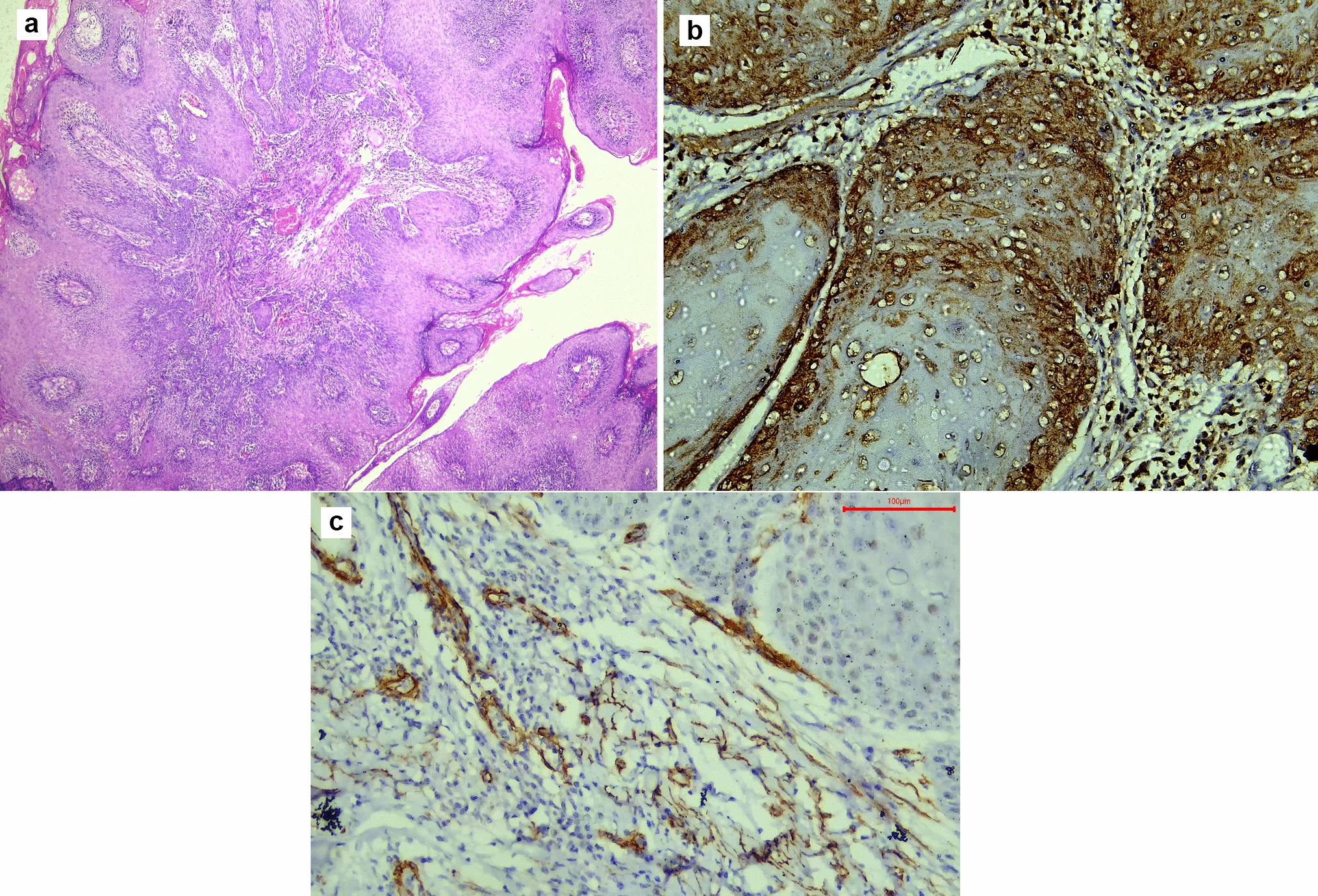
In the following study, the immunohistochemical staining of TGF-β in OPSCC showed cytoplasmic expression in only the basal and parabasal cells of the dysplastic epithelium, with a high percentage of a high and moderate scoring index. These findings match the findings of [33] who found strong basal and parabasal TGF-β expression in the erythematous form of oral lichen planus, which is one of the oral potentially malignant lesions. This may indicate that the pathological behaviour of OPSCC matches that of the oral potentially malignant lesions. They also found that the expression of TGF-β was similar to that of microinvasive carcinoma, which indicates an excellent prognosis.
On the other hand, the immunohistochemical staining of TGF-β in epithelial cells of WDSCC showed significantly the highest expression in the present study, with a high scoring index in all cases. While MDSCC and PDSCC showed focal expression of TGF-β in the epithelium, the weakest scoring index was in the MDSCC group. These findings were in contrast to [34], who found that high-grade tumours and more aggressive clinical behaviour were associated with increased TGF-β. As well [35], contradicted our study as they reported that the expression of TGF-β, as the main inducer of EMT, increases with the advanced stage of cancer, so the percentage of TGF-β expression should be higher in PDSCC. The low percentage of TGF-β expression in MDSCC and PDSCC in the presented study could be attributed to the discohesiveness of the malignant epithelial cells in these groups, leading to a lower area percent of expression than WDSCC in the fixed frame area.
However, the percentage of TGF-β expression in the stroma was highest in PDSCC, followed by WDSCC, OPSCC, and MDSCC; the difference between the groups was insignificant [36] found that the percentage of TGF-β expression in the stroma increased with poor prognosis for cancer., and this was explained by the secretion of TGF-β by the TME, which acts in a paracrine manner and stimulates protumorigenic microenvironmental changes such as the conversion of activated fibroblasts into CAFs, which in turn leads to a higher histological grade, a poorer prognosis and secretion of TGF-β from other cell types, so it is expected to be found in the stroma.
About α-SMA expression in the stroma, it showed weak scattered cytoplasmic expression in the stroma of OPSCC that was only observed beneath the rete ridges. No OPSCC case showed a high scoring index of α-SMA expression in the stroma. These findings were a match for [37] findings in microinvasive carcinoma. As well as [38] findings, which showed that oral potentially malignant lesions had few myofibroblasts and a weak percentage of α-SMA expression in the stroma. This weak and focal α-SMA expression in the stroma indicates a smaller amount of CAFs and activated myofibroblasts, which may indicate a good prognosis for OPSCC.
On the other hand, the expression of α-SMA in the stroma of WDSCC and MDSCC was focal and moderate, while it showed the highest area percent in the stroma of PDSCC, which was significantly different from its expression in OPSCC only. However, the difference in the mean scoring index of α-SMA in the stroma was insignificant among the four groups. These findings were in correlation with [39] who found that there is no significant difference in α-SMA expression comparing different grades of OSCC. On the contrary [40], found that the highest scoring index of α-SMA expression in the stroma was in PDSCC. In this study, α-SMA expression was seen strongly around the invading malignant cells. This was in accordance with [41], who found that α-SMA expression was found strongly around invading nests, indicating that CAFs were aggregated mostly in these areas, which highlights that there is a strong interaction between CAFs and the invading dysplastic cells.
Interestingly, α-SMA staining in the stroma showed the highest expression in the floor of the mouth cases, regardless of the histological grade. This could be explained by [42], who found that OSCC in the floor of the mouth had a worse prognosis, a higher tendency of local invasion, and cervical lymph node metastasis, leading to a high mortality rate compared with OSCC in other sites in the oral cavity, which leads to an incomplete response to treatment and a lower survival rate.
In our study, α-SMA showed nuclear expression in the dysplastic epithelial cells, with cytoplasmic expression in some of the PDSCC cases. This could be explained by the process of EMT, which leads to the formation of myofibroblasts and CAFs from the dysplastic epithelial cells [43]. The nuclear expression of α-SMA was explained by [44], who used correlative confocal fluorescence and transmission electron microscopy to investigate the nuclear expression of α-SMA. They found that α-SMA aggregates mainly in close proximity to the outer nuclear membrane with many deep nuclear invaginations containing α-SMA-rich cytoplasm, giving the false nuclear expression in immunostaining. They thought that the presence of these invaginations related to the state of cellular differentiation, as they may play a role in signal transduction. Furthermore, they proposed that the changes that happen in the shape of the cell and nucleus cause mechanical forces and the direct transmission of forces through the cytoplasmic and nuclear cytoskeletons. In the current study, PDSCC significantly showed the highest score index and area percent of α-SMA in the epithelium. Upon correlating TGF-β expression in epithelium with α-SMA expression in stroma, we found a moderate positive correlation in the WDSCC group only, which means there is a tendency for high TGF-β in epithelium expression to go with high α-SMA in stroma. These findings were in agreement with [45], who found that increased TGF-β expression correlates positively with increased α-SMA expression in the stroma. This could be explained by the role of TGF-β in EMT and the activation of resident fibroblasts into CAFs and myofibroblasts. Finding this moderate positive correlation in the WDSCC group could only be attributed to the high expression of TGF-β in the epithelium of WDSCC. Our results revealed a significant difference in the mean area percent of α-SMA in stroma between cases with tumour budding and those without tumour budding, with cases with tumour budding having a higher mean area percent of α-SMA in stroma. These findings were in agreement with [46], who correlated the presence of tumour budding with the advanced stage of OSCC. The higher the histological grade of OSCC, the more CAFs and myofibroblasts, leading to a high area percent of α-SMA in the stroma.
Moreover, there was a significant difference in the mean area percent of α-SMA in stroma between cases with a low TSR and those with a high TSR, with cases with a low TSR having a higher mean area percent of α-SMA in stroma. This could be explained by [47], who found that a low TSR was associated with high stromal content and widely dispersed tumour cells, hence more CAFs and myofibroblasts leading to a high percentage of α-SMA in the stroma.
From the previously mentioned points, we concluded that OPSCC has more limited invasion and hence favourable prognosis than conventional OSCC. Moreover, expression of α-SMA in the dysplastic epithelium or its surrounding stroma may indicate bad prognosis of OSCC. As well as, OSCC occurring in the floor of the mouth is proposed to have bad prognosis regardless of its histological grade. Regardless of the histopathological grade, the expression of α-SMA in TME is correlated to the presence or absence of tumor budding and low TSR.
Last, we recommend further molecular investigation of OPSCC using a larger sample size, as well as the other variants of OSCC with correlation with the follow-up period. Modify a histopathological grading system for OSCC to include the histopathological negative prognostic factors.


留言 (0)