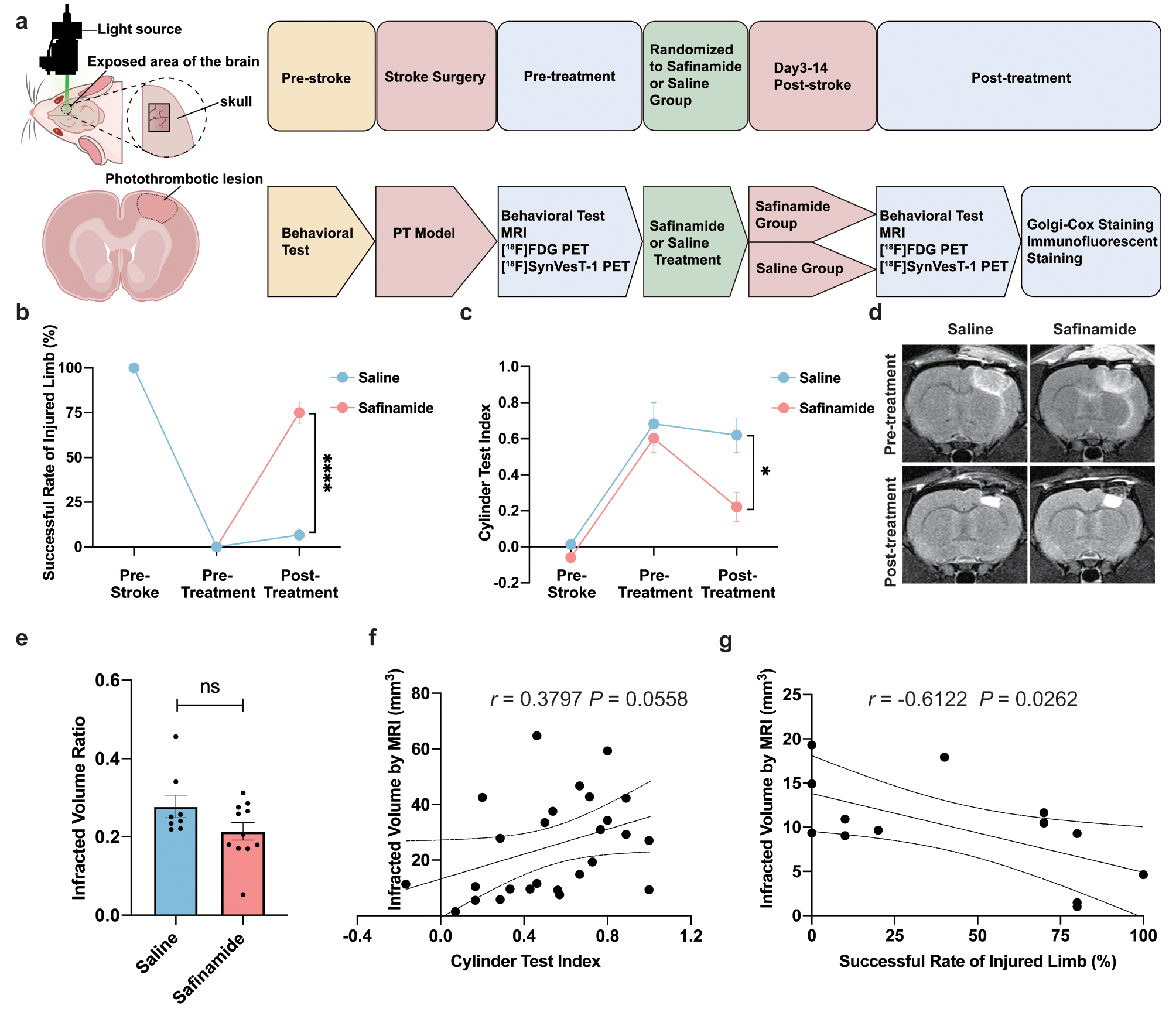
Over the past four decades, the indications for radical prostatectomy in men with prostate cancer have shifted gradually from low and intermediate localised disease to high-grade, high-volume localised and locally advanced stages. While novel surgical techniques, including the introduction of robot-assisted laparoscopic procedures are used increasingly with good reproducible functional and oncological outcomes, the main challenge remains with the inability of the surgeon to identify reliably extra-prostatic cancer cells during surgery to achieve complete excision of the disease. The use of intraoperative fluorescence during surgery to image tissue of interest is not novel and has relied traditionally on using fluorophores administered through direct injections into organs to demonstrate drainage, or vasculature and facilitate lymphadenectomy and precise excision, using Near Infrared (NIR) imaging to detect fluorescence. The contrast agents used in these approaches are neither tissue nor tumour specific. Fluorescence intraoperative imaging using molecular targets to identify specific tissues was therefore developed using a variety of markers conjugated to fluorophores, and innovations in robotic surgical equipment allow now NIR imaging to enhance precision surgery, with the caveat that NIR and white light imaging has not been achievable to date in real-time during surgery [36].
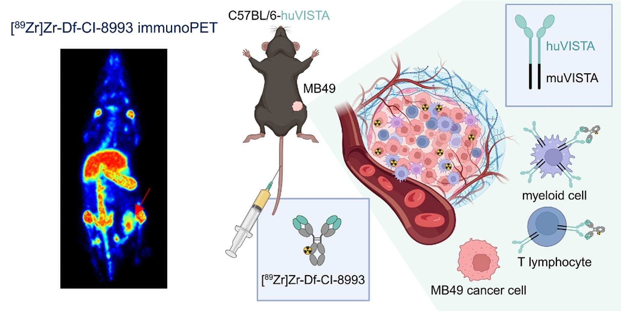
The increasing use of PSMA as a target for imaging and interventions in prostate cancer is transforming the landscape in managing the disease [11, 23, 24, 37, 38]. For intraoperative FMI of cancer, various tumour-targeting molecules, such as antibodies, nanoparticles, proteins, peptides, and small molecules, have been developed. We report herein the pre-clinical development of a novel minibody against Prostate Membrane-Specific Antigen (PSMA), and its first-in-man use after conjugation with the fluorophore IRDye800CW-NHS ester, coupled with an in-house optical system developed to image simultaneously NIR and white light in an attempt to image extra-prostatic cancer cells during RARP. The rationale for the minibody development was to combine the advantages of targeting specificity associated with antibodies, and the fast clearance kinetics associated with smaller molecules. Preclinical studies suggested a speedy clearance and optimal imaging times of the order of 1–2 days.
Extrapolation of doses and clearance times across species of widely differing metabolic rates is, however, not straightforward [39, 40], particularly when the clearance rate of the marker bound to the prostate tumour tissue was unknown. Although we started with the manufacturer-recommended 20 mg dose, we obtained good clinical results with a dose of 5 mg (equivalent to just over 50 µg/kg) or about twice the level considered to be a micro-dose, [41, 42]. There is considerable interest in using micro-dosing with FMI [43, 44]. With modest improvement in imaging sensitivity, micro-dosing should be achievable for laparoscopic/robot-assisted approaches, as it is in wide-field surgery, i.e. non-laparoscopic imaging [45]. Micro-dosing (phase 0) studies have also been suggested for accelerating clinical translation of novel agents [46] and camera sensitivity plays a critical role here. For example, clinical translation of fluorescently labelled bevacizumab [45], has achieved a signal-to-noise ratio of ~ 4:1 for micro-dosing administration. This performance was obtained using cooled cameras although we achieve a comparable signal-to-noise ratio with an uncooled detector, albeit at long exposure times. Clearly, there is scope for further improvement in camera sensitivity. However, to the best of our knowledge there is currently no definition of ‘sufficient sensitivity’ in clinical fluorescence imaging [47]. Fluorescence imaging sensitivity can be increased by reducing the working distance. This increases the excitation power density according to the inverse square law. Unfortunately, while spatial resolution is enhanced as a result of a reduction of the working distance, the field of view is reduced, making surgical navigation more awkward. Working distance could not be usually reduced significantly with our instrumentation since we were using an assistant port for laparoscopy imaging. FMI demands the use of sensitivity high enough to image the low concentrations typically attained by targeted fluorescence agents, in the range of sub-nM to 100 nM. Camera sensitivity is one of the most critical parameters in reaching high signal-to-noise ratio fluorescence detection in short exposure times. In our case, most of the data reported required exposure times of < 500 ms. FMI sensitivity is also determined by the intensity and spectral response of the fluorescence excitation: here we are forced to limit power densities to below 50 mW/cm2 range to eliminate the possibility of tissue heating. Finally, the ability to block excitation light and light from other sources in the detection channel is key; we have used an emission filter with an optical density > 8 at the excitation light wavelength. Furthermore, FMI sensitivity affects the administered dose required. While non-specific, vascular dyes such as indocyanine green (ICG) are often injected in quantities of tens of milligrams when given systemically or ~ 1 mg when given intratumorally [48], reaching concentrations of > 100 nM—2 μM in tissues, targeted agents are used in much lower concentrations [49], since most of the administered dye is cleared from tissues and only a small amount is present in the targeted tissue. Results based on clinical data have shown that due to targeting and clearance, the concentrations of molecularly targeted agents imaged may be five to six orders of magnitude lower than when imaging ICG [48,49,50]. Furthermore, the camera integration time and sensitivity are fixed, while our custom system allows these to be increased over a wide range. More detailed descriptions can be found in the Supplementary information (§S8 and Figures S6 and S7). Finally, the Firefly applies the fluorescence information as an overlay which seems to be associated with an intensity threshold. Further developments to our system, not described here, permit the real-time readout of TBR information and the existence of intensity thresholding may detract from determining accurate TBR values.
Administration of the marker pre-operatively was well tolerated even at the higher doses and did not generate any specific side-effects. In order to develop measurable consistent metrics to define the level of fluorescence in relation to probability of detecting cancer cells, we sought to quantify the TBR used as a measure of the specificity of the uptake of our imaging agent within the target organ. The presence of imaging agent in the background can be secondary to a low retention rate in the target organ, leading to leakage into surrounding tissues or, as in this instance, to potential preferential uptake by other organs (particularly highly vascular structures such as the liver, spleen, gut and kidneys). In vivo intraoperative fluorescence in each evaluable case was aligned with conventional histopathological assessment and fluorescence microscopy studies as illustrated (Figs. 2–7). In some cases, small amounts of cancer tissue were detected using FMI, which would not have been picked up during a standard procedure. This ability of the surgeon to visualise pathological tissue during surgery highlights the overarching promise of this new technology, irrespective of the reagent or optical system used to achieve precision surgery.
We have shown that our detection device is able to provide reliable indication of low TBRs, down to 1.3. Although TBRs, at least in FMI, much below 2 are considered borderline [51], the use of TBRs of < 1.5 has been reported in the literature [52]. One of the limitations of the approach described here is that TBRs are determined post-operatively and this has highlighted the need to provide real-time indications of TBR from selected small areas of fluorescence. Other limitations included the low sample number, the lack of PSMA PET-CT imaging and the extensive learning curve in applying FMI.
In order to emphasise the practical need for TBRs, we have presented only the raw, fluorescence-only images. Images are linearized prior to TBR determinations. This is in contrast to the more usual approach where overlays, linear or non-linear, with or without threshold or otherwise, or heat maps are applied to the fluorescence data prior to display [53]. The high sensitivity of our system is inevitably associated with somewhat higher noise than when higher marker concentrations are used.
The data presented here can only be considered preliminary but suggest that the IR800-IAB2M is a suitable agent for intraoperative identification of extra-prostatic cancer tissue during RARP. Our surgical robot used in this study did not have the advantage of an integrated system to image NIR fluorescence during surgery and had to rely on our in-house optical system described above. A Phase 3 evaluation using a full randomised design is underway. In this evaluation, provision for real-time, on-line metrics has been incorporated in the imaging device although in the medium term it would be desirable to fully integrate this with robot-assisted equipment that uses the next generation of highly sensitive imagers. One of our findings, particularly at higher doses, is that the number of false positives is higher than would be expected. However, intraoperative fluorescence detection always correlated with fluorescence detected from tissue samples in all samples analysed. Clearly, the false positives are due to either (a) inadequate marker clearance, and/or marker accumulation in slow to clear regions or (b) to inadequate specificity of the minibody, or (c) to low levels of PSMA expression in non-prostatic tissues. The latter is known to occur [54, 55], although this depends on the marker epitope. The sensitivity and specificity of IR800-IAB2M appears to compare favourably with other recent reagents tested such as OTL78 (range of average sensitivity and specificity of 33.3%-68.4%, and 52.6%-100% respectively), and IS-002 (Average sensitivity/specificity of 97% and 45% for lymph nodes, 100% and 80% for residual locoregional disease [22, 23]. The use of lower molecular weight [56] markers may be advantageous, though potentially at the expense of specificity. Comprehensive evaluation was not possible in all men. In the early part of the study, we have focused on imaging the pelvic lymph node chain, and not on extra-prostatic non-lymphatic tissue. Results from lymph-node imaging were variable with inconsistencies in the early evaluations. When it was decided to focus on non-lymphatic tissue, we were allowed by our regulatory approvals to study a limited remaining number of patients which we are reporting in more detail herein.
Marker clearance, at least as measured by urine fluorescence, provides only a ‘global’ indication of clearance, and does not reflect the timescales of localised clearance. In particular, clearance from lymph nodes would be expected to be slow, due to the molecular weight of IR800-IAB2M. The apparent lack of agent elimination even at delayed time points in some cases, as well as spatial resolution can pose challenges in tissue margin delineation. We recognise that this represents a limitation of our pilot work, which we will endeavour to overcome by continuing to refine and validate real-time accurate determination of TBRs in the next phase of our evaluation.
It is informative to explore how other commercially available fluorescence guided devices would perform in comparison with our custom system. Of these the Da Vinci® Xi FireflyTM system is commonly used during robotic procedures. Although we did not have access to this during the procedures described here, such a system has recently become available to us. Unfortunately, the Da Vinci fluorescence system is aimed at ICG imaging and uses an excitation wavelength of 805 nm, well away from the optimal 775 nm for the IRDye800 fluorophore. In addition, the Da Vinci system collects fluorescence light only at > ~ 830 nm, at which point the IRDye800 fluorescence is no longer significantly emitting. Furthermore, the camera integration time and sensitivity are fixed, while our custom system allows these to be increased over a wide range. More detailed descriptions can be found in the Supplementary information (§S8 and Figures S6 and S7). Finally, the Firefly applies the fluorescence information as an overlay which seems to be associated with an intensity threshold. Further developments to our system, not described here, permit the real-time readout of TBR information and the existence of intensity thresholding may detract from determining accurate TBR values.
The factors outlined above contribute to our system exhibiting a > 25 × higher sensitivity, for IRDye800 imaging, than that afforded by the Firefly. However, more recent versions of the Firefly system, as used by Nguyen et al. [20] and described therein, do provide significantly enhanced sensitivity. Such systems are likely to be suitable for the procedures described here, though this remains to be established. Furthermore, modifications to optimise the excitation and collection wavelengths are technically straightforward. Other non-robotic devices, such as those used in the work described in [19] are also likely to be appropriately sensitive. However, we did not have access to other laparoscopic fluorescence-capable imaging systems.
In several cases, IHC detection did not match with either intraoperatively detected or microscopy detected fluorescence signals. This highlights the practical difficulties of aligning intraoperatively detected areas of fluorescence with histology and IHC areas. We have found it useful, in the Phase 3 evaluation currently underway to maintain laparoscope working distance and to include an object of known dimensions (e.g. a surgical clip) in the field of view of image on which TBRs are determined. This allows a fixed fluorescence imaging sensitivity to be maintained and to ensure that the areas of fluorescence could be measured and compared with histology findings.
In conclusion, our study demonstrated the pre-clinical development of a PSMA targeted minibody, IR800-IAB2M and its first-in-man utilisation for the intraoperative detection of prostate cancer tissue in lymph nodes and extra-prostatic tissues with favourable outcomes. A larger scale evaluation of the reagent is underway using a randomised design to investigate the benefits of real-time intraoperative fluorescent imaging during RARP in achieving complete and optimal tissue excision for improved oncological and functional outcomes in men with prostate cancer. Findings from our study and others should pave the way for a systematic and simultaneous evaluation of multiple molecularly targeted agents using the most cutting-edge fluorescence platforms integrated in robot-assisted surgical equipment, in order to reach consensus and change future practice based on robust evidence.


留言 (0)