Significant morphological and occasionally immunophenotypic overlap is observed between SCNEC and poorly differentiated RMS originating in different organs. However, in routine practice, this overlap has no significant diagnostic impact given that the anatomic sites affected by these two entities differ significantly in most cases. Additionally, most of alveolar RMS affect children and adolescents, whereas neuroendocrine carcinomas are unexpected or do not occur among this age group, limiting the differential diagnosis. However, topographic overlap between SCNEC and poorly differentiated RMS presenting in adults is noted in a few organs, in particular, in epithelial-lined visceral organs including the sinonasal cavities and the urinary bladder. Several studies have pointed out the frequent expression of pankeratin and neuroendocrine markers in alveolar RMS [12,13,14]. To date, the exact cell of origin of RMS originating within these epithelial-lined organs remained elusive. Likewise, the histogenetic relationship, if any, between these poorly differentiated RMS and SCNEC has not been sufficiently explored.
After the lung, the urinary bladder is the second most common site of origin of SCNEC. In concordance with the literature, SCNEC predominately occurs in older patients with a median age at diagnosis of 67.5 years in this study. However, in contrast to its pulmonary counterpart, SCNEC of the bladder is associated with other histologic subtype/s in around 40–50% of the cases [15]. In our current series, we observed a comparable frequency of urothelial carcinoma and variants as a component in 43.5% of SCNEC.
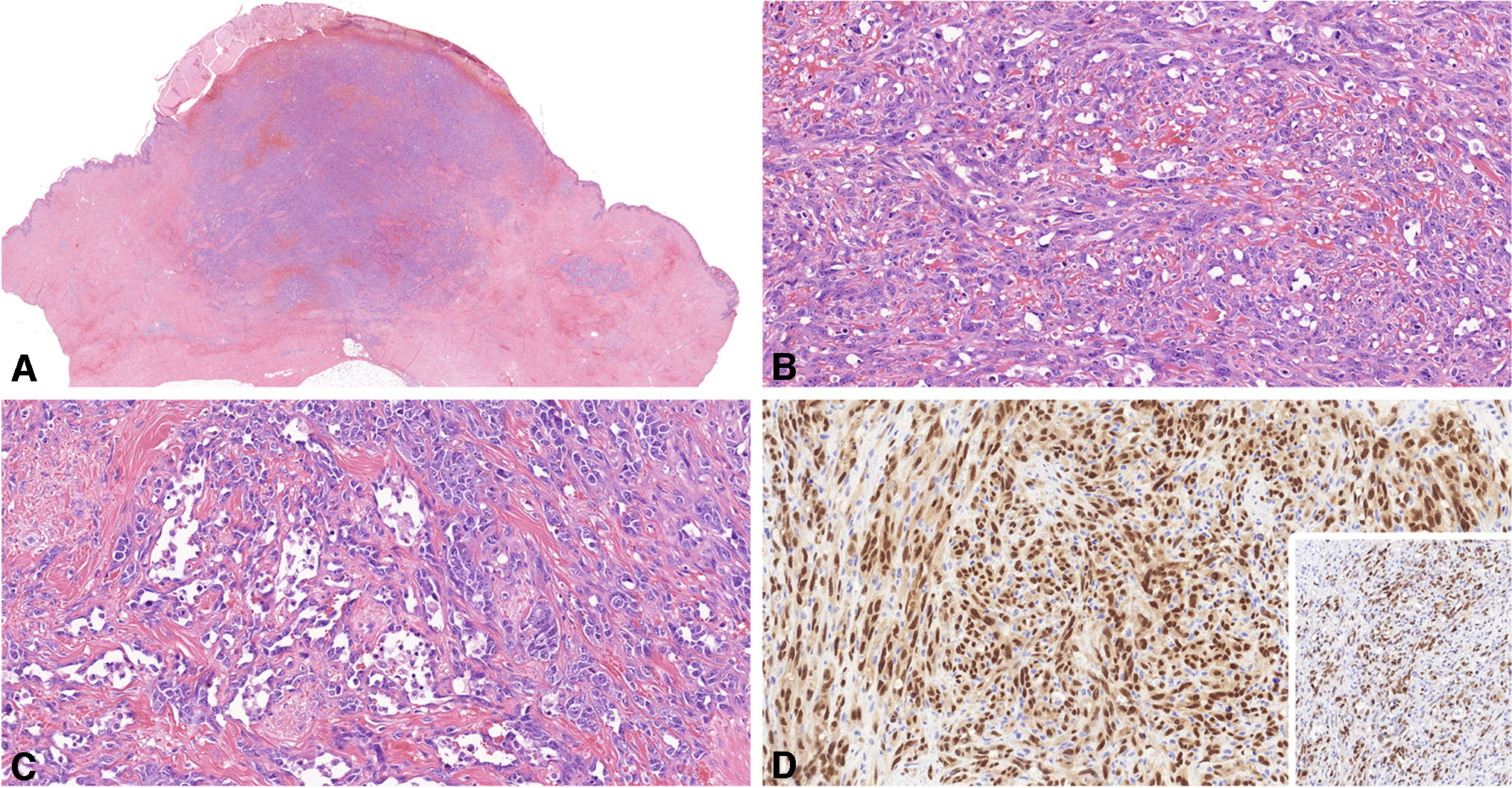
RMS of the urinary bladder is rare, primarily occurring in children and adolescents. There are few adult cases reported in the literature [16]. While most pediatric cases correspond to the embryonal subtype of RMS, bladder RMS in adults and elderly is predominantly poorly differentiated with small cell pattern, recapitulating solid alveolar RMS [7]. Their morphology overlaps significantly with SCNEC, frequently representing pitfalls or diagnostic challenge. The issue is further complicated by frequent variable immunoreactivity of RMS cells with low molecular weight keratins and neuroendocrine markers including CD56, synaptophysin, and, less frequently, chromogranin A. In many cases, the exact diagnosis of SCNEC versus solid small cell RMS of the urinary bladder might be arbitrary and only inclusion of desmin and myogenin and the presence or absence of a urothelial component might facilitate distinction.
Rhabdomyoblastic differentiation of variable extent has been reported in high-grade NEC of different organs [6]. Although considered rare, this phenomenon might be under-diagnosed given that rhabdomyogenic markers are only applied to cases with ambiguous diagnosis or uncertain differentiation and not in clear-cut SCNEC cases. Indeed, in this study, we observed diffuse expression of desmin and myogenin in one case originally reported as SCNEC which justified revising diagnosis to RMS. On the other hand, one of our original RMS cases also presented a high expression of neuroendocrine markers. These findings highlight close phenotypic overlap between SCNEC and RMS in the bladder.
A main characteristic of urinary bladder cancer are mutations in the TERT promoter gene. Hotspot mutations of this promoter were detected in around 70% of bladder cancer cases and were not associated with clinical or pathological parameters [17]. In a recent publication on 132 SCNEC of the bladder, TERT promoter mutations were detected in 68% of cases [18]. We herein report similar frequency in our cohort of 22 SCNEC (68%). In the context of a bladder neoplasm, detection of TERT promoter mutation has emerged as a surrogate marker for urothelial origin. As reported by Priemer et al., the TERT promoter mutational status can differentiate SCNEC of the bladder from those of prostatic origin [19].
In our current study, we found significant morphological and immunophenotypic overlap between SCNEC and RMS of the bladder. The only distinguishing diagnostic differences of RMS are the lack of a urothelial component and the presence of homogeneous rhabdomyoblastic immunophenotype. Our findings suggest a continuum of rhabdomyoblastic differentiation in both entities ranging from virtually absent in classical SCNEC on one end of the spectrum to being the sole pattern in tumors classified as RMS on the opposite end of the spectrum with possible intermediate variants in between, although we have not observed cases with focal or partial rhabdomyoblastic differentiation, but this might be the result of sampling errors. Consistent with this view, none of the four RMS cases tested with targeted RNA sequencing showed any of the gene fusions expected in genuine alveolar-type RMS. The common origin of SCNEC and RMS in the bladder was further strengthened by the high comparable frequency of TERT promoter mutations identified in both entities in our study (68% of SCNEC and 67% of RMS cases, respectively). According to the current literature, TERT promoter mutations are very rare in RMS and were detected in approximately 1.4% of RMS cases not stratified by the organ of origin [20]. On the other hand, > 80% of all alveolar RMS harbor a distinct balanced FOXO1 translocation, which is a characteristic and disease-defining genetic marker [8].
Thompson et al. [14] analyzed 52 alveolar RMS of the sinonasal tract in adults aged ≥ 18 years (mean age, 43) and detected low-molecular weight keratins overall in 54% of cases (CAM5.2 in 50% and AE1/AE3 in 36%). Moreover, the neuroendocrine markers CD56 (100%), synaptophysin (35%), and chromogranin (13%) were frequently expressed. However, despite this significant phenotypic overlap with sinonasal SCNEC, the histogenesis and molecular pathogenesis of adult RMS in these epithelial-lined organs seem quite distinct and site-specific. Notably, FOXO1 rearrangements have been detected by PCR studies in 81% of sinonasal RMS cases with PAX3 as fusion partner in 72.7% of cases [14]. On the contrary, none of our 4 RMS cases and none of four unclassified bladder RMSs in adults studied by Gupta et al. revealed a FOXO1 or other RMS-associated fusion [21]. A fusion involving PPP1R12A (fused to LIN7A or PTPRQ) was detected by Gupta et al. in two RMS cases (one reported as sarcomatoid!). However, these gene fusions have not been reported before, and it is not clear if they drive oncogenesis or represent passenger events. Admittedly, the fusion partners detected by Gupta et al. in two tumors are not included in the TruSight Illumina Panel we used in this study, so the relevance of this fusion needs to be verified in larger future studies.
Treatment of poorly differentiated RMS (including solid alveolar subtypes), in adults typically involves a combination of agents such as cyclophosphamide, vincristine, and doxorubicin [22]. In contrast, standard chemotherapy for SCNEC of the urinary bladder often utilizes platinum-based chemotherapy and etoposide [23]. Accordingly, separating these two disease entities has clear therapeutic relevance. However, distinguishing these two aggressive phenotypic diseases in routine practice is challenging and is significantly influenced by extent of sampling. Thorough sampling and careful histological evaluation to detect minor microscopic urothelial foci (in situ or invasive) is the most reliable clue for verifying a urothelial origin. However, in limited biopsies and in cases with uniform rhabdomyoblastic differentiation, only inclusion of desmin and myogenin in the immunohistochemical panel of any potential SCNEC would facilitate this distinction. Our study indicates that TERT promoter mutations are identified in 67% of assessable RMS cases, representing a potential surrogate for a urothelial origin. Enhanced recognition of RMS cases should help to confirm our hypothesis, and if confirmed, then to address the central question, whether the approach to treat adult RMS of the urinary bladder needs to be revised. Moreover, targeting the urothelial origin through additional modified therapeutic approaches could potentially lead to improved response rates.
In summary, our study confirms the reported significant morphological and immunophenotypic overlap between SCNEC and poorly differentiated RMS of the urinary bladder. We herein add molecular overlap with similar frequency of TERT promotor mutations in SCNEC (68%) and RMS (67%) in our study. The presence of TERT promoter mutations and lack of FOXO1 and other RMS gene fusions in all tested RMS cases are in line with a urothelial origin of most if not all RMS of the bladder in adults. Urinary bladder small round cell RMS in adults probably originates via monomorphic rhabdomyoblastic transdifferentiation in SCNEC and are likely distinct from genuine alveolar RMS in other organs/ soft tissue and bone sites. Our study is however limited by the low number of RMS cases due to rarity of this disease. Accordingly, extended analysis on more RMS cases is needed to further consolidate this notion.


留言 (0)