MORPHOLOGICAL BIOMARKERS PREDICTING EXUDATIVE CONVERSION IN TYPE 1 NONEXUDATIVE MACULAR NEOVASCULARIZATION USING OPTICAL COHERENCE TOMOGRAPHY ANGIOGRAPHY
Purpose:
To investigate the incidence and morphological biomarkers to predict the exudative conversion in eyes with type 1 nonexudative macular neovascularization using swept-source optical coherence tomography angiography.
Methods:
Macular neovascularizations were detected using the retinal pigment epithelium-to-retinal pigment epithelium-fit slab of swept-source optical coherence tomography angiography scan. Depending on whether exudation developed within a year, the eyes were divided into two groups: active and silent. Qualitative and quantitative optical coherence tomography angiography parameters of the two groups were evaluated to discriminate the biomarkers associated with exudative conversion.
Results:
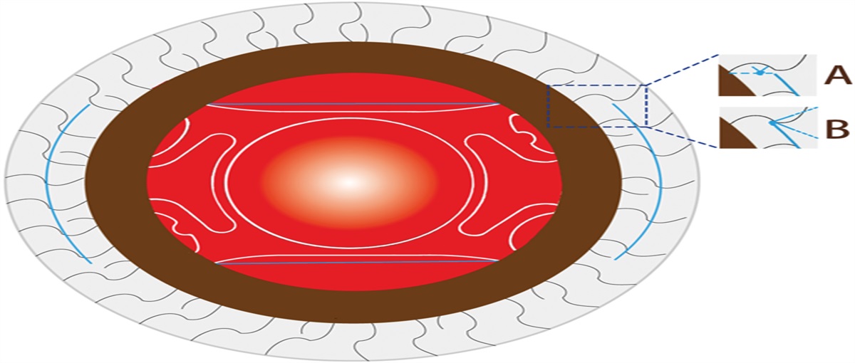
Of the 40 eyes, nine developed exudation within 1 year (incidence rate 22.5%). The active group exhibited a significantly higher “anastomosis and loops” pattern, greater “vessel density,” increased “junction density,” fewer “number of end points,” and lower “lacunarity” compared with the silent group. “Anastomosis and loops” and higher “vessel density” were correlated with the active group in multivariate analyses. A predictive model combining these biomarkers achieved 95% accuracy in predicting exudative conversion.
Conclusion:
At 12 months, the risk of exudation was 22.5%, and “anastomosis and loops” and “vessel density” were useful optical coherence tomography angiography biomarkers for predicting exudative conversion in eyes with type 1 nonexudative macular neovascularization. For eyes with a high risk of exudative conversion, more frequent follow-up is recommended.

留言 (0)