In this study, we evaluated the impact of the sensitivity mode (axial acceptance angle) of the Biograph Vision Quadra LAFOV PET/CT scanner on dynamic imaging with [18F]-FDG to determine Patlak Ki and DV estimates obtained by the indirect or direct Patlak method. Different Patlak start times t*, number and duration of PET data frames were evaluated to optimise the imaging protocol and analysis settings, and to assess the potential for abbreviated scan protocols using a population-based input function.
Indirect Patlak
For the indirect Patlak approach using the full IDIF (0–65 min), only minor differences of 3.4 ± 7.0% (Ki) and 1.2 ± 2.6% (DV) were found between the sensitivity modes. These differences cannot be caused by variations in the IDIF, as the AUC showed excellent agreement, presumably because the number of events for the aortic VOI in each frame was already sufficiently high in the HS mode. Therefore, the difference was caused by the determined activity concentration in the delineated lesion VOI, which could be altered either by the slightly degraded spatial resolution of the UHS mode [23] or by its higher event statistics. Although the largest difference between the two modes is known to be in the centre FOV, no correlation was observed between axial lesion position and differences in Patlak estimates.
However, for the indirect Patlak, the UHS mode was less affected by the number of Patlak frames used to determine Ki estimates, e.g., a deviation for 8 Patlak frames of 2.3% (UHS) and 4.8% (HS) compared to 4 Patlak frames. The rationale for increasing the number of Patlak frames was to have more samples for a more accurate Patlak fit, which is less susceptible to variation in individual fit points. Of note, the bias was negative and fairly constant up to 20 Patlak frames (Fig. 3a), indicating that with 4 Patlak frames the true Ki value might be overestimated. Therefore, 8 Patlak frames were determined as the optimal setting for the indirect Patlak method.
For the abbreviated protocols based on the sPBIF and indirect Patlak, the UHS mode resulted in a lower bias and higher precision for Ki compared to the HS mode, e.g., for 45–65 min p.i. 6.4 ± 8.9% (HS) and 3.8 ± 4.4% (UHS).
Comparison of these results with previous studies on the Biograph Vision Quadra using the HS mode confirms this improvement due to the UHS mode, such as a higher precision of 4.4% compared to 13% (45–65 min p.i.) reported by Sari et al. [20] and a lower bias of 3.8% compared to 5.18% (40–60 min p.i.) reported by Sluis et al. [21]. The more accurate determination of Ki with the UHS mode demonstrated in our work allows the scan protocol to be further shortened while maintaining a higher precision than in the HS mode, e.g. 50–65 min p.i. 4.4 ± 11.2% (UHS) instead of 7.3 ± 20.0% (HS).
In contrast, the impact of sensitivity mode on DV was negligible, e.g., 45–65 min p.i. 14.2 ± 2.7% (HS) and 14.4 ± 2.7% (UHS). The best estimates of DV with sPBIF indirect Patlak were obtained with 8 or more Patlak frames. However, Ki estimates only showed a minor dependency on the number of Patlak frames between 4 and 15 for 45–65 min and 50–65 min. For the shortest scan duration examined, 55–65 min, a stronger dependence on the number of Patlak frames and poor Ki estimates were observed, e.g., -1.9 ± 33.3% (2 frames) and − 20.1 ± 51.6% (4 frames), rendering the use of such short protocols questionable.
Direct Patlak
The indirect Patlak method with ROI kinetic modelling used in this work is a conventional and easy to implement approach to derive tracer kinetics. However, parametric images, which estimate kinetic parameters for each voxel, are more suitable for studying heterogeneous tracer uptake [27]. Although parametric images can be obtained using the indirect Patlak method, in this work we focused on evaluating parametric images obtained using the direct Patlak method. This was done because it has been shown that direct incorporation of the Patlak model into the image reconstruction process allows accurate noise modelling and results in better bias-variance characteristics than those obtained by indirect methods [32, 33]. In particular for the Biograph Vision Quadra, Sari et al. [34] showed that Ki images generated using the direct Patlak method had a twofold higher contrast-to-noise ratio in tumour lesions and yielded 27% higher SNR on average compared to images generated using the indirect Patlak method.
As previously reported for the indirect Patlak with UHS mode, the number of Patlak frames had a smaller impact on Ki estimates as for HS mode. This effect was mitigated for the direct Patlak, e.g., for 6 Patlak frames bias and precision of Ki estimate was − 1.1 ± 2.4% (HS) and − 0.1 ± 1.8% (UHS) compared to 4 Patlak frames. Similarly, for the sPBIF direct Patlak method, only a negligible difference in Ki and DV estimates was observed between the number of Patlak frames. In addition to the reported performance of the UHS and HS mode, another factor is the longer image reconstruction time for the UHS mode. The larger amount of line of responses and event data increases this time, e.g. for the dedicated workstation used in our study from 50 ± 3 min (HS) to 79 ± 3 min (UHS) (average time for a single patient direct Patlak reconstruction including 4 frames for sPBIF generation), which should be considered especially in busy clinical scanning schedules.
The comparison between indirect and direct Patlak showed comparable results for the variation of Ki and DV estimates for scan protocols of 20 min duration: e.g., UHS mode 3.8 ± 4.4% and 2.7 ± 3.4% (Ki) and 14.4 ± 2.7% and 18.2 ± 7.5% (DV) for indirect and direct Patlak, respectively. For shorter scan protocols of 15 min the indirect Patlak showed a lower bias for Ki (4.4 ± 11.2% versus 15.0 ± 10.5%) and better precision for DV (23.1 ± 4.2% versus 2.6 ± 13.9%) compared to the direct Patlak. Of note, these measures are not suitable for assessing which method provides the more accurate Ki and DV estimates. Instead, due to the lack of a ground truth for Ki and DV, they indicate how robust the estimates were within each method for the abbreviated scan durations.
A major advantage of the direct Patlak method is that it yields parametric images with superior contrast in comparison to standard SUV images, e.g., a TBR of 17.2 ± 9.6 (direct Patlak) in comparison to 6.2 ± 3.1 (SUV).
The TBR obtained with direct Patlak and sPBIF was not affected by the sensitivity mode, however degraded towards shorter scan durations 10.6 ± 5.4 (15 min) and with 6.9 ± 3.5 for the 10 min scans did not show any improvement over the TBR obtained by the SUV image.
The Ki and DV estimate images showed excellent agreement and image quality (RC: 0.3 ± 0.3% (Ki), 1.4 ± 0.7% (DV), SSIM: 0.999 ± 0.001 (Ki), 0.997 ± 0.002 (DV), PSNR:75.7 ± 4.1 dB (Ki), 72.4 ± 9.0 dB (DV)) when comparing the sPBIF based method to the IDIF method for a scan duration of 45–65 min.
In comparison to the Ki related results reported by Sari et al. [20] for a 30 min scan duration with t*=35 min, RC was comparable (0.31 ± 0.25% vs. our work 0.3 ± 0.3%) and PSNR indicated an increased image quality (64.03 ± 3.59dB vs. our work 75.7 ± 4.1dB).
It should be noted, that the quantitative indexes SSIM and PSNR are mainly used for image quality assessment for image processing and computer vision, however these metrics are not necessarily well aligned with human perception [35] therefore a conclusion about clinical impact based only on these metrics is questionable.
However, they are a valid measure to detect differences between images in terms of contrast, structure and noise. Therefore, no difference in these measures for the parametric images obtained either in HS or UHS mode is indicated by the small difference in SSIM and PSNR (SSIM difference < 0.1 and PSNR difference < 1dB).
The higher event statistics obtained with the UHS mode are associated with a lower image noise. In addition, the small degradation of the spatial resolution by the increased parallax error due to more oblique line of response facilitates blurring of smaller structures. No differences in TBR were observed in our work, as the impact on the mean values in Ki and DV of the large homogeneous region in the liver was negligible.
The image metrics RC, SSIM and PSNR were determined based on the comparison of sPBIF and IDIF based direct Patlak for the respective mode. Therefore, image noise related to event statistics is present to the same amount in the reference image and no noise induced differences in these metrics were observed.
The estimate images deviated more from the reference measurement with full IDIF towards shorter scan durations. Hence, good agreement and image quality (RC: 6.9 ± 2.3% (Ki), 0.1 ± 3.1% (DV), PSNR: 64.5 ± 3.3 dB (Ki), 61.2 ± 10.6 dB (DV)) could be obtained for an abbreviated scan protocol from 50 to 65 min p.i.
Limitations and outlook
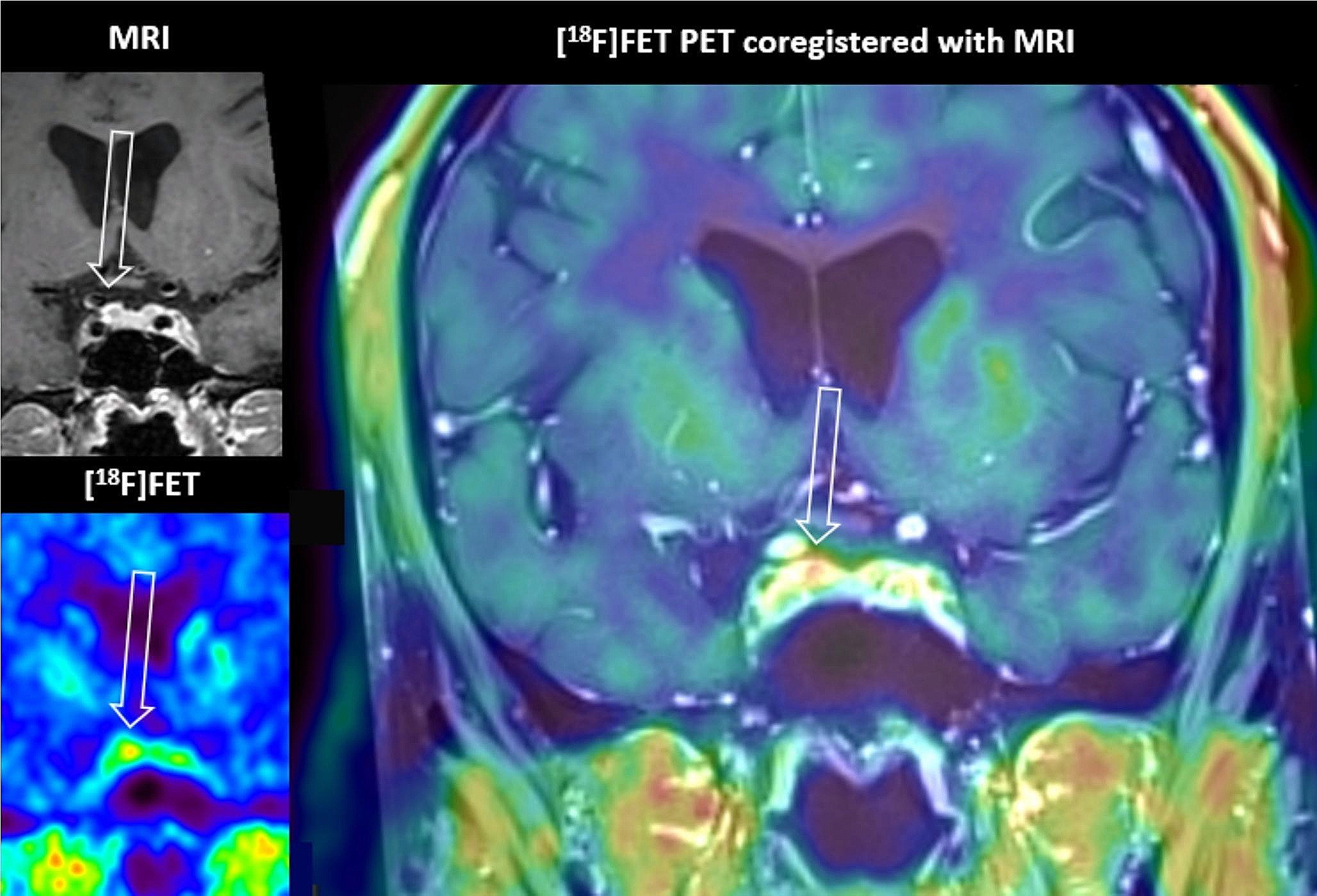
Of note, in Fig. 7a, a halo shaped artefact around the bladder was clearly visible for the Ki estimate (45–65 min, t*=45 min) and less pronounced for the Ki estimate (50–65 min, t*=50 min). This can be explained by the non-linear pattern of the Patlak plot for the bladder and considerable impact of t* on the determination of bladder parametric estimates which results in the Patlak model not being feasible for the bladder [36]. In general, the feasibility of compartmental models for the bladder is questionable, as shown by the inapplicability of one, and two tissue irreversible and reversible compartmental models by Wu et al. [37].
The TBR in Table 4 was 18.0 ± 9.0 and 21.3 ± 11.5 for the IDIF- and sPBIF-based methods, respectively. This difference was caused by an increase in lesion Ki (+ 2.7 ± 3.3% for sPBIF) together with a decrease in Ki determined for liver VOI (-6.9 ± 2.1 for sPBIF). Furthermore, with the Patlak approach in this work, we used a simplified compartmental model for the liver with a single arterial blood input function. Instead, to account for the dual blood supply from both hepatic artery and portal vein, dual blood supply input functions with reversible compartmental models should be considered for a better approximation of liver kinetics [38, 39]. Similarly, a more comprehensive model for the lung should be considered accounting for the effects of regional lung aeration, blood volume, and water on [18F]-FDG uptake [40].
Although in this study scan protocols were already abbreviated to 20 min or less, respiratory and whole-body motion can degrade image quality and quantification accuracy. The impact of motion for total-body PET parametric imaging [41] and advanced methods for patient motion correction of dynamic protocols [42, 43] have been reported. As shown by Sundar et al. [43] by using a diffeomorphic approach for motion correction the volume mismatch across dynamic frames introduced through motion artefacts could be reduced by about 50%. Further by using an advanced parametrization of motion fields in between frames as diffeomorphism, Sun et al. [42] could not only obtain an average improvement in tumor SUVmean of 5.35 ± 4.92% but also for parametric imaging studies a reduction of 11.8% of inter-subject variability in Ki quantification of organs.
We plan to incorporate and access the impact of motion correction for whole-body motion [43] as well as respiratory motion [44] on the 20 min short scan protocols in our future work.
In addition, the number of patients (n = 6) and lesions (n = 26) in this study is limited due to the long PET imaging protocol of 65 min. This complicates the integration of this study protocol into the tight clinical scan schedule and is associated with considerable discomfort for the patients. Additionally, the new Biograph Vision Quadra TB-PET scanner is not yet widely available, which complicates multi-center studies to increase the number of patient scans. Similar to our case, these limitations have also led other groups working on parametric imaging protocols for TB-PET scanners to evaluate their research on a small number of patients and lesions, such as reported by Sari et al. [20], Sluis et al. [21] and Wu et al. [9] with a number of patients (n = 8, 12 and 7) and a number of lesions (n = 34, 20, 26), respectively. Including more datasets for testing is one of our future work and based on our work, we aim to overcome the limitation in statistics by introducing abbreviated scan protocols of less than 20 min into the clinical routine, which we have already started in our institution for a dedicated patient cohort.


留言 (0)