
記住我

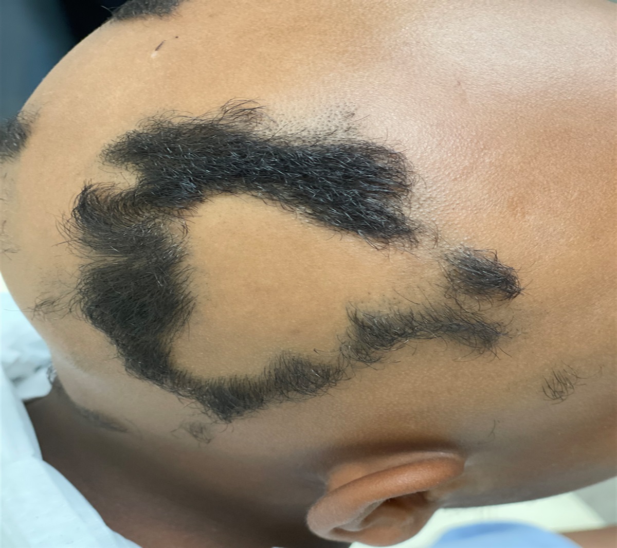


A 9-year-old boy presented to the dermatology clinic with his mother for new-onset hair loss (Figure 1). The patient's mother reports that her son's hair loss has worsened over the past 6 months. He has no ongoing medical problems and takes no medications. There is no family history of hair loss or autoimmune disease. The patient denies any itching or pain associated with the hair loss. Physical examination revealed diffuse hair loss involving approximately 75% of the scalp, accompanied by the presence of irregularly shaped annular patches of hair loss with no evidence of follicular scarring, accentuation, or inflammation.
 FIGURE 1.:
 Irregularly shaped annular patches of hair loss with no evidence of follicular scarring, accentuation, or inflammation.
MULTIPLE-CHOICE QUESTION Trichotillomania Alopecia areata Tinea capitis Secondary syphilis Telogen effluvium ANSWERAlopecia areata
DISCUSSIONAlopecia areata presents as well-demarcated round or oval patches of hair loss. Although the condition can occur at any time, it has been estimated that up to 50% of cases initially present before 16 years old (Afford et al., 2021). Alopecia areata is an inflammatory disorder that is believed to be immunologically mediated with lymphocytes attacking the hair follicle (Pratt et al., 2017). Patients typically note the sudden appearance of one or more sharply circumscribed patches on the scalp or other hair-bearing areas such as the eyebrows or beard. Physical examination will be remarkable for noninflammatory patches of hair loss with characteristic short hairs that taper, referred to as exclamation-point hairs, being present under dermoscopy (Guttikonda et al., 2016). When these hairs are examined under a microscope, telogen hairs with a frayed distal tip will be present (Afford et al., 2021). In more extensive cases, alopecia totalis can occur with the complete loss of scalp hair, and the absence of all scalp and body hair is referred to as alopecia universalis. There is an association of alopecia areata with autoimmune disorders such as vitiligo and Hashimoto's thyroiditis (Zhou et al., 2021). Traditional first-line treatments for this condition include topical corticosteroids, intralesional corticosteroids, methotrexate, and contact immunotherapy. Recently, the FDA has approved the use of Janus kinase inhibitors as a systemic treatment for patients aged 12 years and older in the treatment of severe alopecia areata (Waśkiel-Burnat et al., 2021).
Trichotillomania is caused by the repetitive pulling of one's own hair, either consciously or subconsciously. Trichotillomania can be distinguished from alopecia areata based on findings during the physical examination. Trichotillomania is a clinical diagnosis where physical examination shows the presence of twisted and broken hairs of different lengths among the patches of alopecia (Pratt et al., 2017). In addition, patches of trichotillomania tend to be ill-defined. The “Friar Tuck” sign can be seen in patients with trichotillomania, where the central crown of the scalp shows signs of alopecia whereas the peripheral rim remains relatively unaffected. Treatment centers around education and decreasing the psychosocial stress frequently associated with this disorder.
Tinea capitis is caused by a fungal infection that leads to patchy areas of hair loss on the scalp with associated scale. Frequent black dots are seen on physical examination representing short 1- to 3-mm broken hairs within larger patches of alopecia (Phillips et al., 2017). The combination of these physical findings and the use of fungal culture, or scraping, is essential in differentiating tinea capitis from alopecia areata. In severe, untreated cases, oozing, red plaques known as a kerion occur within areas of hair loss. The gold standard of treatment revolves around selecting the appropriate oral antifungal agent after culture results.
Secondary syphilis can occur in up to 40% of untreated patients. These patients can often present with characteristic moth-eaten patches of hair loss (Kovitwanichkanont & Chong, 2019). Hair regrowth will typically occur within 3–6 months after successful treatment, which revolves around the use of penicillin, which can be administered in various forms.
Telogen effluvium is a form of alopecia that is often secondary to a triggering event or stressor, such as infection, medication, or surgery. Such an event typically predates the onset of hair loss by approximately 3 months. Physical examination of diffuse alopecia accompanied by a positive hair-pull test where more than six hairs can be extracted is consistent with the diagnosis (Pratt et al., 2017). On microscopic examination, these hairs will show a characteristic club shape, with the roots showing no pigment. Spontaneous regrowth typically occurs 6 months after the end of the triggering incident.
REFERENCES Afford R., Leung A. K. C., Lam J. M. (2021). Pediatric alopecia areata. Current Pediatric Reviews, 17(1), 45–54. 10.2174/1573396316666200430084825 Guttikonda A. S., Aruna C., Ramamurthy D. V., Sridevi K., Alagappan S. K. (2016). Evaluation of clinical significance of dermoscopy in alopecia areata. Indian Journal of Dermatology, 61(6), 628–633. 10.4103/0019-5154.193668 Kovitwanichkanont T., Chong A. H. (2019). Superficial fungal infections. Australian Journal of General Practice, 48(10), 706–711. 10.31128/AJGP-05-19-4930 Phillips T. G., Slomiany W. P., Allison R. (2017). Hair loss: Common causes and treatment. American Family Physician, 96(6), 371–378. Pratt C. H., King L. E. Jr., Messenger A. G., Christiano A. M., Sundberg J. P. (2017). Alopecia areata. Nature Reviews. Disease Primers, 3, 17011. 10.1038/nrdp.2017.11 Waśkiel-Burnat A., Kołodziejak M., Sikora M., Stochmal A., Rakowska A., Olszewska M., Rudnicka L. (2021). Therapeutic management in paediatric alopecia areata: A systematic review. Journal of the European Academy of Dermatology and Venereology, 35(6), 1299–1308. 10.1111/jdv.17187 Zhou C., Li X., Wang C., Zhang J. (2021). Alopecia areata: An update on etiopathogenesis, diagnosis, and management. Clinical Reviews in Allergy and Immunology, 61(3), 403–423. 10.1007/s12016-021-08883-0
留言 (0)