
記住我


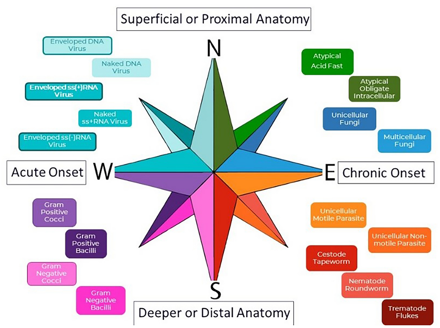

The initial impetus for developing the MedMicroMaps system was to create lecture time in the respiratory infections lecture series to accommodate coverage of the novel SARS-CoV-2 in Fall 2020 by organizing content methodically with consistent themes and paradigm patterns. The MedMicroMaps system uses an arching orientation of cardinal directions north/south/west/east, starting with clinical presentation organized by onset, reading left-to-right/west-to-east for acute (less than 1 week) to chronic (greater than 3 weeks) (Fig. 1, MedMicroMaps compass). The coordinates of top-to-bottom/north-to-south are guides for anatomical location, corresponding to proximal to distal or superficial to deeper organs. The microbe biological classifications are arranged with smallest on top to the largest on the bottom, with viruses on top-left/north-west and helminths on bottom-right/south-east. Furthermore, the microbial colors correspond to staining methods when applicable with Gram-positive bacteria marked as Purple for crystal violet stain and Unicellular Fungi as Blue for lactophenol blue stain. The position of the microbes correlates to disease onset, representing that the microbes in the western quadrants (viruses, typical bacteria) mostly cause acute infections and the microbes in the eastern quadrants (atypical bacteria, fungi, parasites) mostly cause chronic infections.
Fig. 1
MedMicroMaps compass. An interactive guide of infectious diseases on PowerPoint platform was developed for preclinical medical students, using the principles of mind maps and Method of Loci to create a consistent color-coding and spatial patterns arranged on a compass with cardinal directions. North-to-south corresponds to anatomical location and west-to-east corresponds onset of clinical presentation. Color-coded microbes are arranged north-to-south based on smaller to larger, with subset patterns of classification (viruses: DNA on top and RNA on bottom, with border to indicate enveloped and no border for non-enveloped)
The MedMicroMaps sub-sections are organized by organ system and each module starts with a disease presentation for that respective organ system. The key clinical findings align with the process of decision tree logic of clinical reasoning for physicians-in-training, frequently encountered with board-style multiple-choice questions with a clinical vignette [6]. The specific category of disease, ex., acute, upper respiratory infection, has an arrow hyperlink that routes a user to an epidemiological map, listing causative agents of respiratory infections, ranked by severity or incidence, and further clustered by epidemiological factors, ex., elderly versus pediatric, exposure risk, occupation, or medical history (see Supplemental Material, Respiratory MedMicroMaps). The next feature of MedMicroMaps module links the user to the clinical microbiology diagnostic algorithms arranged by biological classifications, based on common patterns for e-learning resources [7]. The Microbe Biology maps use consistent legends with color-coding microbes and pattern organization, ex., DNA on top and RNA on bottom for viruses, Cocci on top and Bacilli on bottom for typical bacteria, Unicellular on top and Multicellular on bottom for fungi and parasites. Lastly, each organ system map has a complete Overview slide to provide consistent outlines for all diseases and a broad guide of the range of infectious agents for a system. The hyperlinks allow the user to toggle back and forth to consider differential diagnoses to include or exclude microbes based on clues provided in a clinical scenario with follow-up laboratory findings.
留言 (0)