Animal Care
All animal protocols were approved by the University of Cincinnati IACUC, Cincinnati, OH. Mice were group-housed in individually ventilated PIV cages maintained on 14 h:10 h light and dark cycles and fed a standard chow diet (Envigo 7912; Harlan Teklad, Madison, WI) with access to water ad libitum unless otherwise specified by individual experimental protocols.
Tg(PDGFB-APPSwInd)J9Lms embryos submitted by Gladstone Institute of Neurological Disease (San Francisco, CA) to Jackson Labs (Bar Harbor, ME) were revived from cryopreservation. Three founder mice were received, and one bred successfully establishing the TgJ9 + mouse line. The Abcg4 KO mice line was generated as previously described (Dodacki et al. 2017). Abcg4 KO mice were bred with TgJ9 + mice to produce Abcg4+/−, J9 + mice. These mice were bred with Abcg4+/−, J9- mice to produce the experimental cohort and ensure no more than a single copy of the J9 transgene was present in any animal.
Genotyping Details
Genomic DNA was isolated from a tail snip. The genotype of Abcg4 allele was determined by PCR using primers (5′-CTGCCCTCCCTTATCAATC-3′) and (5′-TATCACAAGCCAGCCTTCTCGG-3′) to detect a 423 bp fragment for the WT allele, and primers (5′-CTGCCCTCCCTTATCAATC-3′) and (5′- TTGCTCACCATGGTGGCGACCGGTGG-3′) primers were used to detect a 400 bp fragment for the mutant allele. The presence of the J9 transgene was determined by amplifying a 360 bp fragment using primers (PDAPP-F; GGTGAGTTTGTAAGTGATGCC and PDAPP-R; TCTTCTTCTTCCACCTCAGC3). PCR products were run on a 1.5% agarose gel under standard conditions using a 100 bp DNA ladder for size identification. DNA was stained using SYBR Safe DNA gel stain (S33102, Thermo Fisher Scientific, Waltham, MA) and scanned on a gel station (Universal Hood II, Bio-Rad).
Behavioral Testing
Behavioral testing was performed by the University of Cincinnati Mouse Metabolic Phenotyping Center Animal Behavior Core. Mice were assessed at 6–7 months of age and 16–19 months of age.
Open field test: Animals were placed into a novel open field environment (e.g., ~ 36 × 36 in a plastic box or circular field) for up to 30 min. Time spent in the middle and time in the periphery are recorded. Time spent in the periphery near the walls is an indicator of anxiety (Choleris et al. 2001).
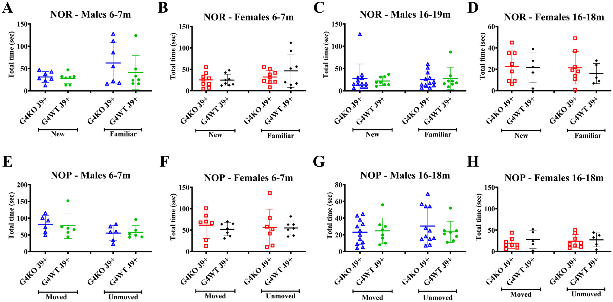
Novel object recognition (NOR): Mice were placed into the open field apparatus, as described above, containing 2 different objects. Animals were given 15 min to explore the objects and returned to their home cages. While the mice were away from the arena, one object was replaced with a different object. The animals were returned to the apparatus 24 h later with one of the former objects and a new object. The difference in the amount of time exploring the new vs. familiar object reflects the memory of the previous experience and the animal’s non-spatial learning (Antunes and Biala 2012).
Novel object placement (NOP): Mice were placed into the open field apparatus, as described above, containing 2 different objects. Animals were given 15 min to explore the objects and returned to their home cages. While the mice were away from the arena, one object was moved to a different location within the apparatus. The animals were returned to the apparatus 24 h later with the same objects, one object in the same location and one object in a different location. The difference in the amount of time exploring the moved vs. unmoved object reflects the memory of the previous experience and the animals’ spatial learning (Antunes and Biala 2012).
Indirect Calorimetry
Mice were individually housed in chambers maintained at 23 °C with 12 h:12 h light:dark cycle for simultaneous measurement of oxygen consumption (vO2, ml/h), carbon dioxide production (vCO2, ml/h), energy expenditure, respiratory exchange ratio (RER), and locomotor activity via indirect calorimetry (TSE Systems, Chesterfield, MO, USA). Energy expenditure was calculated using the simplified Weir equation (H = 1.44 (3.94 VO2 + 1.11 VCO2)), and energy expenditure or heat (H) was expressed as kcal/h. Previously, the instrument was calibrated with gas cylinders containing nitrogen, 1% carbon dioxide, and atmospheric air mixture (oxygen 20.7%, carbon dioxide 0.03%). Data were acquired every 20 min using the LabMaster software (TSE Systems).
Food Intake
Food intake was assessed with a BioDAQ Food intake monitoring system (Research Diets, Inc., New Brunswick, NJ) between the ages 8 and 12 months and 16 and 18 months. Mice were individually housed in BioDAQ cages, which monitored the weight of food in the hopper. Water was provided ad libitum. Cumulative food intake over 3 days was calculated for each mouse in each trial. For each age group, mice were tested twice with a 1-week gap between trials to assess if potential differences in cognitive ability affected the feeding behavior of the mice due to the novel environment of the BioDAQ cage.
Glucose (GTT) and Insulin (ITT) Tolerance Tests
Mice were fasted for 4–6 h and then administered via intraperitoneal injection 2 g/kg glucose for GTT or 0.6 IU/kg human insulin, Humulin R (HI-213; Lilly, Indianapolis, IN) for ITT. Blood glucose was measured with Accu-Chek Nano electronic glucometer (Roche Applied Science, Indianapolis, IN, USA) at 0, 30, 60, 90, and 120 min after intraperitoneal administration of glucose or insulin. If blood glucose dropped below 40 mg/dL, the test was terminated, and the animal was administered glucose.
Measurement of Aß Clearance
A guide stainless steel cannula (22-gauge, Plastics One, Roanoke, VA) was implanted stereotaxically into the right caudate putamen of anesthetized mice. The cannula tip coordinates for placement were 0.9 mm anterior from bregma, 1.9 mm lateral from midline, and 2.9 mm below the surface. Animals were allowed to recover for 4–5 h to allow for some blood-brain barrier recovery, but before substantial inflammatory response develops. Tracer fluid (1.0 μl) containing 50nCi of [14C]-inulin (as reference marker) and 50nCi of [3H]Aβ1–42 was injected with a Motorized Integrated Stereotaxic Injector (iSi) system (Stoelting Co.) into the interstitial fluid (ISF) over 5 min. After injection, the needle was left in place for 5 min. CSF was collected 60 min after injection, and brain tissue from the caudate putamen was collected immediately after.
Thio-S Staining for Plaques
After completing the experiments, the mice were euthanized by CO2 anesthesia followed by thoracotomy to expose the heart. Mice were perfused with chilled PBS followed by 4%PFA via the intracardiac route. The perfused brains were collected and fixed in 4% PFA for another 24 h. Then transferred to 30% sucrose and stored at 4 °C for at least 48 h. Sagittal sections (~ 30 µm) through the hippocampus were obtained and stained with 0.5% ThioS in 50% EtOH for 10 min at room temperature. Sections were rinsed with 50% EtOH, followed by PBS, and mounted on glass slides with aqueous mounting media, then were protected from light until visualized by fluorescence microscopy and the number of plaques counted. Separate sections were additionally stained with AntiNeuN antibody conjugated to Alexa Fluor555 (Millipore Sigma, Cat#MAB377A5) at 1:100 dilution to visualize neuronal structure.


留言 (0)