
記住我


And always with a heart contusion
Arise both doubt and much confusion
– Howard B. Burchell, MD1
Blunt cardiac injury (BCI) encompasses a wide spectrum, from occult and inconsequential contusion to rapidly fatal cardiac rupture. A small percentage of patients present with abnormal electrocardiogram (ECG) or shock, but most are initially asymptomatic. The potential for sudden dysrhythmia or cardiac pump failure mandates consideration of the presence of BCI, including appropriate monitoring and management.
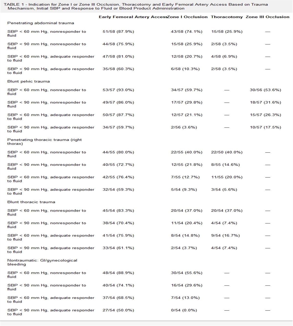
DEFINITIONSThe term myocardial contusion has historically been broadly and nonspecifically applied to BCI, but it is a distinct pathologic entity (see section Pathophysiology hereinafter). Blunt cardiac injury refers to the entire spectrum of cardiac and pericardial injuries resulting from trauma, ranging from asymptomatic myocardial contusion to cardiac rupture.2Significant BCI (SigBCI) is used to identify those injuries requiring specific interventions (e.g., antidysrhythmic, vasoactive, or cardiotonic therapies; mechanical circulatory assistance; surgical repair). The American Association for the Surgery of Trauma Organ Injury Scale classification of cardiac injury was published in 1994 (Fig. 1).3
 Figure 1: Cardiac organ injury scale. Reproduced with permission from Moore et al.3INCIDENCE
The overall incidence of BCI is difficult to ascertain given the variability in diagnostic criteria. If one considers any ECG abnormality or cardiac enzyme elevation to be diagnostic of BCI, the reported incidence may seem relatively high. On the other hand, SigBCI is much less common. For example, Biffl et al.4 studied 359 patients admitted with suspected BCI, of whom 107 (30%) had abnormal ECG or cardiac enzyme elevations. Only 17 patients, 5% of the study population and 16% of those receiving the diagnosis of BCI, had dysrhythmias or cardiac pump failure requiring intervention (i.e., SigBCI). In a 1996 meta-analysis by Maenza et al.,5 there were 170 patients (3.6%) with SigBCI out of 4,681 patients admitted with suspected myocardial contusion. In the National Trauma Data Bank for the years 2017 to 2021, 14,219 patients were diagnosed with BCI among 4.8 million blunt trauma patients, for an overall incidence of 0.3%.6
Autopsy studies have identified BCI as a contributing factor in a large percentage of prehospital deaths. In a Los Angeles County autopsy study, 96 of 304 blunt trauma fatalities (32%) had BCI, including 78% of those who died at the injury scene.7 In a study of 61 fatal falls from height, cardiac injuries were found in 33 (54%) of patients. In nearly half of the BCI cases, the cardiac injuries were the cause or a contributing factor to death.8
PATHOPHYSIOLOGYThe most common injury mechanisms associated with SigBCI are motor vehicle crashes (MVCs), pedestrians struck by automobiles, motorcycle crashes, and falls.7–9 In the autopsy study of Teixeira et al.,7 48 (50%) of the BCI victims had been involved in an MVC, and 35% were pedestrians struck by automobiles. Another 9% were involved in motorcycle crashes, and 3% had fallen. Most commonly, there is either a direct blow to the chest with a concussive force transmitted to the heart, impact of the heart with the sternum, or compression between the sternum and spine. The wall of the right ventricle (RV), given its anterior orientation and proximity to the sternum, is most often affected. Before widespread use of automotive safety devices such as shoulder harnesses and airbags, a damaged steering wheel was a marker of potential BCI. The “seatbelt syndrome” was described in the 1980s, with cautions about the risk of BCI related to seatbelt use and an association with sternal fractures.10,11 Sternal fractures are not in and of themselves associated with BCI;12–14 in fact, they may be protective, absorbing force that might otherwise be transmitted to the heart.15,16
Blast injuries or lateral chest wall trauma can also lead to BCI, with transmission of concussive forces and lacerations from fractured rib fragments. Deceleration injuries (e.g., after high-speed MVC or fall from height) can cause tearing at points of fixation such as the pericardium or atriocaval junction. Sudden increase in venous pressure transmitted from the abdomen or periphery may cause cardiac rupture (atria in particular) or valve injury via hydraulic forces. Given the higher pressures in the left heart, the mitral and aortic valves are at greater risk of damage compared with the pulmonary and tricuspid valves.
The severity of cardiac injury generally relates to the severity of the applied force. The timing of impact is important as well; for example, impact during end-diastole is believed to create higher risk for valve, septum, or ventricular free wall rupture, whereas atrial rupture is more likely to occur near end-systole when valves are closed.9 Depending on the timing in the cardiac cycle, even a relatively minor blow can trigger a lethal dysrhythmia (i.e., commotio cordis). The pathophysiology of graded impact injury to the heart was evaluated by Baxter et al.17 in a standard Langendorff preparation. They found that complete electrical arrest occurred immediately following impact in all hearts, with time to recovery correlating directly with the magnitude of injury. A similar graded decrease in ventricular contractility was observed. In all hearts, coronary artery blood flow was decreased immediately after impact but normalized by 20 minutes. Importantly, they observed no delayed ventricular arrhythmias after 24 hours. This finding corroborates other animal studies18,19 and is consistent with clinical experience, in which delayed complications are rare.
A myocardial contusion may be identified grossly by a hemorrhagic appearance of the epicardial surface. Histologically, it is marked by patchy myocardial cellular necrosis confined to specific muscle bundles, with infiltrates of polymorphonuclear leukocytes.20 In contrast to an area of infarction, which is typically associated with coagulation necrosis, the transition zone between affected and healthy tissue is more abrupt in the contusion injury. The contused area heals with patchy scar interspersed with normal myocardium, indicative of normal or enhanced circulation.19,20
Doty et al.19 and Tenzer20 both distinguished cardiac concussion from myocardial contusion based on a lack of myocardial cellular damage in the concussive injury. However, cardiac concussion is not a benign entity because it can trigger a range of dysrhythmias, including commotio cordis. Doty et al.19 wrote of the concussive blow, “This phenomenon occasionally finds useful purpose in cardiac resuscitation as the ‘precordial thump.’”
Commotio cordis deserves special mention, as it was thrust into the public eye in January 2023, when the on-field cardiac arrest of Buffalo Bills football player Damar Hamlin was witnessed by an estimated 21 million television viewers. The prevailing theory was that it was due to commotio cordis.21 Commotio cordis is triggered by a precordial blow delivered during a narrow window during ventricular repolarization, between 30 and 15 milliseconds before the peak of the T wave.22 The blow is believed to stimulate premature ventricular depolarization from the activation of K+ ATP channels.21 Of note, in their commotio cordis studies, Link et al.22 found no evidence of myocardial contusion on histologic examination in any animals.
DIFFERENTIAL DIAGNOSISAside from chest wall pain due to trauma, many patients with BCI are initially asymptomatic. In the patient with ECG abnormality or shock, it is important to differentiate BCI from acute coronary syndrome, heart failure exacerbation, or other nontraumatic cardiac problem. It is also important to determine whether a cardiac event led to the traumatic event. Any cardiac symptoms warrant aggressive diagnostic evaluation.
DIAGNOSTIC APPROACHA major focus of the BCI literature over the past few decades has been the appropriate diagnostic evaluation and monitoring of the asymptomatic patient at risk for BCI. Unfortunately, the interpretation of the body of work is confounded by the lack of consensus on the diagnostic criteria for BCI. Given the benign inconsequential nature of many injuries, it is not the diagnosis of BCI per se that is important; rather, it is the identification and prompt treatment of SigBCI. Many SigBCIs are clinically apparent early in the patient's course, with new ECG changes, dysrhythmias, or signs of cardiac dysfunction. However, they may manifest later: in one series, 35% of SigBCI did not become evident until more than 6 hours after presentation.4 A variety of diagnostic modalities may be used.
Physical ExaminationPatients with benign myocardial contusion may have signs of chest wall trauma but typically have no specific signs or symptoms. Any murmur or abnormal pulse examination requires additional study.
ElectrocardiogramThe ECG is the cornerstone of evaluation of the patient with possible BCI, and it is recommended at the time of presentation in all patients with significant thoracic trauma.12 Sinus tachycardia or other nonspecific ECG changes are relatively common and do not correlate with BCI or SigBCI. An abnormal ECG (e.g., new bundle branch block, premature ventricular contractions, ST segment or T-wave changes) does not necessarily predict SigBCI either, but it warrants further evaluation. Conversely, a normal ECG is reassuring but does not completely rule out BCI.23–26 Right-sided ECG (i.e., V4R) and signal-averaged ECG do not improve the prediction of SigBCI.24,27 Continuous ECG monitoring is recommended for the patient being admitted with possible BCI, as dysrhythmias may occur in a delayed manner. In the study of Biffl et al.,4 SigBCI manifested as late as 22 hours after presentation.
Cardiac EnzymesOne of the most heavily debated areas in the management of BCI is the role of cardiac enzymes—what to measure, when to measure it, and what to do with the result. The value of creatine phosphokinase and its myocardial band isoenzyme levels was questioned over 30 years ago.4,28,29 Given its poor predictive value for SigBCI, routine measurements are not recommended.12,30 In 1992, Mair et al.31 recommended cardiac troponin (Tn) T as a superior biomarker for BCI, citing its sensitivity, cardiospecificity, and release kinetics, as well as availability of a commercial-use kit. It heralds even small amounts of myocardial necrosis, increases a few hours after the onset of myocardial damage, and remains elevated for several days. Troponin I and TnT are both measured to assess myocardial damage in acute coronary syndromes, and they appear to be equivalent in their clinical utility. However, neither has been demonstrated to reliably predict SigBCI.
The 2012 Eastern Association for the Surgery of Trauma guidelines12 recommended serial measurement of TnI but offered no evidence supporting such a recommendation. In the authors' experience, serial testing of asymptomatic patients seems to only lead to more resource utilization (e.g., echocardiograms, cardiology consultations, prolonged observation). A useful indication for Tn measurement is to rule out BCI, allowing discharge from the ED in patients who do not otherwise require admission.12,23 High-sensitivity cardiac Tn assays have been developed in an attempt to make earlier, more accurate diagnoses of myocardial infarction (MI) and other damage. It remains to be seen whether any of these assays may predict SigBCI.32,33
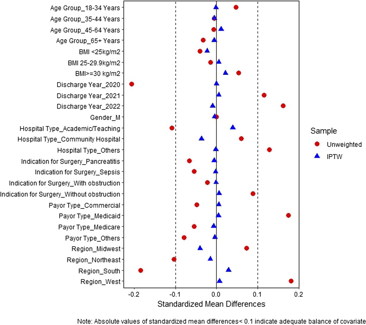
EchocardiographyEchocardiography is an excellent tool for evaluating overall cardiac performance and identifying specific wall motion abnormalities, pericardial tamponade, and structural abnormalities such as valvular and septal injuries (Fig. 2).34,35 As such, it is a critically important test in the patient with SigBCI or other cardiac problem. However, it is not recommended for routine screening and is not necessary in the asymptomatic patient with isolated cardiac enzyme elevation or nonspecific/benign ECG changes.
 Figure 2: Echocardiographic images demonstrating traumatic atrial septal defect (panel 1), ruptured papillary muscle and flail mitral valve leaflet (panel 2), and color flow Doppler of mitral regurgitation (panel 3). Reproduced from Menaker et al.34Cardiac Computed Tomography or Magnetic Resonance Imaging
The 2012 Eastern Association for the Surgery of Trauma guidelines recommended computed tomography (CT) or magnetic resonance imaging to help differentiate acute MI from BCI and determine the need for cardiac catheterization or anticoagulation.36,37 Imaging with CT is performed liberally in patients with chest trauma, and it will often detect pericardial tears, pericardial effusion, and potentially other cardiac injuries.38 Multidetector-row computed tomography with ECG-gating capability, as well as multidetector-row computed tomography angiography, represent technological advances that enhance evaluation of coronary anatomy and myocardial perfusion. There are clear advantages of CT over magnetic resonance imaging, and it may help differentiate between BCI and acute MI.38 However, we submit that the differentiation is better made by a clinician and recommend cardiology consultation if there is such a diagnostic dilemma.
Radionuclide ImagingDoty et al.,19 citing the difficulty of BCI diagnosis based on ECG and cardiac enzymes, recommended radionuclide imaging to detect even mild injuries. However, it was subsequently determined that this test was not able to reliably predict SigBCI, so there is currently minimal role for this modality in BCI evaluation.
OVERVIEW OF SIGBCI Electrical DisturbancesCardiac dysrhythmias and conduction blocks may occur as a result of BCI or in association with other thoracic trauma; they may also represent underlying cardiac disease.39 The type of disturbance in BCI is dependent on the injured portion of the heart.40 The most common ECG abnormality in BCI is sinus tachycardia;41 other dysrhythmias occur in 1% to 6% of patients, with atrial fibrillation being the most common.39,42 Atrial fibrillation occurs commonly in critically ill and injured patients for a variety of reasons, and the presence of atrial fibrillation alone does not indicate a patient has BCI. Hadjizacharia et al.42 demonstrated that the incidence of atrial fibrillation after thoracic trauma is the same as after abdominal and head trauma. Supraventricular tachycardia and ventricular dysrhythmias are less common but do occur following BCI. Ventricular fibrillation (VF) is typically seen in commotio cordis; sustained ventricular tachycardia is very uncommon in the absence of coronary artery or other structural heart disease.43 Bundle branch block is another ECG abnormality seen in BCI.41 Because of the proclivity for injury to the RV, a right bundle branch block is the most common conduction disturbance, followed by first-degree atrioventricular nodal block.43,44
Cardiac Hypokinesis/Pump FailureA hallmark finding in BCI is a hypokinetic segment of myocardium, which can compromise cardiac output. This may be demonstrated on various imaging studies, but echocardiography is a reliable diagnostic tool.
Pericardial LacerationPericardial lacerations may bleed, creating pericardial effusion. If large enough, a tear allows herniation of the heart, which can significantly compromise cardiac function to the point of cardiac arrest (Fig. 3).45 If pericardial injury is combined with a cardiac laceration, there can be exsanguination into the pleural or peritoneal cavity without pericardial tamponade. Fulda et al.9 reported a series of 59 patients who presented with cardiac or pericardial rupture: isolated cardiac chamber rupture in 37, isolated pericardial tear in 17, and both in 5 patients. Of the 22 with pericardial tears, 6 had cardiac herniation. In one series of patients suffering fatal falls,8 the most frequent cardiac injury was pericardial tearing, found in 15 patients (45%); additional cardiac injuries were found in 14 of the 15 with pericardial lacerations.
 Figure 3: Pericardial rupture with cardiac herniation. Reproduced from Heelan Gladden et al.45Septal Rupture
Septal injuries from blunt chest trauma have been described in both the intra-atrial and intraventricular septae, but they are uncommon injuries.34,46,47 In the autopsy series from Los Angeles County, 10 of 96 patients (10%) with BCI were found to have an injury to the septum.7
Valve RuptureSignificant injury to cardiac valves will result in valvular regurgitation. The most commonly injured cardiac valve is the aortic valve, followed by mitral, tricuspid, and pulmonary valves.48,49 The aortic valve cusps may be torn or avulsed when a sudden increase in intrathoracic pressure leads to increase in aortic pressure, usually in early diastole. Acute severe cardiac failure with fulminant pulmonary edema may ensue, but a milder injury may present with syncope or angina. The characteristic decrescendo diastolic murmur may not be present; echocardiography is the most reliable way to identify this injury.43 Compression of the heart in early systole during isovolumetric contraction may result in tears of mitral valve leaflets or rupture of papillary muscles or chordae tendinea. A holosystolic murmur will be heard and acute heart failure may ensue, with shock and pulmonary edema. Less severe injuries may present more insidiously, with development of heart failure symptoms over weeks to months. Tricuspid valve injuries can occur when there is a sudden increase in intrathoracic pressure during systole, causing avulsion of papillary muscles.45 An association of traumatic tricuspid injury and heart block has been described.50 The associated heart block may range from first degree to complete heart block requiring transvenous pacing.50
Coronary Artery InjuryCoronary artery dissection, occlusion, laceration, and aneurysm formation have all been described and may result in symptoms ranging from angina to sudden death.51–55 Typical ST-segment elevations will be seen on ECG, and revascularization is required.
Cardiac Wall RuptureCardiac rupture is the most severe form of BCI, and the vast majority are lethal within minutes (Fig. 4).8 Patients who reach the hospital alive will typically have a pericardial effusion or tamponade, and the cardiac rupture is diagnosed intraoperatively. In 1864, Morel-Lavallee56 described a murmur that sounds like a splashing mill wheel, calling it “bruit de moulin”; however, it is rarely reported.9 Many cardiac ruptures are associated with a pericardial laceration, so there is free pleural or peritoneal bleeding. In the autopsy study of Teixeira et al.,7 transmural rupture was present in 61 (64%) of the deceased patients with BCI. The distribution was right atrium in 30%, RV in 27%, left ventricle (LV) in 19%, and left atrium in 17%, with multiple chambers ruptured in 26% of patients. In a study of fatal falls from height, cardiac rupture occurred in right atrium and RV in 39% each, left atrium in 18%, and LV in 9%.8 The size of the transmural tear was generally larger with higher heights (<1 cm when falling <15 m; more extensive and irregular when higher than 15 m). Higher heights are also associated with ventricular rupture (RV usually >15 m and LV >25 m).8
 Figure 4: Transmural tear of the RV. Reproduced from Türk et al.8Commotio Cordis
Commotio cordis is the third leading cause of sudden cardiac death among young healthy persons, generally occurring during participation in sports. Baseball accounts for one-half of cases, followed by softball and football (11% each).21 Overall, fewer than 20 cases are reported in the United States each year.21,57 Studies from the 1970s to 1990s reported survival in the 10% to 15% range, but Maron and colleagues58 reported 58% survival in recent years, attributed to awareness and public cardiopulmonary resuscitation education. Link et al.22 studied the delivery of regulation baseballs at 20 to 70 mph and found the energy of impact to be an important factor in the creation of commotio cordis, with impacts of 40 mph most likely to trigger VF. While VF was less common with velocities >40 mph, the investigators found higher rates of ST-segment elevation, bundle branch block, transient heart block, and structural injuries (papillary muscle tears, myocardial rupture) associated with 50 to 70 mph blows.
MANAGEMENT OF THE PATIENT WITH POSSIBLE BCIThe initial evaluation of the injured patient should follow the principles outlined in the American College of Surgeons' Advanced Trauma Life Support course.59 In the patient with shock, all etiologies should be considered. This is particularly true among patients who have sustained thoracic trauma, in whom cardiogenic and cardiac compressive problems are more likely. Physical examination, ECG tracing, and extended focused assessment with sonography for trauma should identify life-threatening problems such as pericardial tamponade and hemodynamically significant dysrhythmias (Fig. 5).
 Figure 5:
 Management algorithm for BCI.
The Hemodynamically Abnormal Patient Pericardial Tamponade and Cardiac RuptureIn the patient in shock with pericardial tamponade or the patient in extremis following thoracic trauma, immediate resuscitative thoracotomy is indicated to decompress the pericardium and control the source of bleeding. Cardiac chamber rupture may be discovered in this setting. Surgical repair offers the only hope of survival of patients with blunt cardiac rupture. Upon recognition of the rupture, trans-sternal extension (i.e., bilateral anterolateral, or “clamshell” thoracotomy) is recommended to enhance exposure including access to posterior tears. Small lacerations may be controlled digitally while suture repair is performed. Atrial or vena caval injuries may be controlled with a Satinsky clamp to facilitate repair. Larger wounds, or those in the ventricle, may be controlled with temporary balloon occlusion, but care must be taken to avoid extending the tear with the balloon. The patient may require temporary caval inflow occlusion to perform large repairs.
Pericardiocentesis is discouraged as primary therapy for tamponade; however, there may be a role in the patient with marginal hemodynamic stability and delayed access to surgical intervention. The presence of pericardial blood under pressure may cause subendocardial ischemia and lead to recalcitrant dysrhythmia; thus, decompression with placement of a drainage catheter is recommended to maintain hemodynamic stability until surgery can be performed. Ultrasound guidance is recommended for pericardiocentesis to enhance success and avoid complications such as cardiac laceration. Median sternotomy is reasonable in the patient with a high likelihood of isolated cardiac injury, but it is not as versatile or as expedient as anterolateral thoracotomy; thus, it is not recommended for the patient in severe shock. Large pericardial tears should be repaired to avoid cardiac herniation; a patch graft may be required. Small tears may be left alone or repaired primarily.9
Significant DysrhythmiasSignificant dysrhythmias should be treated expediently per Advanced Cardiac Life Support protocols, with cardioversion in unstable patients.60 Atrial fibrillation is treated with β-antagonists or calcium channel blockers for rate control. Supraventricular tachycardia may be slowed with vagal maneuvers or atrioventricular nodal blockade (e.g., adenosine). β-Blockade can be effective in recurrent cases. Ventricular dysrhythmias, particularly VF, demand immediate defibrillation to restore a normal rhythm. Survivors of commotio cordis will usually arrive in the hospital with a perfusing rhythm and only need monitoring for recurrent problems. Patients with sustained ventricular tachycardia should be evaluated for coronary artery disease or other structural heart disease.43 Most cases of conduction disturbances are transient and benign, but in the setting of complete heart block, pacemaker placement may be necessary to maintain adequate heart rate and prevent hemodynamic instability.61
Acute Cardiogenic ShockAcute circulatory failure may be due to primary cardiac failure or structural lesions such as valve, septum, or coronary artery injuries. Patients with pump failure will require some combination of volume loading, inotropic medication, or mechanical circulatory assistance. Invasive hemodynamic monitoring is required in most such cases. In more severe cases of pump failure, short-term mechanical circulatory assist devices may be needed. Percutaneous placement of certain devices allows for rapid deployment in the unstable patient. Device options include the intra-aortic balloon pump,62 percutaneous transvalvular microaxial support devices (Impella), and extracorporeal membrane oxygenation (ECMO).63 With the need to anticoagulate patients on ECMO, bleeding and thrombosis remain the most common complications with the utilization of ECMO in the trauma patient. Surgically placed centrifugal pumps may also be considered.
Acute aortic valve injury will require emergent surgical repair, as mechanical circulatory support devices are contraindicated. In contrast, acute severe mitral valve injury may be managed with aortic balloon counterpulsation and vasodilator therapy until surgical repair can be performed.43 Coronary artery injury will likely require a revascularization procedure.
The Hemodynamically Normal PatientIn the hemodynamically normal patient, 12-lead ECG should be obtained, but echocardiography may be performed selectively. We generally reserve echocardiography for stable patients in whom there is some evidence of organ hypoperfusion such as persistent metabolic acidosis, oliguria, orthostatic changes in vital signs, and hyperlactatemia.
Electrical DisturbancesTreatment of dysrhythmias and conduction abnormalities in BCI is the same as in the non-BCI patient, in accordance with Advanced Cardiac Life Support guidelines (see previous section). Attention should be paid to replacement of electrolytes such as potassium and magnesium and correction of metabolic disturbances and hypoxia. Sinus tachycardia is a physiologic and compensatory response to trauma and should not be treated with β-antagonists; the underlying cause should be sought and addressed.
Pericardial EffusionAn effusion in the absence of tamponade physiology needs to be investigated but does not require emergent thoracotomy. Timely intervention is recommended, as tamponade could develop or there could be ongoing hemorrhage into the pleural or peritoneal cavity. If the patient remains stable, subxiphoid pericardial window may be performed. A large amount of blood or ongoing bleeding requires sternotomy or thoracotomy. If a small amount of blood clears with irrigation, that is reassuring. Thoracoscopy may be considered if there is a hemothorax, to rule out a pericardial laceration and cardiac bleeding into the pleural space.
Cardiac HypokinesisEnsuring appropriate volume status and inotropic support is the primary management strategy for cardiac hypokinesis after BCI. Invasive hemodynamic monitoring may be helpful in managing volume status and guiding the selection of vasoactive agents.
Trauma patients with BCI may require urgent or emergent noncardiac surgery. If the patient can wait until hemodynamic stability, that is preferable; otherwise, surgery can proceed with appropriate hemodynamic monitoring. The anesthesiologist must be made aware of the risks of dysrhythmia and cardiac dysfunction so that management can be tailored to treat potential cardiogenic shock rather than presumed hemorrhagic shock. Pulmonary artery catheterization may be warranted in certain circumstances.
Valve/Septum/Coronary Artery InjuriesEven if initially hemodynamically normal, patients with these injuries usually require surgical management. Some patients have insidious worsening of physiology. Cardiothoracic surgery consultation is recommended.
Persistent ECG ChangesPatients with persistent sinus tachycardia or other new ECG changes or any patient who requires admission for another reason should be monitored with telemetry for 24 hours. Serial cardiac enzymes and echocardiography are not recommended. If a SigBCI manifests, it is treated as described previously.
The Asymptomatic Patient With Nonspecific ECG ChangesThe asymptomatic patient may be discharged from the emergency department if their ECG has normalized or has nonspecific changes. Because SigBCI may manifest later and a normal ECG cannot rule out BCI with certainty, Tn should be measured 8 hours postinjury. If it is normal, the patient can be safely discharged from the emergency department.12,23 If Tn is elevated, the patient should be kept for observation. Serial Tn measurements are not recommended because they are not predictive of SigBCI; similarly, echocardiography is unnecessary and not recommended, as it will not change management in the asymptomatic patient. The patient should be monitored with telemetry for 24 hours.
OUTCOMES AND LONG-TERM MANAGEMENTThe long-term prognosis after BCI is dependent on the presence and type of SigBCI. Structural injuries are very uncommon, and surgery may be curative. However, significant myocardial contusion, or coronary artery injuries, may lead to chronic heart failure or other issues. Doty et al.19 described four patients with ventricular aneurysms after severe myocardial contusion, but there have been only a few other case reports that have documented this outcome.64–66 In fact, the data regarding the long-term outcomes of BCI are limited. In one prospective study, patients who were diagnosed with BCI were followed up at 6 months by phone call.67 All patients reported normalization at follow-up with no mortalities, malignant arrhythmias, or heart failure. Of note, of 12 patients found to have an abnormal radionuclide scan (right ventricular ejection fraction less than 40% and/or wall motion abnormality), only 25% (3 of 12) were contacted for follow-up.67 Another prospective study evaluated 12 patients who sustained a myocardial contusion 12 months prior and compared them to 12 matched patients who sustained blunt chest trauma without myocardial contusion. The two groups were indistinguishable with regard to ECG, RV, and LV function.68 More recently, a prospective study evaluated patients with blunt thoracic trauma at 3 and 12 months. Of those with a myocardial contusion and wall motion abnormalities, 10 of 17 had persistent wall motion abnormalities at 3 months and only 4 of 17 at 12 months. In addition, with exercise testing, there were no ECG abnormalities, and none had limitations from a cardiac source at follow-up.69
Postpericardiotomy syndrome is an inflammatory process involving the pleura and pericardium. It can present with fever, chest pain, pericardial rub, ECG changes, and pericardial effusion. It is generally effectively treated with anti-inflammatory agents.70
While individuals with BCI generally have a good long-term outcome, it is important to note that research on the long-term prognosis of BCI is limited. There currently is not a consensus on the best long-term follow-up strategy. Based on the current literature, routine cardiac follow-up is probably not necessary in the asymptomatic patient. Follow-up echocardiography is not necessary in the patient who has had clinical resolution of BCI signs or symptoms. Its use should be individualized among patients who have undergone surgical or catheter-based interventions or who have persistent cardiac issues.
AUTHORSHIPAll authors contributed in the literature review, interpretation of the literature, and drafting and critical revision of the manuscript.
DISCLOSUREConflicts of Interest: Author Disclosure forms have been supplied and are provided as Supplemental Digital Content (https://links.lww.com/TA/D417).
REFERENCES 1. Burchell HB. Unusual forms of heart disease. Circulation. 1954;10:574–579. 2. Mattox KL, Flint LM, Carrico CJ, Grover F, Meredith J, Morris J, et al. Blunt cardiac injury. J Trauma. 1992;33:649–650. 3. Moore EE, Malangoni MA, Cogbill TH, Shackford SR, Champion HR, Jurkovich GJ, et al. Organ injury scaling. IV: thoracic vascular, lung, cardiac, and diaphragm. J Trauma. 1994;36:299–300. 4. Biffl WL, Moore FA, Moore EE, Sauaia A, Read RA, Burch JM. Cardiac enzymes are irrelevant in the patient with suspected myocardial contusion. Am J Surg. 1994;168:523–528. 5. Maenza RL, Seaberg D, D’Amico F. A meta-analysis of blunt cardiac trauma: ending myocardial confusion. Am J Emerg Med. 1996;14:237–241. 6. American College of Surgeons Committee on Trauma. National Trauma Data Bank Participant Use File (2017–2021). Chicago, IL: American College of Surgeons; 2023. 7. Teixeira PGR, Georgiou C, Inaba K, Dubose J, Plurad D, Chan LS, et al. Blunt cardiac trauma: lessons learned from the medical examiner. J Trauma. 2009;67:1259–1264. 8. Türk EE, Tsokos M. Blunt cardiac trauma caused by fatal falls from height: an autopsy-based assessment of the injury pattern. J Trauma. 2004;57:301–304. 9. Fulda G, Brathwaite CE, Rodriguez A, Turney SZ, Dunham CM, Cowley RA. Blunt traumatic rupture of the heart and pericardium: a ten-year experience (1979–1989). J Trauma. 1991;31:167–173; discussion 172-3. 10. Muwanga CL, Cole RP, Sloan JP, Bruce E, Dove AF, Dave SH. Cardiac contusion in patients wearing seat belts. Injury. 1986;17:37–39. 11. Hamilton JR, Dearden C, Rutherford WH. Myocardial contusion associated with fracture of the sternum: important features of the seat belt syndrome. Injury. 1984;16:155–156. 12. Clancy K, Velopulos C, Bilaniuk JW, Collier B, Crowley W, Kurek S, et al. Screening for blunt cardiac injury: an Eastern Association for the Surgery of Trauma practice management guideline. J Trauma Acute Care Surg. 2012;73:S301–S306. 13. Wiener Y, Achildiev B, Karni T, Halevi A. Echocardiogram in sternal fracture. Am J Emerg Med. 2001;19:403–405. 14. Athanassiadi K, Gerazounis M, Moustardas M, Metaxas E. Sternal fractures: retrospective analysis of 100 cases. World J Surg. 2002;26:1243–1246. 15. Yilmaz EN, van Heek NT, van der Spoel JI, Bakker FC, Patka P, Haarman HJ. Myocardial contusion as a result of isolated sternal fractures: a fact or a myth? Eur J Emerg Med. 1999;6:293–295. 16. Roy-Shapira A, Levi I, Khoda J. Sternal fractures: a red flag or a red herring? J Trauma. 1994;37:59–61. 17. Baxter BT, Moore EE, Synhorst DP, Reiter MJ, Harken AH. Graded experimental myocardial contusion: impact on cardiac rhythm, coronary artery flow, ventricular function, and myocardial oxygen consumption. J Trauma. 1988;28:1411–1417. 18. Liedtke AJ, Allen RP, Nellis SH. Effects of blunt cardiac trauma on coronary vasomotion, perfusion, myocardial mechanics, and metabolism. J Trauma. 1980;20:777–785. 19. Doty DB, Anderson AE, Rose EF, Go RT, Chiu CL, Ehrenhaft JL. Cardiac trauma: clinical and experimental correlations of myocardial contusion. Ann Surg. 1974;180:452–460. 20. Tenzer ML. The spectrum of myocardial contusion: a review. J Trauma. 1985;25:620–627. 21. Peng T, Derry LT, Yogeswaran V, Goldschlager NF. Commotio Cordis in 2023. Sports Med. 2023;53:1527–1536. 22. Link MS, Wang PJ, Pandian NG, Bharati S, Udelson JE, Lee MY, et al. An experimental model of sudden death due to low-energy chest-wall impact (commotio cordis). N Engl J Med. 1998;338:1805–1811. 23. Velmahos GC, Karaiskakis M, Salim A, Toutouzas KG, Murray J, Asensio J, et al. Normal electrocardiography and serum troponin I levels preclude the presence of clinically significant blunt cardiac injury. J Trauma. 2003;54:45–51. 24. Fulda GJ, Giberson F, Hailstone D, Law A, Stillabower M. An evaluation of serum troponin T and signal-averaged electrocardiography in predicting electrocardiographic abnormalities after blunt chest trauma. J Trauma. 1997;43:304–312; discussion 310-2. 25. García-Fernández MA, López-Pérez JM, Pérez-Castellano N, Quero LF, Virgós-Lamela A, Otero-Ferreiro A, et al. Role of transesophageal echocardiography in the assessment of patients with blunt chest trauma: correlation of echocardiographic findings with the electrocardiogram and creatine kinase monoclonal antibody measurements. Am Heart J. 1998;135:476–481. 26. Salim A, Velmahos GC, Jindal A, Chan L, Vassiliu P, Belzberg H, et al. Clinically significant blunt cardiac trauma: role of serum troponin levels combined with electrocardiographic findings. J Trauma. 2001;50:237–243. 27. Walsh P, Marks G, Aranguri C, Williams J, Rothenberg SJ, Dang C, et al. Use of V4R in patients who sustain blunt chest trauma. J Trauma. 2001;51:60–63. 28. Baxter BT, Moore EE, Moore FA, McCroskey BL, Ammons LA. A plea for sensible management of myocardial contusion. Am J Surg. 1989;158:557–562; discussion 561-2. 29. Fabian TC, Cicala RS, Croce MA, Westbrook LL, Coleman PA, Minard G, et al. A prospective evaluation of myocardial contusion: correlation of significant arrhythmias and cardiac output with CPK-MB measurements. J Trauma. 1991;31(5):653–660 discussion 659-60. 30. Kyriazidis IP, Jakob DA, Vargas JAH, Franco OH, Degiannis E, Dorn P, et al. Accuracy of diagnostic tests in cardiac injury after blunt chest trauma: a systematic review and meta-analysis. World J Emerg Surg. 2023;18(1):36. 31. Mair J, Dienstl F, Puschendorf B. Cardiac troponin T in the diagnosis of myocardial injury. Crit Rev Clin Lab Sci. 1992;29:31–57. 32. Masseli F, Masseli M, Dolscheid-Pommerich R, Stoffel-Wagner B, Schiller W. Comparison of perioperative high-sensitive troponin T and troponin I assays in cardiac surgery. Heart Lung Circ. 2022;31:407–414. 33. Nestelberger T, Boeddinghaus J, Lopez-Ayala P, Kaier TE, Marber M, Gysin V, et al. Cardiovascular biomarkers in the early discrimination of type 2 myocardial infarction. JAMA Cardiol. 2021;6:771–780. 34. Menaker J, Tesoriero RB, Hyder M, Sikorski R, Scalea TM. Traumatic atrial septal defect and papillary muscle rupture requiring mitral valve replacement after blunt injury. J Trauma. 2009;67:1126. 35. Chirillo F, Totis O, Cavarzerani A, Bruni A, Farnia A, Sarpellon M, et al. Usefulness of transthoracic and transoesophageal echocardiography in recognition and management of cardiovascular injuries after blunt chest trauma. Heart. 1996;75:301–306. 36. Malbranque G, Serfaty JM, Himbert D, Steg PG, Laissy JP. Myocardial infarction after blunt chest trauma: usefulness of cardiac ECG-gated CT and MRI for positive and aetiologic diagnosis. Emerg Radiol. 2011;18:271–274. 37. Southam S, Jutila C, Ketai L. Contrast-enhanced cardiac MRI in blunt chest trauma: differentiating cardiac contusion from acute peri-traumatic myocardial infarction. J Thorac Imaging. 2006;21:176–178. 38. Co SJ, Yong-Hing CJ, Galea-Soler S, Ruzsics B, Schoepf UJ, Ajlan A, et al. Role of imaging in penetrating and blunt traumatic injury to the heart. Radiographics. 2011;31:E101–E115. 39. Ismailov RM, Ness RB, Redmond CK, Talbott EO, Weiss HB. Trauma associated with cardiac dysrhythmias: results from a large matched case-control study. J Trauma. 2007;62:1186–1191. 40. Yousef R, Carr JA. Blunt cardiac trauma: a review of the current knowledge and management. Ann Thorac Surg. 2014;98:1134–1140. 41. Marcolini EG, Keegan J. Blunt cardiac injury. Emerg Med Clin North Am. 2015;33:519–527.
留言 (0)