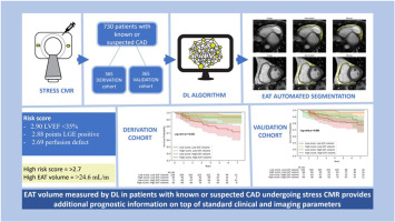
DEep LearnIng-based QuaNtification of Epicardial Adipose Tissue predicts MACE in patients undergoing stress CMR
Stress cardiac magnetic resonance (CMR) is a powerful diagnostic tool with remarkable accuracy in identifying functionally obstructive lesions and strong predictive power for cardiovascular events in patients with known or suspected coronary artery disease (CAD) 1.
Stress-induced myocardial ischemia and the presence of late gadolinium enhancement (LGE) have emerged as key predictive factors. They demonstrate a significant correlation with increased mortality rates and risks of adverse events. Conversely, normal stress CMR results are associated with a good prognosis for at least 3.5 years 1.
Adipose tissue located between the myocardium and the visceral layer of the pericardium without an intervening fascial plane is termed epicardial adipose tissue (EAT) 2. A strong link has been established between EAT and the development of CAD and even acute coronary syndromes over the past two decades 3.
However, if EAT quantification has an additional prognostic value in patients undergoing stress CMR is not yet known.
EAT has increasingly been characterized using non-invasive imaging. Echocardiography provided the first method to quantify EAT by measuring its thickness along the free wall of the right ventricle 4. Subsequently, three-dimensional (3D) techniques for assessing EAT have been proposed, such as CMR and cardiac computed tomography (CCT) 5. The volume of EAT measured at CCT has been shown to have prognostic value 6 alongside CCT known ability to predict events based on coronary artery characteristics 7.
The ability of magnetic resonance to characterize tissue makes it a standard imaging modality for body fat quantification 8. CMR also provides excellent visualization of the visceral and parietal pericardium 9, which enables precise volumetric quantification of EAT.
With CMR, volumetric assessment of EAT can be performed semi-automatically by tracing the pericardial contours in diastole from the atrioventricular groove to the apex. Then, the software calculates EAT volume based on the sum of the EAT areas of all images, taking into account slice thickness and intersection gaps 10. However, these measurements are time-consuming and cannot easily be incorporated into clinical routine assessments with CMR. This has led to the development of artificial intelligence applications that enable EAT to be quantified faster [11], [12].
Our study aims to assess the prognostic significance of EAT volume in patients undergoing clinically indicated stress CMR through the utilization of a newly developed deep learning (DL) algorithm, specifically designed for the fully automated segmentation of EAT.


留言 (0)