
記住我


In the not-too-distant past, the zenith of micropropagation technology was epitomized by axillary shoot production. However, a discernible surge in scholarly interest in somatic embryogenesis has been noted, particularly since the mid-1980s (Bornman 1993). Currently, the cultivation of somatic embryos in vitro and their subsequent transformation into artificial or synthetic seeds stands as the forefront of micropropagation potential, presenting an efficacious mechanism for vegetative propagation (Conger et al. 1989). As delineated by Gray, the capability to vegetatively propagate exemplary individuals with efficiencies comparable to traditional seed propagation holds the promise of revolutionizing crop production, encompassing both traditionally seed-propagated and vegetatively propagated plants through the controlled cultivation and conversion of somatic embryos in vitro (Gray and Conger 1985; Gray 1989).
Within the array of in vitro cultivation methods, somatic embryogenesis stands out as a pivotal advancement in plant tissue culture (Egertsdotter et al. 2019; Simonović et al. 2020). This innovation has facilitated large-scale propagation and the creation of biotechnological instruments aimed at augmenting both the quantity and quality of plantation forestry (Egertsdotter et al. 2019). The combination of somatic embryogenesis and cryopreservation forms the foundation for diverse varietal forestry practices (Fehér 2019; Castander-Olarieta et al. 2022). Plant cells have the potential to undergo somatic embryogenesis when appropriately stimulated with plant growth regulators, specific incubation conditions, and supplementation of the culture medium. Somatic embryogenesis in plants involves two pathways: direct and indirect. In direct somatic embryogenesis, embryos form directly from isolated cells, bypassing the formation of callous tissue (Fehér 2019). On the other hand, indirect somatic embryogenesis involves the formation of a callus as a preliminary stage before the development of somatic embryos. It is worth mentioning that not all plant cells possess this morphogenic capacity. Therefore, identifying the factors influencing this response has proven to be a challenging task (Ramírez-Mosqueda 2022).
A previous study demonstrated the establishment of a micropropagation protocol for T. stans through the utilization of thidiazuron. The combined use of BA and naphthaleneacetic acid (NAA) along with thidiazuron (TDZ) in T. stans cultures yielded favorable and synergistic outcomes, resulting in heightened rates of shoot induction and proliferation (Hussain et al. 2019a, b).
Another investigation conducted by Hussain et al. (2019a, b) explored the influence of meta-topolin on in vitro organogenesis in T. stans L., assessed genetic fidelity, and performed phytochemical profiling on both wild and regenerated plants (Hussain et al. 2019a, b). However, the application of somatic embryogenesis in T. stans has not been explored, instigating the present authors’ interest in this study. To achieve the induction of somatic embryos of T. stans, a primary or conditioning static medium having 1.0 mg L−1 2,4-D for the redetermination and reprogramming stage was applied (Fig. 1), followed by a secondary induction medium containing low auxin and cytokinins ratio of 2,4-D and BA (1:1 to 1:5) where the unfolding of the developmental sequence induction of embryogenesis was observed.
Figure 1.
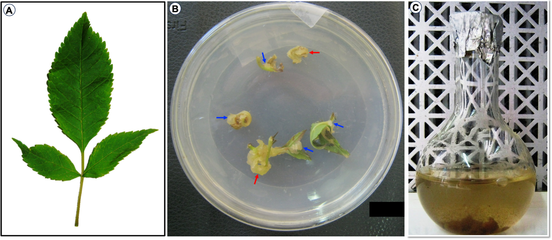
Tissue culture of Tecoma stans (L.) Juss. ex Kunth. (A) Fresh collected leaf. (B) Callus was induced from the leaves, nodes, and internodes of the explants grown on solid Murashige and Skoog (MS) medium containing 1.0 mg L−1 2,4-dichlorophenoxyacetic acid. After 3 to 4 wk, a friable light yellowish-white callus was formed. (C) Cell suspension culture was initiated by transferring the callus into liquid medium supplemented with 1.0 mg L−1 6-benzyladenine. The red arrow indicates the induced callus resulting from the cut areas of the internodes in contact with the MS medium. The blue arrow indicates the induced callus at the scored areas of the leaves.
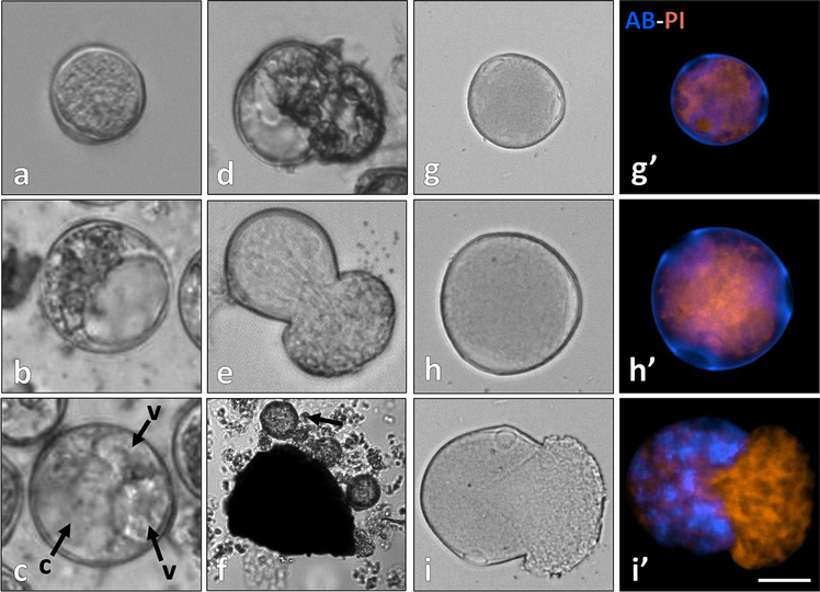
Several stages of pre-embryonic and embryonic somatic embryos were detected; however, the 1:2 ratio of 2,4-D and BA growth regulators (Table 1) gave the best results of the number of mature somatic embryos (16 ± 2 torpedo embryos per volume) compared to the rest of the tested hormone concentrations. Microscopical examination showed (Fig. 2) isolated somatic cells and clusters, earlier embryogenic stages (one-cell proper and two-cell proper, quadrant stage, octant, dermatogen, early globular, and early heart-stage embryos). Moreover, mature globular, heart, cotyledon, and torpedo stages were also identified (Figs. 3 and 4). The torpedo stage further developed into the vegetative phase when utilizing a combination of 2,4-D and BA (in a ratio of 1:2). This concentration is deemed optimal for regulating cell growth and completing the somatic embryo growth cycle (Fig. 4). Namde and Wani (2014) have studied the effects of explants and plant growth hormones, 2,4-D, kinetin, and BA, on T. stans culture. Of interest, the 2:2 ratio of 2,4-D and BA gave 100% callus induction frequency, which was in accordance with the present study results upon using 2.0 mg L−1 BA.
Table 1. Effect of plant growth regulator on the production of somatic embryos (torpedo stage) of Tecoma stans (L.) Juss. ex Kunth leaves cell suspension cultures harvested at day 27 of culture incubation under the specified conditionsFigure 2.
Phase-contrast microscopical examination of the cell suspension culture of Tecoma stans (L.) Juss. ex Kunth in Murashige and Skoog liquid medium supplemented 1.0 mg L−1 2,4-dichlorophenoxyacetic acid and 2.0 mg L−1 6-benzyladenine at day 14 of culture incubation. (A) Cell division, isolated cells, and clusters. (B) Earlier embryogenic stages (one-cell proper). (C) Pre-embryonic stages (two-cell embryo and two-cell proper). (D, E) Four-cell embryo; quadrant stage. (F) Octant (red arrow), dermatogen, and early globular stages (blue arrow). (G, H) Early heart-stage embryos (× 40).
Figure 3.
Phase-contrast microscopical examination of (A) globular, (B) late heart, and (C) cotyledon stages of Tecoma stans (L.) Juss. ex Kunth embryos induced in Murashige and Skoog liquid medium supplemented with 1.0 mg L−1 2,4-dichlorophenoxyacetic acid and 2.0 mg L−1 6-benzyladenine, at day 21 of culture incubation (× 20).
Figure 4.
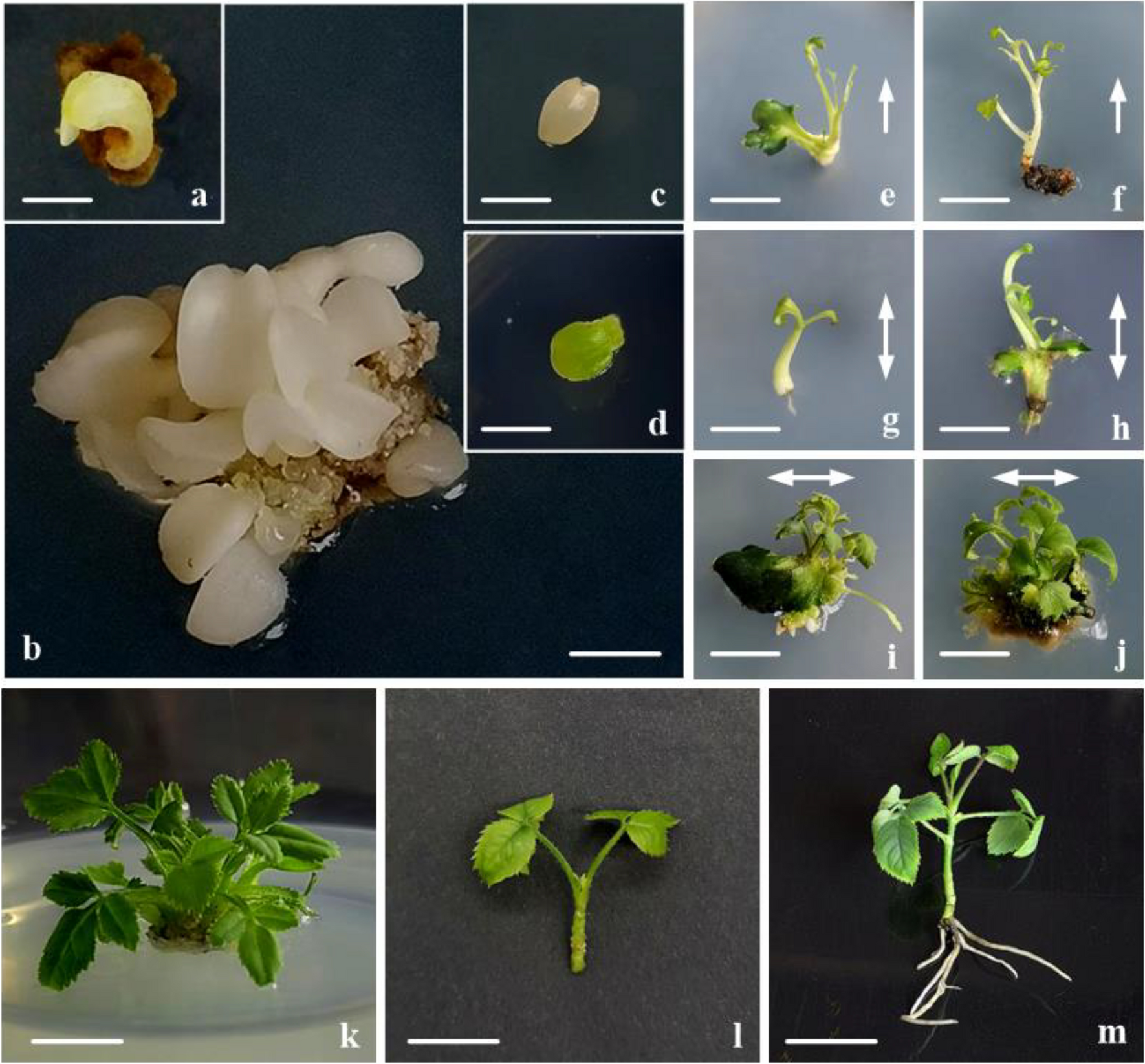
Macroscopical examination of somatic embryonic stages and ectopic proliferation of Tecoma stans (L.) Juss. ex Kunth induced in Murashige and Skoog (MS) liquid medium under 2.0 or 5.0 mg L−1 6-benzyladenine (BA) with 1.0 mg L−1 2,4-dichlorophenoxyacetic acid (2,4-D). (A) Embryonic phases (torpedo, heart, and globular stages denoted as t, h, and g respectively) were observed at the interface of the leaf explant. (B, C) Mature torpedo stages harvested at day 27 of suspension culture incubation. (D) Ectopic proliferation on the leaf explants. (E, F) Mature embryos harvested at day 31. (G) Vegetative phase grown on plain solid MS medium at day 38. Scale bar = 2 mm. BA2, 2.0 mg L−1 6-benzyladenine; BA5, 5.0 mg L−1 6-benzyladenine.
留言 (0)