
記住我


Fibular osteotomy is a standard procedure, frequently performed as part of deformity correction surgery of the leg. Complications related to fibular osteotomy are well described.1–5 The most feared complication related to fibular osteotomy is peroneal nerve palsy.2–5 The incidence of peroneal nerve palsy is related to the level where the osteotomy is performed. Nevertheless, even when performed between the distal and middle third of the fibula, an area that is considered to be relatively safe, injury to the extensor hallucis longus (EHL) was still described.2 Another potential complication is nonunion.
In our opinion, these complications are mostly related to technical errors and bone thermal injury. During the last 15 years, to avoid complications related to fibular osteotomy, we developed a specialized technique.
The aim of this study was to describe our technique developed to diminish complications related to fibular osteotomy.
TECHNIQUEAfter receiving approval from the Institutional Review Board, we retrospectively reviewed the medical records of all patients who underwent fibular osteotomy as part of leg deformity correction and/or leg lengthening surgery between the years 2006 and 2019. We found 168 patients who underwent 210 fibular osteotomies at the level of the middle third of the fibula. There were 102 males and 66 females with a mean age of 14 years (range: 3 to 66 y).
Operative TechniqueFibular osteotomy is performed as the initial step of deformity correction surgery. The patient is positioned supine with a bump under the buttock on the operated side to prevent the foot from rolling outwards. After preparation and draping, a sterile tourniquet is applied. The preferred area for osteotomy is between the middle and distal third of the fibula, which is the safest region. At this level, the fibula is quite superficial and usually can be easily palpated. A roughly 3 cm skin incision is done, followed by deeper dissection between peroneal muscles at the front and gastrocnemius-soleus muscle at the back. Sharp dissection using scissors reveals the fibula. Two small Hohmann retractors are inserted subperiosteally anterior and posterior to the fibula. To prevent thermal necrosis, we predrill the fibula with an Ilizarov 1.8 wire from lateral to medial, as well as from anterior to posterior. The drill holes are positioned in a line of anticipated osteotomy (Fig. 1). The number of drill holes depends on the size of the fibula, though, we aim to place a drill hole every 2 to 3 mm. The osteotomy is completed with an osteotome directed from anterior to posterior (Figs. 2, 3). The choice of this direction reduces the likelihood of peroneal artery injury, which is located just medial to the fibula, next to the tibiofibular interosseous membrane. In addition, this course avoids injuring the peroneal nerve branch that innervates the EHL, which is another serious complication occasionally associated with fibular osteotomy.3 The tourniquet can be released at this stage, if not required for further surgical steps. The skin is closed in layers (subcutaneous layer and skin).
 FIGURE 1:
 First, drill holes are created in line with planned osteotomy (black arrow pointing to a magnified view).
 FIGURE 2:
 Next, osteotomy is completed using osteotome in anterior to posterior direction (black arrow pointing to diagram that clarifies the direction of the osteotome).
 FIGURE 3:
 A magnified view of the osteotomy site after drill holes were placed showing again the position of the osteotome.
EXPECTED OUTCOMESThere were no complications related to fibular osteotomy; no peroneal nerve injury, as well as no EHL branch injury, and no intraoperative bleeding after completion of the osteotomy and tourniquet removal. Healing time of the osteotomy was somewhat variable based on the additional procedures that were performed. Union rates were up to 8 weeks in patients who did not have concomitant lengthening procedures (average 5.5 wk). In patients who had lengthening, fibular union was seen no later than 8 weeks after the end of lengthening (average 5 wk). No delayed unions or nonunions have been observed. A clinical example of fibular osteotomy as part of a leg-lengthening procedure is shown in Figure 4.
 FIGURE 4:
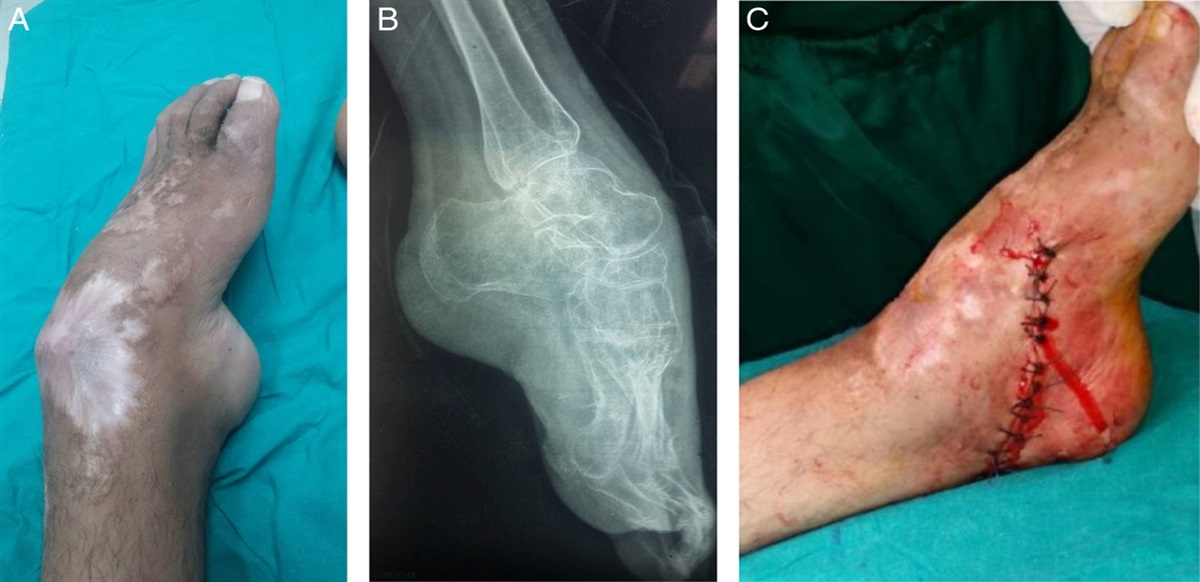
 Clinical example. An 11-year-old patient who had fibular osteotomy as part of a tibial lengthening procedure. A, completed osteotomy at surgery. B, One month after surgery distraction at the fibular osteotomy site can be seen. C, One year after surgery, there is complete remodeling at the osteotomy site.
COMPLICATIONS AND DISCUSSIONOsteotomy of the fibula is a common procedure in deformity correction surgery and leg lengthening. The osteotomy can be performed in different regions of the fibula. Complication rates are directly related to the level of the osteotomy. Wootton et al3 recommended not to perform fibular osteotomy in zones II to III (from just below the fibular head to 15 cm distal to this level) due to a high incidence of neurological complications (21 out of 105 patients, 20% incidence).
We routinely perform osteotomy of the fibula between the middle and distal third; however, even at this level, complications are not uncommon, and primarily related to injury to the peroneal artery and the peroneal nerve.
Low-heat osteotomy using predrilling, followed by completion of the osteotomy of the fibula by an osteotome in the anterior to posterior direction, is a safe, simple, and straightforward procedure. This method has become the standard technique in our practice for the last 15 years. Since applying this technique, no complications related to the fibular osteotomy have been observed in our practice.
REFERENCES 1. Sachs O, Katzman A, Abu-Johar E, et al. Treatment of adolescent Blount disease using the Taylor spatial frame with and without fibular osteotomy: is there any difference? J Pediatr Orthop. 2015;35:501–506. 2. Dilawaiz NR, Quick TJ, Eastwood DM. Focal dome osteotomy for correction of tibial deformity in children. J Pediatr Orthop B. 2005;14:340–346. 3. Wootton JR, Asworth MJ, McLaren CA. Neurological complications of high tibial osteotomy as causative factor: a clinical and anatomical study. Ann R Coll Surg Engl. 1995;77:31–34. 4. Gibson M, Barnes M, Allen M, et al. Weakness of foot dorsiflexion and changes in compartment pressures after tibial osteotomy. J Bone Joint Surg Br. 1986;68-B:471–475. 5. Curley P, Eyres K, Brezinova V, et al. Common peroneal nerve dysfunction after high tibial osteotomy. J Bone Joint Surg. 1990;72:405–408.
留言 (0)