Bacterial strains, growth conditions and reagents
All tested bacterial strains were procured from the American Type Culture Collection (ATCC) and the Korean Collection for Type Cultures (KCTC). Clinical isolates of A. baumannii were acquired from the Kyungpook National University Hospital National Culture Collection for Pathogens (KNUH-NCCP). DH5α was employed for the cloning process, while SoluBL21™ was utilized for protein purification. Cultures of all bacterial strains were cultivated in Luria Bertani (LB; MBcell #MB-L4488) broth at 37 °C, with agitation at 200 rpm. Ampicillin was supplemented at 100 μg/ml concentration when deemed necessary. Recombinant protein expression was induced by adding isopropyl-B-D-thiogalactopyranoside (IPTG; Biosesang #I1006) at the specified concentrations.
Purification of recombinant endolysin PA90 or Tha-PA90
The plasmid encoding thanatin-fused PA90 (Tha-PA90) was generated through polymerase chain reaction (PCR), utilizing the pAS033 plasmid as a template. The pAS033 plasmid was designed to overexpress cell-penetrating peptide DS4.3-fused pA90 on the pET21a backbone [26]. Notably, the DNA sequence encoding the Thanatin peptide (GSKKPVPIIYCNRRTGKCQRM) was incorporated into the primer sequences (Additional file 1: Table S1, underlined). Following sequencing analysis to confirm the construct, the resultant plasmid, pAS036, was introduced into SoluBL21™ for the subsequent purification of Tha-PA90.
Cultures of SoluBL21™ carrying either pET21a::PA90 (pAS025) [26] or pAS036 were cultivated in 1.5 L of LB broth supplemented with ampicillin (100 μg/ml) at 37 °C with vigorous aeration at 200 rpm. Once the optical density at 600 nm reached 0.6, IPTG was introduced into the culture to induce protein expression. Following a 5-h incubation at 37 °C in a shaking incubator, the cells were harvested by centrifugation at 5000 × g for 10 min at 4 °C. Subsequently, cell lysis was achieved via sonication, utilizing the resuspended pellet in 100 ml of lysis buffer (comprising 20 mM Tris–HCl, pH 8.5, 0.5 M NaCl, and 10 mM imidazole). After eliminating unbroken cells through centrifugation at 14,000 × g for 30 min at 4 °C, the supernatant fraction was filtered through a 0.4 μm-pore size filter (GVS #FJ25ASCCA004FL01). The subsequent step involved applying each collected fraction to a 5 ml HisTrap HP column (Cytiva #17,524,802), installed in an ÄKTA fast protein liquid chromatography (FPLC) system (Cytiva, USA), and controlled using UNICORN 5.1 software. The resin-bound endolysins were eluted using a gradient of imidazole concentration (10 mM–0.5 M) in a buffer of 20 mM Tris–HCl, pH 8.5, and 0.5 M NaCl. Subsequently, the eluted fraction was diluted in 20 mM Tris–HCl, pH 8.5 buffer for application to a 5 ml HiTrap SP column (Cytiva #17–1152-01) for further purification. Elution from the HiTrap SP column was conducted using 20 mM Tris–HCl, pH 8.5 buffer containing 0.6 M NaCl. Finally, the fractions obtained were dialyzed overnight at 4 °C in a 20 mM Tris–HCl, pH 7.5, and 150 mM NaCl solution, utilizing Snakeskin™ dialysis tubing (Thermoscientific #68,700). Filtration through a 0.4 μm-pore size filter (GVS #FJ25ASCCA004FL01) was performed to remove aggregated proteins. The quantification of each endolysin was determined using the Bradford assay kit (Biorad #5,000,006), with bovine serum albumin (BSA; Promega, #R396A) serving as the standard.
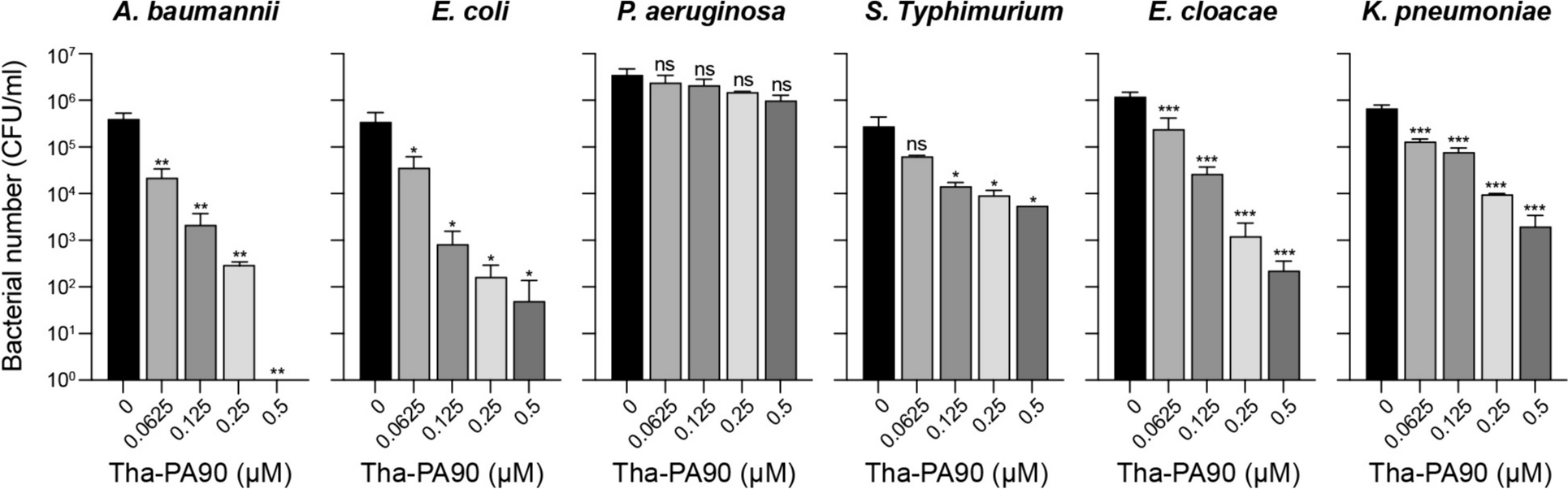
Determination of the lytic spectrum of Tha-PA90
The antibacterial activity of Tha-PA90 was assessed through a CFU reduction assay, employing A. baumannii, Escherichia coli, Pseudomonas aeruginosa, Salmonella Typhimurium, Enterobacter cloacae, and Klebsiella pneumoniae as the target strains. Bacterial cultures were allowed to reach the mid-exponential growth phase of OD600 = 0.8 to initiate the assay. Subsequently, the cells were harvested by centrifugation at 3500 × g for 5 min. The harvested cells were adjusted to 1 × 10⁶ CFU in a 20 mM HEPES solution at pH 7.4 and mixed with varying concentrations of Tha-PA90 (0, 0.0625, 0.125, 0.25, and 0.5 μM) in a 96-well plate. Following a 2-h incubation at 37 °C, the cells were diluted in 1 × phosphate-buffered saline (PBS) and placed onto LB agar plates to facilitate the enumeration of surviving bacteria.
Permeability test by 1-N-phenylnaphthylamine uptake assay
Bacterial membrane permeability was assessed via a 1-N-phenylnaphthylamine (NPN) uptake assay [27]. A. baumannii ATCC 19606 strain was cultured in LB until the optical density at 600 nm reached 0.4. The harvested cells were washed and resuspended in a 5 mM HEPES solution at pH 7.2. The CFU of the cells were adjusted to approximately 1 × 10⁸ CFU/ml. These cells were added to the wells of a 96-well black plate containing a mixture of 50 μL of 40 μM NPN (Sigma-Aldrich #104,043) and 50 μL of 2 μM Tha-PA90. PA90, thanatin peptide (AnyGen, South Korea), or a combination of thanatin peptide and Tha-PA90 was also tested for comparative purposes. As a positive control, 1 mM EDTA was used, while the buffer containing only NPN was the negative control. The plate was incubated at 37 °C for 30 min. Fluorescence was measured at an excitation wavelength of 350 nm and an emission wavelength of 420 nm using a microplate reader (TECAN Infinite M PLEX). The NPN uptake factor was calculated by dividing the fluorescence value of the cell suspension with the buffer containing only NPN after subtracting the fluorescence value of the cell suspension.
Detection of internalized Tha-PA90 in mammalian cells by western blot analysis
Adenocarcinomic human alveolar basal epithelial cells A549 were cultured in Dulbecco’s modified Eagle’s medium (DMEM; HyClone #SH30243.01) supplemented with 10% fetal bovine serum (FBS; Welgene #S001-01, South Korea) and 1% penicillin–streptomycin (Gibco #15,140,122) at 37 °C in a 5% CO2 incubator. A549 cells were seeded at 1 × 10⁵ cells/well density and treated with 1, 5, or 10 μM of Tha-PA90 at 37 °C for 30 min. The same concentrations of PA90 were also applied for comparison. After washing with 1 × Dulbecco’s phosphate-buffered saline (DPBS) 5 times to eliminate external proteins, whole cell lysates were obtained by lysing the cells using 0.05% triton X-100 (v/v of 1 × DPBS). Centrifugation at 13,000 × g for 20 min was performed to remove cell debris, and the supernatant was filtered through a 0.4 μM-pore-size syringe filter (GVS #FJ25ASCCA004FL01). The filtered supernatant was subjected to 10% SDS-PAGE, and the gel was subsequently transferred to a PVDF membrane (GE Healthcare #10,600,022). The membrane was blocked with 5% skim milk (w/v in TBST) at room temperature (RT) for 30 min. Internalized endolysins were detected using an anti-his antibody (Thermofisher #MA1-21,315-1MG) as the primary antibody. Beta-actin was detected using an anti-beta-actin antibody (Cell Signaling #4967S). After incubation at RT for 1 h, the membranes were washed three times with 1 × TBST and incubated for 1 h with HRP-conjugated secondary antibodies: anti-mouse IgG for anti-his (Sigma-Aldrich #12–349) and anti-rabbit IgG for anti-beta-actin (Cell signaling #7074S). Following another round of washing with 1 × TBST, signals were detected using ECL Western Blotting Substrate (Thermo Scientific #32,209) on an iBright imaging system (Thermo Scientific).
Evaluation of cytotoxicity of Tha-PA90 using LDH assay
The effect of Tha-PA90 on A549 cells was assessed utilizing the lactate dehydrogenase (LDH) PLUS Cytotoxicity Assay kit (DYNEBIO #GBL-P1000). A549 cells were cultured in a 24-well plate, with an initial seeding density of 1 × 10⁵ cells per well, and maintained at 37 °C in a 5% CO2 humidified incubator. Following washing with 1 × DPBS, the cells were exposed to varying concentrations of Tha-PA90 (0, 2.5, 5, and 10 μM) for 24 h in DMEM. Mammalian cell lysis buffer served as the positive control. After incubation, the supernatant was transferred into a new 96-well plate and combined with 100 μL of the LDH test reagent. This mixture was subsequently incubated for 30 min, and the addition of stop buffer stopped the reaction. Absorbance was measured at 490 nm using a microplate reader (TECAN Infinite M PLEX). Cytotoxicity was expressed as a percentage, with untreated cells serving as the negative control for the calculation.
In vivo efficacy of Tha-PA90 in mouse A. baumannii infection model
All mouse experiments were conducted according to the guidelines established by the Institutional Mouse Use and Care Committee of Hankuk University of Foreign Studies (HUFS-2021–0003). Seven-week-old female BALB/c mice were procured from Raonbio (South Korea). Cyclophosphamide monohydrate (CPM; 3 mg/mouse; Glentham, #GK2037-1G) was administered via the intraperitoneal route 4 days and 1 day before bacterial infection. A. baumannii ATCC 19606 was cultured in brain heart infusion (BHI) media at 37 °C for 18 h with agitation at 200 rpm. Bacterial cells were harvested by centrifugation at 3,500 × g for 5 min and resuspended in 1 × PBS. The CPM-pretreated mice were injected intraperitoneally with 1 × 10⁸ CFU of A. baumannii. PA90 or Tha-PA90 at a dosage of 600 μg per mouse was intraperitoneally administered 1 h post infection. In the control experiment (mock), mice were injected with 1 × PBS. Throughout the experiment, the health and survival of the mice were monitored.
The mice were euthanized under anesthesia using a combination of ketamine (200 mg/kg) and xylazine (10 mg/kg) at 10 h dpi. Each organ, including the heart, spleen, liver, kidney, and lung, was aseptically isolated and homogenized in 1 × PBS containing 0.05% Triton X-100, utilizing a T10 ULTRA-TURRAX homogenizer (IKA). Homogenate samples were plated on LB plates containing trimethoprim (5 μg/ml; Sigma #T7883). After overnight incubation at 37 °C, the bacterial numbers were enumerated and expressed as CFU/g of organ or CFU/ml of blood.
Measurement of cytokine levels in A. baumannii-infected mouse upon Tha-PA90 treatment
Serum was obtained by subjecting blood samples from A. baumannii-infected mice, treated with Tha-PA90 as previously described, to centrifugation for 30 min at 3000 × g at 4 °C. The levels of IL-6 and TNF-alpha in the serum were quantified using the Mouse IL-6 Uncoated ELISA Kit (Invitrogen #80–7064-88) and the Mouse TNF-alpha Uncoated ELISA Kit (Invitrogen # 88–7324-88), respectively.
For the isolation of total RNA from mouse liver, which had been infected with A. baumannii and treated with Tha-PA90 as mentioned earlier, the TaKaRa MiniBEST Universal RNA Extraction kit (TaKaRa #9767) was utilized according to the manufacturer’s instructions. The concentration and purity of the total RNA were determined using a spectrophotometer (DeNOVIX DS-11 FX). Subsequently, the isolated RNA was transcribed into cDNA using the Dyne cDNA Synthesis Kit (Dyne Bio # DYRT1120). Expression levels were assessed via qPCR analysis, employing a real-time PCR cycler (QIAGEN Roter-Gene Q) and TOPreal™ qPCR 2 × PreMIX (Enzynomics #RT500M). The primer sequences used for the reaction are presented in Table S1. Relative expression levels were determined using the ΔΔCt method, where ΔΔCt = ΔCt (experimental group)—ΔCt (control group). Cycle threshold (Ct) values were normalized to the Ct value of GAPDH, and changes relative to the mock were plotted. All experiments were conducted in triplicate.
Statistical analysis
Data analysis was conducted using GraphPad Prism software version 9.3.0. A two-tailed Student’s t-test assessed the differences between the two groups, while survival experiments were analyzed using the log-rank (Mantel–Cox) test. All data are presented as the mean ± SD, and differences were considered to be statistically significant when P < 0.05.


留言 (0)