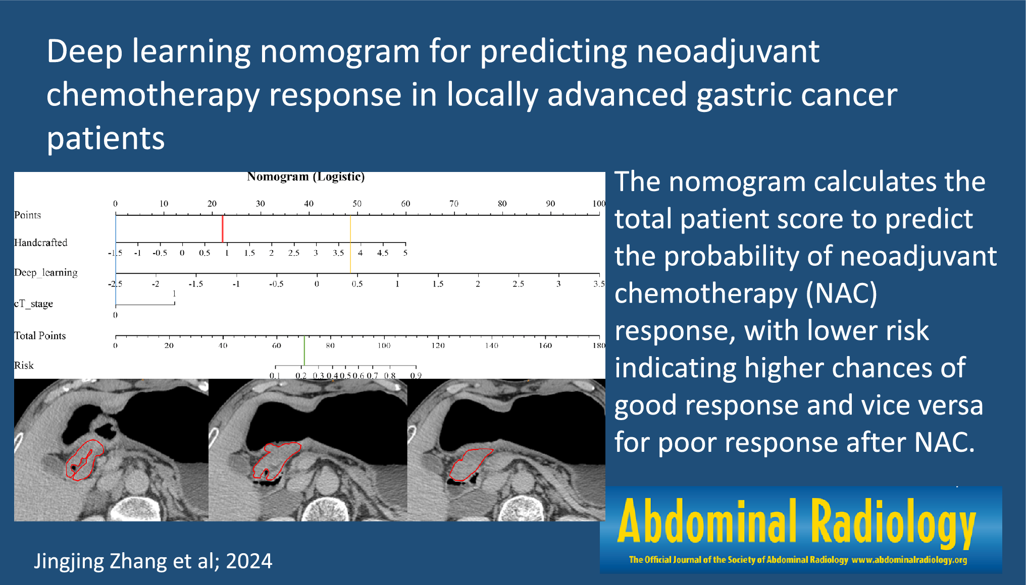
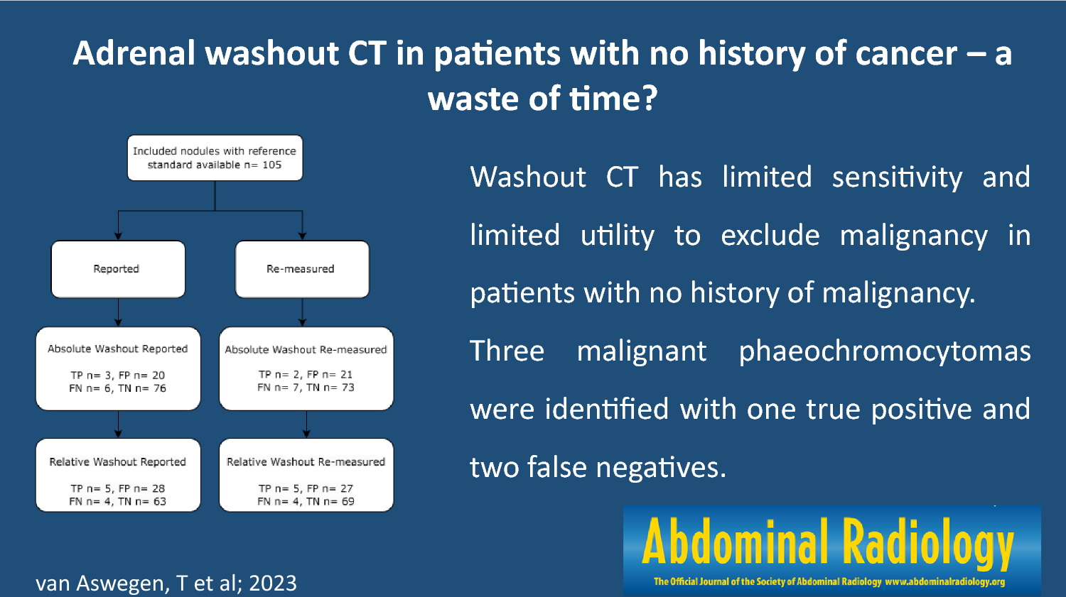
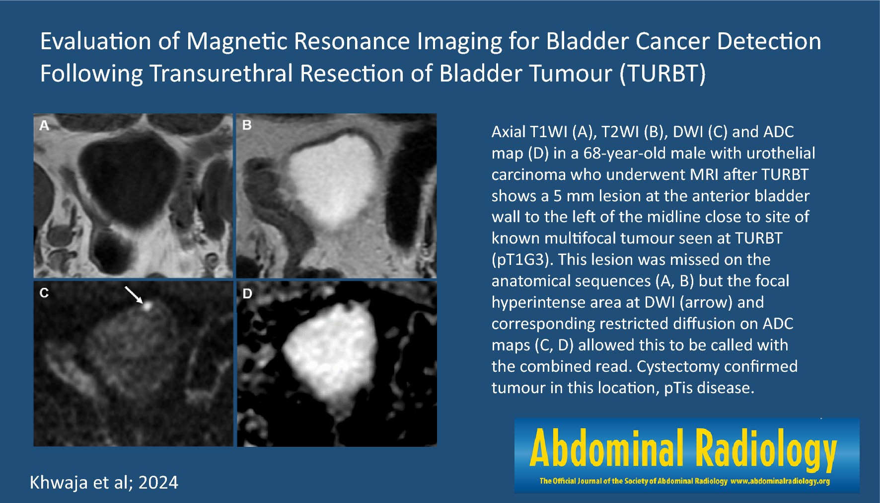
Patients
This study was approved by the Human Research Committee of our hospital’s institutional review board and the requirement for written informed consent was waived by the board because of the retrospective design. This study was conducted in accordance with the Health Insurance Portability and Accountability Act of 1996. Patients with histopathologically confirmed bladder SCNEC who underwent surgical excision or biopsy at two Japanese institutions were studied from August 2010 to August 2023. We found 250 patients with histopathologically confirmed UC at a single Japanese hospital during the same time period. We randomly selected 80 patients with UC who underwent preoperative CT and MRI because we examined patients with UC ten times as many as bladder SCNEC. This study included 10 patients with bladder SCNEC (age range, 53–86 year; median age, 77 year) and 80 patients with UC (age range, 47–87 year; median age, 72 year). The patient characteristics of bladder SCNEC are shown in Table 1.
Table 1 Patient characteristics of bladder SCNECCT Imaging
All patients had CT imaging using an eight-slice CT scanner (LightSpeed Ultra; GE Healthcare, Milwaukee, WI, USA), a 16-slice CT scanner (LightSpeed 16; GE Healthcare, Milwaukee, WI, USA), 64-slice CT scanner (SOMATOM go top; Siemens Healthcare, Erlangen, Germany), or a 64-slice CT scanner (Brilliance CT 64; Philips Healthcare, Best, The Netherlands). All 90 patients had axial unenhanced CT images obtained and 47 patients had axial contrast-enhanced CT images (seven SCNECs and 40 UCs). Contrast-enhanced CT images were obtained 65–100 s after an intravenous injection of 100-mL nonionic iodine contrast material was initiated. Axial and coronal multiplanar reconstruction images were reconstructed with a section thickness ranging from 2.5 to 5 mm and no overlap.
MRI protocols
MRI was performed using a 1.5-T unit (Intera Achieva 1.5 T Pulsar; Philips Healthcare, Best, The Netherlands), a 1.5-T unit (SIGNA Explorer; GE Healthcare, Milwaukee, WI, USA), a 3.0-T unit (Intera Achieva 3.0 T Quasar Dual; Philips Healthcare, Best, The Netherlands), or 3.0-T unit (DISCOVERY MR750w; GE Healthcare, Milwaukee, WI, USA). All MRI images were obtained with a section thickness of 4–5 mm, an intersection gap of 1 to 2 mm and a field of view of 23 × 23 to 30 × 30 cm. Axial and coronal or sagittal oblique T2-weighted fast spin-echo (TR/TE, 2,586–6,086/90–120 ms), axial T1-weighted spin-echo (TR/TE, 498–789/10 ms), and axial diffusion-weighted single shot spin-echo echo-planar (TR/TE, 4,000–4,800/68–80 ms; b-value = 0 and 1,000 s/mm2) images were obtained in 88 patients (eight SCNEC and 80 UC).
Imaging analysis
All images were independently assessed by two radiologists with 24- and 10-years post-training experience in urogenital imaging, and any disagreements were resolved by consensus. The clinical information and pathological diagnosis were blinded by the reviewers.
First, the maximum diameter and height of the tumor were quantitatively measured. Number (single or multiple), location (dome, right lateral, left lateral, trigone, anterior, or posterior), configuration (pedunculated or broad-based), margins (smooth or irregular), arising in bladder diverticulum, non-neoplastic bladder wall thickening, surrounding fat stranding, lymphadenopathy, and calcification were qualitatively evaluated. If multiple lesions were found, the largest tumor alone was assessed. The acute (≤ 90°) and obtuse (> 90°) angles between the tumor surface and the adjacent bladder wall were used to characterize pedunculated and broad-based lesions, respectively. Irregular margins included spiculated, serrated, and needle-like margins. Arising in bladder diverticulum was defined as a bladder cancer localized within the bladder diverticulum. Non-neoplastic bladder wall thickening was defined as smooth and uniform bladder wall thickening excluding the bladder cancer. Surrounding fat stranding was defined as abnormal increased fat attenuation adjacent to the bladder cancer on CT. A lymph node in the pelvis with a short-axis diameter of more than 8 mm was characterized as lymphadenopathy. Subsequently, CT attenuation (Hounsfield Unit [HU]) of the solid component on unenhanced and contrast-enhanced CT was assessed by positioning the region of interest (ROI) above the tumor.
Second, MRI was used to identify the clinical T category based on the American Joint Committee on Cancer TNM Staging System for Bladder Cancer, eighth edition in 2017. Homogeneity and signal intensity on T1- and T2-weighted images were qualitatively evaluated and signal intensity of the tumor was compared with that of the iliopsoas muscle (low, iso-, or high signal intensity).
Third, the signal intensity ratio on T1- and T2-weighted images and the apparent diffusion coefficient (ADC) value of the solid component were evaluated. A reviewer with 10-year post-training experience in urogenital imaging designated the ROI in the solid component and iliopsoas muscle on the T1- and T2-weighted images and recorded these signal intensities. The ratio of the solid component to the intensity of the muscle signal was computed. ADC values of the solid component were also assessed on ADC maps by positioning ROI on the tumor. ROIs on ADC maps were placed on the solid component as extensively as possible inside the tumor while omitting stalk areas using T2- and contrast-enhanced T1-weighted images.
Finally, the presence and signal intensity of stalk on T2-weighted images and inchworm signs on diffusion-weighted images were evaluated. The signal intensity of the stalk was divided into three categories: low, high, and mixed low and high signal intensity relative to the tumor. The inchworm sign was defined as hyperintense bladder cancer with a hypointense submucosal stalk [11].
Statistical analysis
All statistical analyses were performed with EZR (Saitama Medical Center, Jichi Medical University, Saitama, Japan), which is a graphical user interface for R (The R Foundation for Statistical Computing, Vienna, Austria). More precisely, it is a modified version of R commander designed to add statistical functions frequently used in biostatistics [12]. The Mann–Whitney U test was used to compare quantitative data (age, maximum diameter, height, CT attenuation, signal intensity ratio, and ADC value) between bladder SCNEC and UC. Fisher’s exact test was used to compare the qualitative outcomes (number, location, configuration, margins, arising in bladder diverticulum, non-neoplastic bladder wall thickening, surrounding fat stranding, lymphadenopathy, calcification, clinical T category, homogeneity and signal intensity on T1- and T2-weighted images, stalk, and inchworm sign) between bladder SCNEC and UC. p values of < 0.05 were considered significant. κ statistics was used to assess the interobserver variability of qualitative assessments. Kappa values of 0.81 to 1.00 exhibit almost perfect agreement; 0.61 to 0.80—substantial agreement; 0.41 to 0.60—moderate agreement; 0.21 to 0.40—fair agreement; and 0.01 to 0.20—slight agreement [13].


留言 (0)