
記住我


GDE2 is expressed in the developing and postnatal spinal cord and brain [20, 31, 32]; however, its expression in the adult brain has not been determined. To assess GDE2 expression in the adult mouse brain, we performed RNAscope for Gde2 transcripts in coronal sections from 4-month-old WT mice. We detected Gde2 expression in major brain areas including the thalamus (Th) and caudate putamen (CP) (Fig. 1A, B), with particularly strong expression in the medial habenula and the medial and anterior amygdala (Fig. 1C–F), which are areas associated with regulation of emotional and motivational aspects of behavior including fear/anxiety responses [33, 34]. Gde2 is also expressed throughout the hippocampus, a region implicated in learning and memory (Fig. 1G–J) [35, 36]. In addition, Gde2 transcripts are detected in the cortex with enriched expression in deep cortical layers 4–6 (Fig. 1K, L). Similar to previous studies using younger mice [31, 32], Gde2 expression in the cortex is mainly localized to neurons with some expression in non-neuronal cells (Fig. 1M, N). This aligns with previous expression and transcriptomic data showing Gde2 expression in neurons, mature oligodendrocytes, and endothelial cells [37, 38].
Fig.1
Gde2 mRNA expression in the adult brain. A–N Exemplar images of RNAscope detection of Gde2 transcripts (white) in the adult brain. Nuclei are marked by DAPI staining (blue), and NeuN antibody staining (green) marks neurons. Boxed areas in A and B are magnified in panels (C, E, K) and (D, F, L), respectively. Boxed areas in G and H are magnified in panels I, J. Hatched lines in K and L mark cortical layers. Scale bar: (A, B): 500 µm; (C–L): 250 µm; (M, N): 50 µm. CP: Caudate Putamen, Th: Thalamus, CTX: Cortex, HPF: Hippocampal Formation
General health of Gde2KO miceSimilar to previously published work [18], Gde2KO mice were viable, fertile, and born at the expected Mendelian frequencies. Male and female Gde2KO mice were born in about equal numbers. Gde2KO mice showed no obvious differences in health or appearance from WT mice. However, upon weighing, male Gde2KO mice consistently have slightly lower body weight compared to WT mice (~ 10%, Additional file 1: Fig. S1A). This difference in body weight between genotypes decreases with age (Additional file 1: Fig. S1A). No difference in body weight was observed for female mice (Additional file 1: Fig. S1B).
Gde2KO mice exhibit hyperactivityAnimals were first tested in the Open Field (OF) task at 7-months of age (Additional file 1: Fig. S1C). At the 7-month time point, WT and Gde2KO mice showed no significant difference in the distance traveled in the OF chamber (Fig. 2A–C, Additional file 1: Fig. S2A–C). We next tested these same mice at 16-months of age to see if any age-progressive phenotypes emerged (Additional file 1: Fig. S1C). At this time point, Gde2KO mice showed significantly increased locomotion, and this hyperactivity was the most pronounced at the beginning of testing (Fig. 2D, E). Importantly, despite increased novelty-induced activity, Gde2KO males demonstrated significant habituation as testing progressed (Additional file 1: Fig. S2D, E). Female Gde2KOs showed a trending increase in activity at both ages; however, these differences did not reach significance (Additional file 1: Fig. S2A, C, D, F).
Fig. 2
Hyperactivity phenotype in Gde2KO mice. A Schematic of OF test. B–E Analysis of OF test. B, C Total distance (B) and its dynamics (C) during 30 min of testing in the OF task for WT and Gde2KO mice at 7 month time point. No effect of genotype or its interactions were detected (three-way mixed design ANOVA, Ps > 0.05). D, E The same measures and analysis as in B and C, respectively, for 16-month-old mice. Gde2KO mice demonstrated higher motor activity that was partially ameliorated during the duration of testing as indicated by significant effect of genotype (F(1,52) = 20.92, P < 3.0E−5) and genotype × block interaction (F(8,416) = 2.15, P < 0.031). F Schematic of 2 trial Y maze test. G–J Analysis of Y maze task (G, H). Total distance (G) and dynamics (H) during Y maze testing in 7-month-old mice. Gde2KO mice were more active than WT mice during trial 1 (one arm closed) (ANOVA, effect of genotype F(1,63) = 5.95, P < 0.018) and particularly during trial 2 (all 3 arms open) (F(1,63) = 39.97, P < 1.0E-5). I, J The same measures and analyses as in G and H, respectively, for 16-month old mice. Gde2KO mice were more active than WT mice in both trials (F(1,47) > 17.75, P < 1.0E−4). K Schematic of plus maze. L The distance traveled (L) and its dynamics (M) by 7-month-old WT and Gde2KO mice. The higher motor activity of Gde2KO mice was confirmed by ANOVA (effect of genotype, (F(1,70) = 10.35, P < 0.002). All graphs are means ± SEM; P > 0.05; *P < 0.05, **P < 0.01, ***P < 0.001, and ****P < 0.0001. See Additional file 2: Table S1 for statistical details. Schematics in A, F, and K created in BioRender.com
Hyperactivity in Gde2KO mice was also evident in the two-trial Y maze at 7- and 16-months of age and in both sexes (Fig. 2F–J, Additional file 1: Fig. S2G–L). In this task, mice explore a Y-shaped maze across two 5-min trials. In the first trial, they are allowed to explore two of the arms. In the second trial, a novel third arm is opened for exploration (Fig. 2F, see “Methods”). Notably, at 7-months of age, the between-genotype differences in distance traveled were more pronounced in trial 2 than in trial 1, indicating increased hyperactivity in Gde2KOs upon the opening of the new arm in the second trial (Fig. 2G, H). At 16-months of age, increased activity of Gde2KO mice was observed in trial 1 and did not increase further in trial 2 (Fig. 2I, J).
In the plus maze task, Gde2KO animals at 7-months were tested to see if hyperactivity was novelty induced (Additional file 1: Fig. S1C). Mice were placed in a plus shaped maze with open and closed arms and were allowed to explore for 10 min (Fig. 2K; see “Methods”). Compared to WT, Gde2KO mice travel more distance over time compared with WT in both open and closed arms (Fig. 2L, M). The between-genotype difference is observed in both males and females (Additional file 1: Fig. S3A–C). Notably, we see a significant difference in distance traveled by Gde2KOs during the first minute of the test when the environment is most novel (Fig. 2M). We further analyzed the distance traveled by mice in the plus maze task in blocks of 5 min to match the block durations of the OF analysis. Gde2KOs still demonstrate hyperactivity when analyzed in 5-min blocks of time (Additional file 1: Fig. S3D). This observation is in agreement with the hyperactivity phenotypes observed in the Y maze task at 7 months.
Taken together, Gde2KO mice display hyperactivity at younger ages mainly in response to new situations/environments; however, aged Gde2KO mice show hyperactivity across all tasks measuring distance traveled regardless of novelty. Importantly, despite the hyperactivity observed in Gde2KO mice, the extent of the hyperactivity was moderate and not debilitating as the motor exploration in these mice showed preserved habituation across all tests.
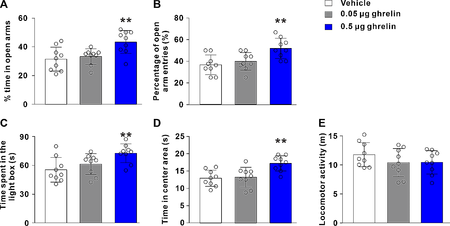
Normal anxiety behavior in Gde2KO miceMice normally avoid open spaces such as the center area of the OF and the open arms in the plus maze. Thus, distance traveled and time spent in these areas can be used as a measure of anxiety levels [39, 40]. Gde2KO mice were tested at 7 months and 16 months in the OF test to determine if they had an age-progressive anxiety phenotype. At both time points, male and female Gde2KO mice show no difference in the percent distance traveled in the center of the OF arena (Fig. 3A–D; Additional file 1: Fig. S4A–F). Additionally, no difference in the percent time spent in the center was found between genotypes at either age (Additional file 1: Fig. S4G–K). In line with these observations, WT and Gde2KO mice spent an equivalent amount of time in the open arms of the plus maze (Fig. 3E, F) and showed no significant difference in distance traveled in the open arms whether analyzed in 1- or 5-min blocks of time (Additional file 1: Fig. S4L–N). Since Gde2KO mice in the OF task showed no increased anxiety phenotype between time points, the plus maze task was not repeated at the later age. In sum, these observations suggest that loss of GDE2 has no effect on anxiety levels.
Fig. 3
Anxiety, startle response, and PPI assessment in Gde2KO mice. A–D Analysis of anxiety phenotype in OF test. A, B Total percent distance traveled in the center of OF setup (A) and its dynamics (B) at 7 months. No effect of genotype or its interactions were detected (three-way mixed design ANOVA, P > 0.05). C, D The same measurements and analysis as in A and B, respectively, for 16-month-old mice (ANOVA, P > 0.05). E, F Total percent distance traveled in the open arms of the plus maze (E) and its dynamics (F) at 7 months. No effect of genotype or its interactions were detected (ANOVA, Ps > 0.05). G Schematic of PPI task setup (left) and illustration of prepulse and pulse stimulus delivery above the corresponding startle response (middle and right). H–J Combined startle response (H) and the dynamics separated by males (I) and females (J) when the 120 dB pulse stimulus was delivered. Gde2KO mice had lower startle amplitude than WT mice (ANOVA, effect of genotype F(1,70) = 12.02, P < 0.0009). Male Gde2KO mice showed the most notable startle response decrease at the start of the test (Fisher LSD post-hoc P < 0.05). (K-M) Mean %PPI for all prepulse levels (K) and %PPI at each prepulse stimulus intensity are shown for males (L) and females (M). The three-way mixed design ANOVA test revealed a significant effect of genotype × sex interaction (F(1,70) = 6.09, P < 0.02), with female Gde2KO mice having significantly lower %PPI (Fisher LSD post-hoc P < 0.05). All graphs are means ± SEM; ns, P > 0.05; *P < 0.05, and ***P < 0.001. See Additional file 2: Table S1 for statistical details. Schematic in G created in BioRender.com
Gde2KO mice show differences in startle response and PPIStartle response and sensorimotor gating can be assessed using the startle reflex and pre-pulse inhibition (PPI) to acoustic stimuli. In this test, animals exhibit a startle reflex in response to a sound stimulus (pulse). Their startle reflex is typically dampened by playing a lesser prepulse ahead of the main pulse. This dampening of the startle response is indicative of sensorimotor gating [41].
To test if loss of GDE2 affects these behaviors, we placed 7-month-old mice in a startle chamber and played a series of pulses or pulses preceded by a prepulse (Fig. 3G; Additional file 1: Fig. S1C; see “Methods”). Gde2KO mice exhibit a significantly reduced startle amplitude in response to the sound stimulus (Fig. 3H). The most significant difference in startle response for male Gde2KOs was at the start of the test and was less pronounced when the startle amplitude decreased due to habituation (Fig. 3I). Gde2KO females show a significant decrease in startle response (Additional file 1: Fig. S5A) that was not modulated by habituation (Fig. 3J, trending decrease not significant after Bonferroni correction).
No overall differences in PPI were observed between WT and Gde2KO mice (Additional file 1: Fig S5B); however, when analyzed separately for each sex, there was a significant reduction in PPI for female Gde2KO mice (Fig. 3K). This reduction in PPI for females was observed as a main effect of genotype without any interaction of genotype and level of prepulse (Fig. 3M). Male Gde2KO mice do not exhibit any changes in PPI (Fig. 3K, L). We examined the correlation between startle activity and PPI to see if the difference in startle response between genotypes could be related to the levels of PPI. These phenotypes appear independent, with startle reactivity accounting for less than 36% of the variability in PPI in all cases (Additional file 1: Fig. S5C). Since a startle and PPI phenotype was evident at the 7-month time point in Gde2KO mice, we did not retest the animals in this task at a later time point. Based on these observations, we conclude that Gde2KO mice of both sexes have reduced startle reactivity, with female Gde2KOs showing additional deficits in PPI.
Aged Gde2KO mice show differences in social motivation and spatial working memoryGDE2 is expressed at high levels in the medial habenula (Fig. 1C, D), an area associated with encoding social behaviors [33]. Accordingly, we used the 3-chamber social preference test to evaluate social motivation in the absence of GDE2 (Fig. 4A). Briefly, one chamber contained a wire enclosure with a stimulus mouse, and the other chamber contained a wire enclosure with a toy. After a habituation trial where both chambers were empty, the mouse being tested was placed in the middle chamber, and social preference was assessed based on time spent around the stimulus mouse compared to the toy (Fig. 4A; see “Methods”). Mice were tested at 11-months in the social motivation task (Additional file 1: Fig. S1C, see “Methods”). While WT mice spent significantly more time with the mouse than the toy, Gde2KOs showed no preference between the two (Fig. 4B, Additional file 1: Fig. S6A). This suggests that Gde2KO mice have reduced social preference compared to WT animals. This loss of social preference is seen in both male and female Gde2KO mice (Additional file 1: Fig. S6B–E). During the social motivation trial, Gde2KOs showed no differences in motor activity that could confound interpretation (Fig. 4C). However, during habituation, there was an increase in the activity of Gde2KO mice, particularly females, in the center compartment but not in the other two compartments (Additional file 1: Fig. S6F–J). Since Gde2KO mice exhibited a decrease in social preference at the 11-month time point, we did not repeat the task at 16 months.
Fig. 4
Gde2KO mice exhibit social motivation and spatial working memory deficits. A Schematic of social motivation test. The stimulus mouse and toy are only present during the social motivation trial. B Total time spent in mouse and toy areas during social motivation trial at 11 months. A significant effect of genotype is seen (three-way mixed design ANOVA, F(1,67) = 5.58, P < 0.022), with WT mice showing an increase in time spent by the mouse area compared to the toy area (Fisher LSD post-hoc P < 0.0003) while Gde2KO mice showed no difference in time spent between the two areas (Fisher LSD post-hoc P > 0.05). C Total distance traveled during the duration of the social motivation trial in each compartment. No effect of genotype or its interactions were detected (ANOVA, Ps > 0.05). D–G Y Maze spatial memory analysis. D, E Total percent time spent in novel vs. new arm during trial 2 (D) and dynamics at 7 months. WT and Gde2KO mice spent significantly more time in the novel arm compared to the old arm (ANOVA, effect of arm, F(1, ≥ 23) > 18.58, P < 0.0003) with the largest difference during the first minute. F, G The same measures and analyses as in D and E, respectively, for 16-month-old mice. Again, WT and Gde2KO mice spent significantly more time in the novel arm compared to the old arm (ANOVA, effect of arm, F(1, ≥ 23) > 38.95, P < 0.0001). WT mice show a preference for the novel arm throughout the 5-min trial while Gde2KO mice only show a preference for the novel arm at the start. All graphs are means ± SEM; ns, P > 0.05; *P < 0.05, **P < 0.01, ***P < 0.001, and ****P < 0.0001. See Additional file 2: Table S1 for statistical details. Schematic in A created in BioRender.com
We further tested the spatial working memory of Gde2KO mice using the two-trial Y maze described earlier (Fig. 2F; see “Methods”). In this paradigm, the time spent in the novel arm in trial 2 is indicative of spatial working memory. At 7-months, both WT and Gde2KO mice prefer to spend time in the novel arm rather than the old arm (Fig. 4D, Additional file 1: Fig. S1C). As is typical, both genotypes show a strong preference for the novel arm during the first minute of the test (Fig. 4E), and these effects were not modified by sex (Additional file 1: Fig. S7A–D). We tested the mice again at 16-months, since memory deficits can be an age progressive phenotype (Additional file 1: Fig. S1C). WT and Gde2KO mice still prefer the novel arm over the old arm on average (Fig. 4F). WT mice retain their preference for the novel arm throughout the task; however, Gde2KO mice lose this preference after the first 2 min (Fig. 4G). These results suggest that 16-month-old Gde2KO mice have impaired performance in this spatial working memory test compared to WT mice of the same age. These deficits were also observed when males and females were analyzed separately (Additional file 1: Fig. S7E–H). Taken together, aged Gde2KO mice show abnormal social and spatial preferences in the time scales associated with working and/or short-term memory. Accordingly, we performed further tests to assess memory and cognition in Gde2KO mice.
Short- and long-term spatial memory impairment in Gde2KO males in the Morris water mazeThe Morris Water Maze (MWM) test was used to assess learning and spatial memory in Gde2KO mice [42, 43]. In this task, mice were made to swim in a pool that contained a hidden platform in one quadrant (Fig. 5A; see “Methods”). Since the MWM task is stressful for the mice and we had not seen memory deficits in Gde2KO mice at the 7-month time point in the Y maze, we tested the mice only at the 16-month time point (Additional file 1: Fig. S1C). The mice were trained over the course of two days to locate the platform in quadrant one (Fig. 5A; see “Methods”). During training trials, Gde2KO mice showed no differences in distance traveled or latency to reach the platform (Fig. 5B; Additional file 1: Fig. S8A), and there was no effect of sex on performance (Additional file 1: Fig. S8B, C). These results suggest that Gde2KO mice are capable of learning the platform location.
Fig. 5
Spatial memory deficits in MWM for Gde2KO male mice. A Schematic of MWM task. Mice were tested over 4 days: during the first 2 days, the platform was in quadrant 1. During the last 2 days, the platform was moved to quadrant 3. B Total distance mice traveled before reaching the platform during non-probe trials. No effect of genotype or its interactions were detected (three-way mixed design ANOVA, Ps > 0.05) (C–E) Analysis of percent time spent in the central area of each quadrant during the probe trial at the end of day 2. WT and Gde2KO mice exhibit differences in time spent across quadrant areas (C, ANOVA, effect of area, F(3, ≥ 78) > 15.19, P < 0.0001). However, WT mice and not Gde2KO mice spent significantly more time in quadrant 1 compared to quadrant 2 (Fisher LSD post-hoc, P < 0.009 and P > 0.05, respectively). Only male WT mice spent significantly more time in quadrant 1 compared to quadrant 2 (E, Fisher LSD post-hoc, P < 0.006), while female WT mice showed no preference between quadrants (D, Fisher LSD post-hoc, P > 0.05). F–H The same measures and analyses as in C–E, respectively, for the probe trial at the start of day 3. WT and Gde2KO mice showed differences in time spent across quadrant areas (F, ANOVA, effect of area F(3,78) > 12.91, P < 0.0001). Only WT mice spent significantly more time in quadrant 1 compared to quadrant 2 (Fisher LSD post-hoc, P < 0.024). Specifically, only male WT mice spent significantly more time in quadrant 1 compared to quadrant 2 (H, Fisher LSD post-hoc, P < 0.039), while female WT mice showed no preference between quadrants (G, Fisher LSD post-hoc, P > 0.05). The dotted lines at 16% represent the expected time spent in each area due to chance. All graphs are means ± SEM; ns, P > 0.05; *P < 0.05, and **P < 0.01. See Additional file 2: Table S1 for statistical details. Schematic in A created in BioRender.com
We next tested Gde2KO mice for their ability to remember the platform location after platform removal. During the short delay probe trial at the end of day 2 (see scheme in Fig. 5A), we assessed short-term spatial memory by analyzing the percent time that mice spent in each quadrant area. Both genotypes prefer the correct platform location (quadrant area 1) at higher than chance level; however, only WT mice could accurately distinguish between the first and second quadrants (Fig. 5C). The inability of Gde2KOs to discern the first and second quadrants suggests that they have decreased short-term recall of the platform location. This effect is mainly seen in males rather than females (Fig. 5D, E).
We assessed long-term spatial memory by measuring how well mice remembered the platform location after an overnight delay (~ 24 h). Using data collected during the long delay probe trial at the start of day 3 (see scheme in Fig. 5A), we found that similar to the short delay probe trials, WT mice spent significantly more time in the correct quadrant area (quadrant area 1) than the neighboring quadrant area 2 (Fig. 5F). In contrast, Gde2KO mice did not show a preference between these two quadrants (Fig. 5F). Thus, Gde2KO mice have less precise long-term recall of the platform location than WT mice. This pattern is significant only in males and not females (Fig. 5G, H).
We then measured the ability of animals to learn a new platform location and decrease their preference for the old location of the platform by moving the platform to quadrant 3 on the third day of the MWM test (see scheme in Fig. 5A). Over the course of days 3 and 4, both genotypes learned the new location of the platform (Additional file 1: Fig. S8D) and spent less time around the quadrant area that contained the previous platform location (Additional file 1: Fig. S8G). No sex differences were observed in this reversal portion of the task (Additional file 1: Fig. S8E, F, H, I).
These collective observations show that Gde2KO mice, particularly males, fail to recall the specific location of the platform in short- and long-term memory probe trials, while still preserving a general memory of the platform’s location. However, their ability to successfully learn new information is not affected. These findings provide evidence that Gde2KO mice have short and long-term spatial memory impairments.
Loss of GDE2 affects cued fear memory and secondary contextual fear acquisitionGDE2 is expressed in the amygdala (Fig. 1D, E), which is implicated in fear acquisition and memory [34]. Accordingly, we assessed these behaviors using a cued and contextual fear conditioning (FC) test (Fig. 6A, see “Methods”). During the acquisition trial, mice were introduced to context 1 (see scheme in Fig. 6A), where mice received a mild shock (US) after hearing a tone (CS). This was repeated three times throughout the trial, so that mice learned to associate the CS and context with the US. Mice were tested at the 11-month time point to determine if a memory deficit developed after the 7-month time point when no memory deficit was apparent (Additional file 1: Fig. S1C). Gde2KO mice of both sexes showed no differences in contextual or cued fear acquisition in context 1 compared with WT controls (Fig. 6B, C; Additional file 1: Fig. S9A–D). We next tested animals for contextual fear memory 24 h after the acquisition trial. In this trial, mice were put back in context 1 but received no CS or US (see scheme in Fig. 6A). We found no significant difference in context 1 fear memory between WT and Gde2KO mice of either sex (Fig. 6D, E; Additional file 1: Fig. S9E, F).
Fig. 6
Immediate cued fear memory and secondary contextual fear acquisition deficits in Gde2KO mice. A Schematic of the FC paradigm. Mice are placed in Context 1 on day 1 and at the start of day 2. In trial 1 (fear acquisition), a tone is played before a shock is delivered to the animals 3 separate times during the test. During trial 2 (contextual fear testing), the mice are placed in the same context, but they receive no sound or shock. During trial 3 (Cued memory testing), the mice are placed in a different box, and only the tone is played while no shock is delivered. B, C Percent time WT and Gde2KO mice spent freezing in the ITI (B) and during the CS (C) throughout the fear acquisition trial. No effect of genotype or its interactions were detected (three-way mixed design ANOVA, Ps > 0.05). D, E Percent time spent freezing (D) and dynamics (E) during the contextual fear testing trial. No effect of genotype or its interactions were detected (three-way mixed design ANOVA, Ps > 0.05). F Percent time spent freezing during the CS in trial 3. No effect of genotype or its interactions were detected (three-way mixed design ANOVA, Ps > 0.05). G Quantification of time spent freezing during the ITI between tone deliveries during trial 3 revealed a significant decrease in time spent freezing by Gde2KO mice as the test progressed (ANOVA, effect of genotype (F(1,56) = 6.16, P < 0.016) and genotype x ITI interaction (F(3,168) = 3.0857, P < 0.0288). All graphs are means ± SEM; ns, P > 0.05; and **P < 0.01. See Additional file 2: Table S1 for statistical details. Schematic in A created in BioRender.com
We then tested cued fear memory by placing mice in context 2 and playing the CS without any shock (see scheme in Fig. 6A). Gde2KO mice, on average, had no s
留言 (0)