
記住我


The study was approved by the Ethics Committee of the Canton of Vaud, Switzerland (No. 2022–01989) and by the Ethics Committee of the Ludwig-Maximilians-Universität Munich, Germany (No. 19–438).
A total of 23 angiographies with polyethylene glycol 200 (PEG 200) and Accupaque®300 were performed during the periods of 2012–2013 and 2020–2022. The PM angiographies on the cadavers (♂17, ♀6; mean age 56 years (16–96 years)) were performed prior to medico-legal autopsies, which were performed on behalf of the public prosecutor. The corpses had a mean height of 170 cm (144–192 cm) and a mean weight of 73 kg (48–121 kg), with a body mass index (BMI) of 19.7—39.1 kg/m2 (median 24.4 kg/m2).
The MPMCTAs were performed at the University Centre of Legal Medicine (CURML), Lausanne, Switzerland, and at the Institute of Legal Medicine, Munich, Germany. Before each angiography, a native CT (Lausanne: until 2014: CT 8 LightSpeed, from 2015 – 30/09/2020: CT 64 LightSpeed, from 01/10/2020: CT HD750 GE Healthcare, Milwaukee, WI, USA; Munich: CT 64 LightSpeed, all GE Healthcare, Milwaukee, WI, USA). Blood samples were taken from the femoral vein for toxicological analysis. Several perfusion parameters were then tested based on the existing protocol by Grabherr et al. The corresponding CT angiography images were evaluated at the different phases in order to assess image quality, vessel opacification and organ enhancement. As suggested by Grabherr et al., the Virtangio® device was used as the injection device. The volume for the different body sizes was not adapted, as in the protocol of Grabherr et al. [6].
After MPMCTA, a complete medico-legal autopsy was performed in each case according to local and international standards.
The selection of the cases took place based on the preautopsy known information. We included cases, were we assumed the causes of death would be natural and cases without obvious signs of putrefaction. Causes of death were natural in 11 cases, mainly of cardiac origin (n = 10; one ruptured splenic artery aneurysm). Non-natural causes of death included two cases of drowning, one case of asphyxia and five cases of polytrauma. In four cases, the cause of death could not be determined after autopsy.
The condition of the corpses with respect to possible decomposition was assessed radiologically using the radio alteration index (RAI) [21]. We found a median RAI of five (range 0–44). Thus, most of the cadavers showed little or no evidence of decomposition.
Computed tomography was performed with the following parameters:
Field of view 500 mm, voltage 120 kV, modulated current from 200–400 mAs, tube rotation 0.8 s; pitch 0.998; slice thickness 1.25 mm for the arterial and venous phase and, until 30/04/2021, 2,5 mm for the dynamic phase. After that date, also the dynamic phase was performed with a slice thickness of 1.25 mm.
Liquids used:
Polyethylene glycol (PEG) 200 g/mol (Sigma-Aldrich®, Merck KGaA, Darmstadt, Germany) is a liquid, water-soluble polymer. It is hygroscopic, non-toxic and biodegradable.
Accupaque® 300 is a clinical contrast agent manufactured by GE Healthcare (GE Healthcare Buchler GmbH & Co.KG, Braunschweig, Germany). It is water soluble and its radiopaque ability is due to its high iohexol (iodine) content.
Before performing the first MPMCTA with a PEG-Accupaque mixture, angiographies were performed on three forensic cadavers (groups 1 and 2) to confirm the suitability of this mixture for forensic postmortem angiography. Following these successful tests, a thorough verification of the mixing ratio for Polyethylene Glycol 200 and Accupaque® 300 was carried out.
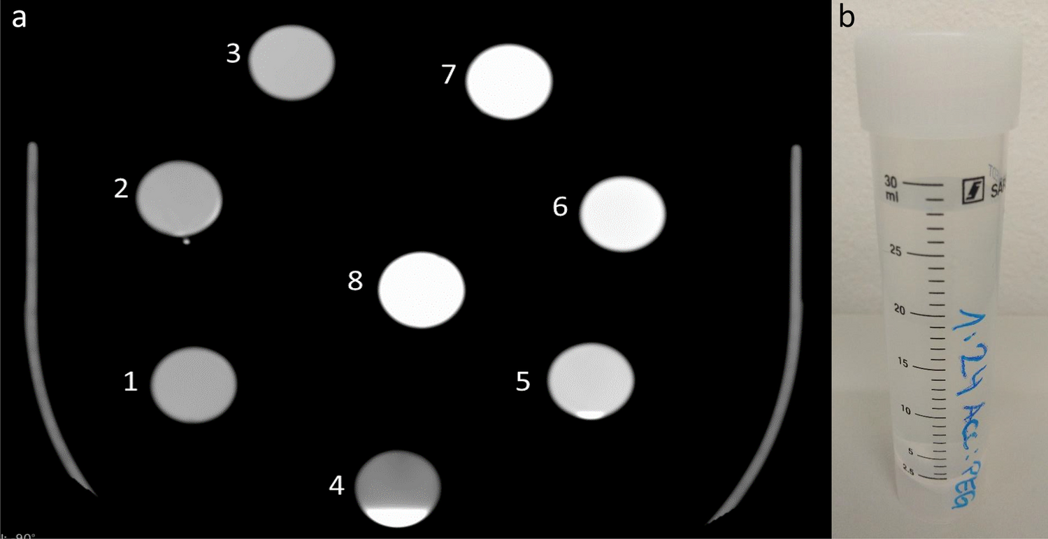
We first measured the Hounsfield units (HU) of PEG alone, which were approximately 60. The two substances were then mixed in tubes, in an extracorporeal setting, with different mixing ratios, and the HU of the different mixtures were then measured on the CT images. Accupaque® 300: PEG ratios from 1:8 to 1:24 in eight steps (1:8, 1:10, 1:12, 1:15, 1:18, 1:20, 1:22, 1:24) were tested in tubes in an extracorporeal setting.
As a preliminary result, Hounsfield units between 520 (ratio 1:24) and 1300 HU (ratio 1:8) were measured for the specified mixing ratios (Fig. 1a & b). A value of approximately 700 HU was previously determined to represent the best image quality for a voltage of 120 KVp. This value was obtained with a mixing ratio of Accupaque® 300: PEG 200 of 1:15.
Fig. 1
a) Tested mixing ratios in the tubes with 120 KVp, WL: 315, WW1229 (1: 1:24 & 520HU; 2: 1:22 & 540HU; 3: 1:20 & 600HU; 4: 1:18 & 330HU above, 4800HU below; 5: 1:15 & 720HU; 6: 1:12 & 880 HU; 7: 1:10 & 1070HU; 8: 1:8 & 1300HU). b) An example of the test tube for the 1:24 mixing ratio(No.1)
Polyethylene glycol itself is non-toxic to humans and nature. Therefore, residues of PEG 200 and also of the clinically used contrast agent Accupaque 300 could be disposed via the respective Institute of Forensic Medicine. There are also no concerns when burying a corpse after PMCTA with PEG-Accupaque, especially as a large part of the contrast agent mixture is already removed from the corpse during the autopsy.
PM angiographyAfter a native (no contrast injection) whole-body CT scan a complete external examination of the body was performed. The femoral vessels (artery and vein) were then prepared for cannulation, as described by Grabherr et al. 2008 and 2011 [6, 22].
PEG 200 and Accupaque® 300 were then mixed in the required quantities. In addition, 200 ml of the mixture was always added to fill the pump system. In order to obtain an adequate protocol, we adjusted the injection volumes in the different phases.
The arterial, venous and dynamic phases, together with the CT scans were performed according to the protocol of Grabherr et al. [6]. Finally, we divided our 23 cases into four groups (Table 1).
Table 1 Tested angiography parametersAll MPMCTAs were performed with the flow rates established by Grabherr et al. [6]. It was shown that with a flow rate of 800 ml/min for the arterial and venous phase and of 200 ml/min for the dynamic phase a very good image quality could be achieved, and no adaptation seemed to be necessary.
In four cases no dynamic phase was performed. Three of these cases were the first cases in group 1 and 2, one case was a rupture of the abdominal aorta and therefore no dynamic phase was performed.
Image qualityThe images of the different phases were evaluated in terms of image quality. The image quality contains the opacity of the different structures, visualization of previously defined vessels and the possibility of a diagnostic assessability. The images available in DICOM format were analyzed at the CURML, site of Lausanne using the program "AW Server 3.2 Ext. 4.0" and at the Forensic Institute in Munich using the program "Osirix Version 11.0".
To assess the image quality, the filling status of the vessels during the different phases was evaluated separately for the following vessels and organs: superior sagittal venous cerebral sinus (Sinus sag.), transverse venous cerebral sinus (Sinus trans.), bilateral jugular vein (Vv. Jug.), bilateral common carotid artery (Aa. Caro.), right ventricle of the heart (R ventricle), left ventricle of the heart (L ventricle), right coronary artery (RCA), left circumflex coronary artery (LCX), left anterior descending coronary artery (LAD), left pulmonary artery (LPA), right pulmonary artery (RPA), right femoral vein (V. fem. R), right femoral artery (A. fem. R), left femoral vein (V. fem. R), left femoral artery (A. fem. L) and the brachial arteries on both sides (Aa. brach.). The aorta and the vena cava were not evaluated separately because we assumed that these vessels would be filled when the downstream vessels were filled with contrast medium.
Femoral vessels were graded according to the side of cannulation (grading of vessels contralateral to the injection side). All other vessels were graded according to filling status. Some of the vessels were very small in diameter, such as the coronary arteries. Therefore, it was not possible to develop a suitable ROI (region of interest) for all vessels with the same diameter. Instead, the vessels were tracked with the cursor and the Hounsfield units were measured.
Whether the vessel was completely (1), partially (2) or not filled (3) was assessed in each case and for each phase. A vessel was considered 'partially filled' if approximately 50–80% of the vessel was opacified. If the vessel was less than 50% filled, it was recorded as not filled.
Opacification of organs (brain, liver, stomach, spleen, kidneys) was defined as complete (1), partial (2) or not opacified (3). It was documented whether the organ was overfilled [1 +], defined as an overload of contrast agent and leakage from the vessels into the surrounding organ tissue. The presence of a layering artefact was also an aspect of the assessment (Fig. 2). Layering is defined by the presence of two different fluids visible in one vessel. This artefact is an image produced by the fact that the contrast agent mixture used does not mix with the resting blood. In angiographies using PEG200 as a carrier substance the blood is found on top of the contrast agent mixture in the vessels, especially in the ascending aorta (described in the supine position of the body), preventing the filling of the right coronary artery.
Fig. 2
So-called layering in ascending aorta, provokating a non-opacification of the right coronary artery (green arrowheads). The left coronary artery was opacified (green arrow). (axial view; WL: 300, WW: 1500)
Image quality assessmentRadiological assessment of image quality, filling of the various vessels and enhancement of organ structures was performed by two forensic pathologists with six to 18 years of experience in forensic radiology. For the possible over-enhancement of some organ structures, a ROI of approximately 1 cm2 with Hounsfield units of 200 or more was considered significantly overfilled.
留言 (0)