Experimental animals
TRAIL-R knockout mice (Trail-r−/−, C57BL/6 background) were provided by Prof. Henning Walczak (UCL Cancer Institute, University College London, United Kingdom) [14]. Wild-type C57BL/6 Trail-r+/+ (female, 6–7 weeks old) and Trail-r−/− (female, 6–7 weeks old) mice were maintained and used in accordance with the guidelines of the Association for Assessment and Accreditation of Laboratory Animal Care at the National Taiwan University Medical Center. All experimental protocols were approved by the Animal Ethics Committee of the National Taiwan University Medical Center (IACUC Approval No: 20190025).
Cells
CD4+ T cells were isolated from murine spleens using an EasySep Mouse CD4+ T cell Isolation Kit (STEMCELL Technologies, catalog no. 19852; Vancouver, BC, Canada). Murine CD4+ T cells and Jurkat T cells (American Type Cell Culture [ATCC]: TIB-152) were cultured in RPMI-1640 medium (Invitrogen, Carlsbad, CA), containing 10% fetal calf serum (Invitrogen), penicillin (10 units/mL; Invitrogen), and streptomycin (10 µg/mL; Invitrogen). EL4 T cells (ATCC: TIB-39) were cultured in DMEM medium (Gibco, Grand Island, NY) containing 10% horse serum (Gibco), penicillin (10 units/mL; Invitrogen), and streptomycin (10 µg/mL; Invitrogen).
Plasmids, siRNAs, and transfection
To generate expression plasmids for flag-tagged full-length (1–381; TRAIL-RWT-FLAG) or death domain-truncated TRAIL-R mutant (1–273; TRAIL-R△DD-FLAG), the coding sequence for full-length and death domain-truncated derivatives of mouse TRAIL-R were isolated using RT-PCR, and the amplified product was ligated in-frame into pcDNA3.1 vector. The resulting plasmids were confirmed using DNA sequencing. The primers used for TRAIL-RWT-FLAG were 5′-CGTCAGATCCGCTAGCATGGACTACAAGGACGACGATGACAAGGAGCCTCCAGGACCCAGCA-3′ and 5′-GTCGACTGCAGAATTCTCAAACGCACTGAGATCCTC-3′; the primers for TRAIL-R△DD-FLAG were 5′-CGTCAGATCCGCTAGCATGGACTACAAGGACGACGATGACAAGGAGCCTCCAGGACCCAGCA-3′ and 5′-GTCGACTGCAGAATTCTCACTTCAGGTCGTCAGCTGAGT-3′. TRAIL-RWT-FLAG or TRAIL-RΔDD-FLAG plasmids were transfected into EL4 T cells using electroporation. Briefly, EL4 cells (5 × 10⁵ cells) were gently mixed with the plasmid (10 µg) in an electroporation buffer (Neon Transfection System Kit, catalog no. MPK10096; Invitrogen), transferred to an electroporation cuvette, and electroporated using the Neon Transfection System (Invitrogen), following the manufacturer’s protocols. SHP-1 siRNA (catalog no. sc-29479), and control siRNA (catalog no. sc-37007) were purchased from Santa Cruz Biotechnology (Dallas, TX). For each transfection, 1 μg of siRNA duplex mixed with 5 μL siRNA transfection reagent (catalog no. sc-29528; Santa Cruz Biotechnology) was incubated with murine CD4+ T cells (2 × 10⁵ cells), according to the manufacturer’s protocol.
Antibodies and reagents
Recombinant mouse TRAIL protein (catalog no. Z03367) was purchased from GenScript (Piscataway, NJ). Stimulatory anti-CD3 (catalog no. 100253) and anti-CD28 (catalog no. 102121) antibodies were purchased from BioLegend (San Diego, CA). The antibodies used for flow cytometry, including anti-CD69-FITC (catalog no. 104506) and anti-IL-2-PE (catalog no. 503808), were also purchased from BioLegend. Anti-CD25-PE (catalog no. 553866) antibody was purchased from BD Biosciences (Franklin Lakes, NJ). For immunoblotting and immunoprecipitation, anti-LCK (catalog no. 2752S), anti-ZAP70 (D1C10E, catalog no. 3165S), anti-p-ZAP70 (Y319/Y352, catalog no. 2717S), anti-PLCγ1 (D9H10, catalog no. 5690), anti-p-PLCγ1 (Y783, catalog no. 2821S), anti-p-LAT (Y255, catalog no. 45170), anti-PKCθ (E117Y, catalog no. 13643), anti-p-PKCθ (T538, catalog no. 9377), anti-p-IKKβ (S176/180, catalog no. 2697S), anti-JNK (catalog no. 9252), anti-p-JNK (T183/Y185, catalog no. 9251), anti-p38 mAPK (catalog no. 9212), anti-caspase 8 (11G10, catalog no. 9748S), anti-p-SHP-1 (Y564, catalog no. 8849S), anti-SHP-2 (D50F2, catalog no. 3397S), anti-p-SHP-2 (Y580, catalog no. 3703S), anti-CBL (catalog no. 2747S), anti-CSK (C74C1, catalog no. 4980), and anti-FLAG (DYKDDDDK Tag Antibody, catalog no. 2368) antibodies were purchased from Cell Signaling Technology (Danvers, MA). Anti-p-LCK (Y394, catalog no. 933102), anti-ERK1/2 (catalog no. 686901), and anti-p-ERK1/2 (T202/Y204, catalog no. 675506) antibodies were purchased from BioLegend. Anti-p-CD3ζ (Y142, catalog no. abx012434) antibody was purchased from Abbexa (Cambridge, UK). Anti-LAT (catalog no. PA5-82419) antibody was purchased from Invitrogen. Anti-TRAIL-R (catalog no. 3062) was purchased from BioVision (Milpitas, CA). Anti-CD3ζ (G3, catalog no. ab11281), Anti-IKKβ (EPR6043, catalog no. ab124957), anti-Caspase 3 (EPR18297, catalog no. ab184787), and anti-SHP-1 (catalog no. ab227503) antibodies were purchased from Abcam (Cambridge, UK). Anti-p38 MAPK (catalog no. 14064–1) and anti-p-p38 MAPK (T180/Y182, catalog no. 28796–1) antibodies were purchased from Proteintech (Rosemont, IL). Anti-flotillin-1 antibody (catalog no. 610821) was purchased from BD Biosciences (Franklin Lakes, NJ). Anti-β-actin (clone C4, catalog no. MAB1501) antibody was purchased from Merck Millipore (Billerica, MA). For confocal imaging, Lck-Cy3 was purchased from Bioss (catalog no. BS-2649R; Woburn, MA). Anti-ZAP-70-FITC (catalog no. 313404) was purchased from BioLegend. Anti-ganglioside GM-1 (catalog no. ab23943) was purchased from Abcam. All primary antibodies used for immunoblotting were used at 1:1000 dilutions. The immunoprecipitation antibodies were used at a dilution of 1:200. The pan-caspase inhibitor Z-VAD-FMK (catalog no. ab120487) was purchased from Abcam. All the antibodies used for the immunoblotting and immunoprecipitation were listed in Supplementary Table S1.
Immunoblotting analysis
Total cell lysates were prepared on ice using PhosphoSafe Extraction Reagent (catalog no. 71296; Merck Millipore). The protein extracts were fractionated on SDS–polyacrylamide gel electrophoresis (SDS-PAGE) and transferred onto polyvinylidene difluoride membranes. The membranes were probed with individual primary antibodies, followed by incubation with horseradish peroxidase-conjugated secondary antibodies. The horseradish peroxidase substrate reaction was performed using a chemiluminescent reagent (catalog no. WBKLS0500; Merck Millipore), and the chemiluminescent signal was detected using iBright Imaging Systems (Invitrogen).
Immunoprecipitation
Cell extracts were incubated with individual antibodies and protein G-agarose beads (catalog no. M16-266; Merck Millipore) in lysis buffer (50 mM Tris [pH 8.0], 150 mM NaCl, 1% Triton X-100, 0.5% deoxycholate, 0.1% SDS, 2 µg/mL leupeptin, 5 µg/mL aprotinin, 1 mM phenylmethylsulphonyl fluoride, 1 mM dithiothreitol, and 1 mM Na3VO4) with continuous rotation at 4 °C for 3 h. Immunoprecipitates were washed three times with lysis buffer before use in the immunoblotting analyses.
Lipid raft isolation
Cell extracts were incubated with 0.2% Triton-X, 50 mM HEPES, 100 mM NaCl, 5 mM EDTA, 1% Ser/Thr protein kinase inhibitor, 1% Thr protein kinase inhibitor, and 1% protease inhibitor, for 1 h on ice. The lysate was diluted (1:1) with 80% ice-cold sucrose in 150 mM NaCl, 5 mM EDTA, and 25 mM MES; transferred to Ultra-Clear centrifuge tubes (Beckman Coulter, Brea, CA); and overlaid with ice-cold 30% sucrose followed by ice-cold 5% sucrose. The sucrose gradients were centrifuged at 200 000 × g in a Beckman Coulter SW41Ti rotor for 22 h at 4 °C. Twelve 375 μL fractions were collected and SDS-PAGE sample buffer was added to the harvested fractions before immunoblotting analyses.
Flow cytometry
For the cell proliferation assay, cells were stained with carboxyfluorescein diacetate succinimidyl diester (CellTrace CFSE kit, catalog no. C34570; Thermo Fisher Scientific, Waltham, MA) with indicated treatment for 96 h followed by flow cytometry. For the intracellular staining of IL-2, the cells were pre-incubated with monensin (GolgiStop™, catalog no. BDB554724; BD Biosciences) for 4 h, followed by fixation and permeabilization with BD Cytofix/Cytoperm (catalog no. 554714; BD Biosciences) and staining with the indicated antibodies for 30 min on ice before flow cytometric analysis. For the apoptosis assay, cells were stained with a PE-Annexin V/7-AAD apoptosis detection kit (catalog no. 640934; BioLegend). The ratio of Annexin V+ cells to total cells indicated the percentage of apoptotic cells. All data were acquired using a FACSCanto II (BD Biosciences) and analyzed with FlowJo ver.10.8.1 software (BD Life Sciences, Ashland, OR).
Enzyme-linked immunosorbent assays
The levels of IL-2 (ELISA MAX™ Deluxe Set Mouse IL-2, catalog no. 431004; BioLegend), IL-4 (ELISA MAX™ Deluxe Set Mouse IL-4, catalog no. 431104; BioLegend), and IFN-γ (ELISA MAX™ Deluxe Set Mouse IFN-γ, catalog no. 430804; BioLegend), from the cell lysates were analyzed by enzyme-linked immunosorbent assays (ELISAs) as described by the manufacturer (BioLegend).
Confocal microscopy
Cells (2.0 × 10⁶) were incubated with anti-CD3 (3 µg/mL) and anti-CD28 (2 µg/mL) antibodies in the presence or absence of TRAIL (10 µg/mL) for 30 min at 37 °C. After washing with phosphate-buffered saline (PBS) at 4 °C, the cells were fixed with 4% formaldehyde for 1 h, permeabilized with 0.5% Triton X-100 in PBS for 15 min, and stained with the indicated antibodies. After washing with 1% bovine serum albumin in PBS, the cells were mounted on glass slides using a drop of Corning Matrigel matrix (catalog no. 354234; Corning Life Sciences, AZ). Images were obtained using a Leica SP8 STED confocal microscope (Wetzlar, Germany) with a 100 × objective lens. The laser sources used were a 405 nm diode laser and a 470–670 nm white-light laser.
Liquid chromatography–tandem mass spectrometry (LC–MS/MS) and data analysis
Mass spectrometry was performed as described previously [15, 16]. Briefly, protein samples were separated using SDS-PAGE and stained with Coomassie Brilliant Blue. The protein bands were then excised and digested with trypsin. The resulting peptide mixtures were analyzed using an LTQ Orbitrap Elite Hybrid Mass Spectrometer (Thermo Fisher Scientific, Waltham, MA). The peptide data were analyzed using Mascot MS/MS Ion Search (Matrix Science, Boston, MA) under the following conditions: peptide mass tolerance, 20 parts per million; fragment MS/MS tolerance, 1.2 daltons; allowed up to one missed cleavage; peptide charges, 2+, 3+, and 4+.
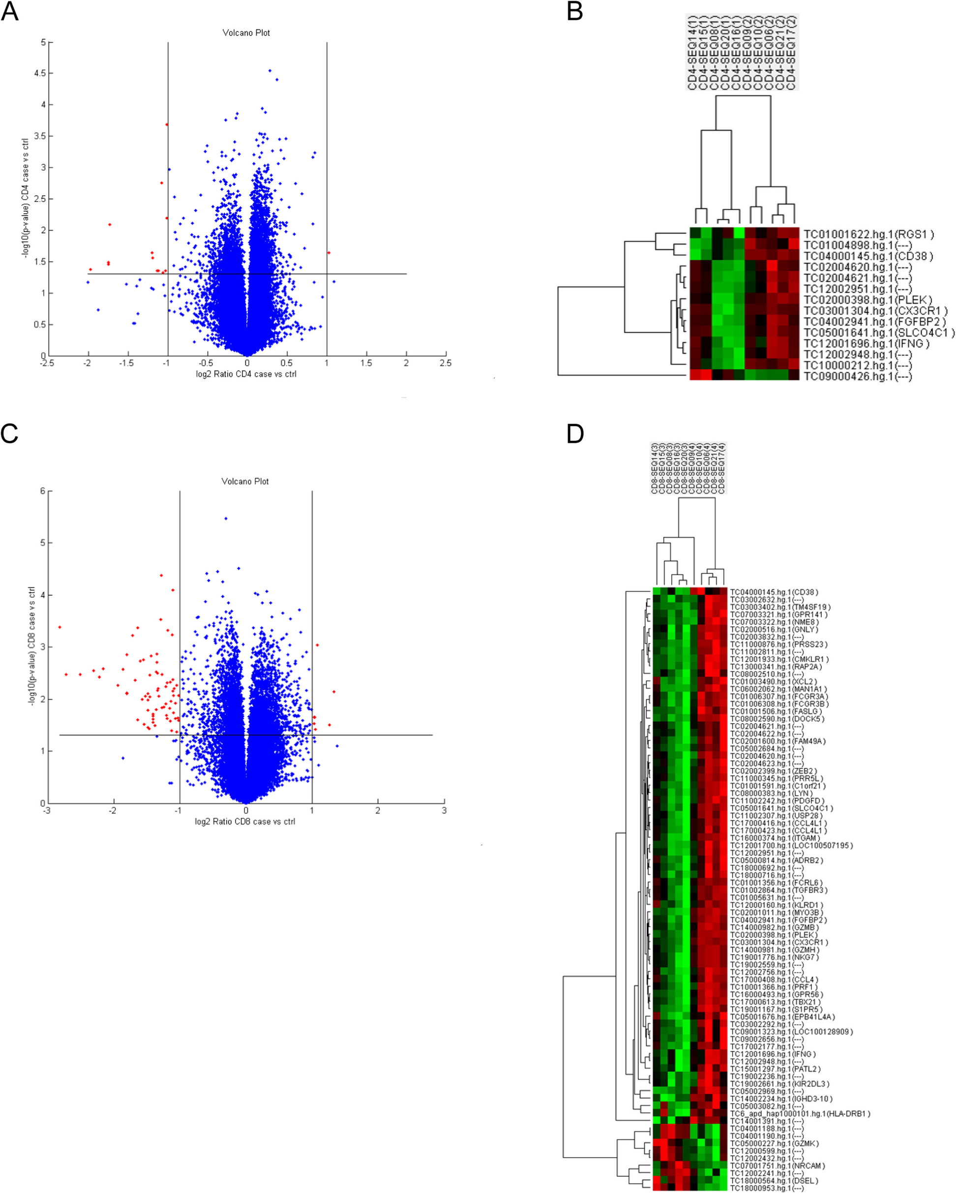
RNA sequencing (RNA-seq) analysis
Sample preparation and RNA-seq analysis were performed as previously described [13]. Briefly, total RNA from CD4+ T cells was extracted using TRIzol reagent (catalog no. 15596026; Invitrogen), and 1 µg of total RNA was used for constructing sequencing libraries. RNA libraries for RNA sequencing (RNA-seq) analysis were prepared using the SureSelect XT HS2 mRNA Library Prep Kit (Agilent, Santa Clara, CA). Library samples were analyzed on an Illumina NovaSeq 6000 sequencer (150 bp paired-end reads and dual index configuration). Quality control was performed using Trimmomatic software (v0.36). Reads were aligned to the mouse reference GRCm38 using HISAT2. Feature counts were normalized using the DESeq2 package (v1.28.1). Principal component (factoextra ver.1.0.3; free software) and Gene Ontology enrichment analyses (clusterProfiler ver.3.6; free software) were performed using R. The data generated in this study were deposited in the Gene Expression Omnibus of the National Center for Biotechnology Information [17] and are accessible through the accession number GSE222519 (https://www.ncbi.nlm.nih.gov/geo/query/acc.cgi?acc=GSE222519).
Statistical analysis
Densitometric analysis of the immunoblotting results was performed using Image-Pro Plus software (Media Cybernetics). All statistical analyses were performed using the Prism 9.0 (GraphPad Software, San Diego, CA). Group differences were analyzed using non-parametric Mann–Whitney U test or two-tailed Student’s t test. Statistical significance was defined at p < 0.05.


留言 (0)