The findings of this prospective study shed light on the intricate landscape of revision rhinoplasty, specifically when augmented by the precision associated with costal graft integration [10]. The predominant indications for revision surgery, including nasal obstruction and dissatisfaction with nasal appearance, underscore the complexity of cases encountered in this series [11]. The quantifiable improvements in functional outcomes, as measured by the Nasal Obstruction Symptom Evaluation (NOSE) scale, reflect the tangible success of revision rhinoplasty with costal grafts. The significant reduction in NOSE scores at 3 months postoperatively emphasizes the efficacy of the surgical interventions in alleviating nasal obstruction symptoms. Simultaneously, the enhanced aesthetic satisfaction, as captured by the Rhinoplasty Outcome Evaluation (ROE) questionnaire, reinforces the transformative impact on patients’ subjective experiences of their nasal appearance.
The study’s outcomes align with and contribute to the evolving literature on revision rhinoplasty [12]. The use of costal grafts has been previously associated with improved structural support and durability, corroborating existing evidence in the field [13, 14]. The observed complications underscore the need for a nuanced approach, emphasizing the importance of careful patient selection and comprehensive postoperative care to mitigate adverse events [15].
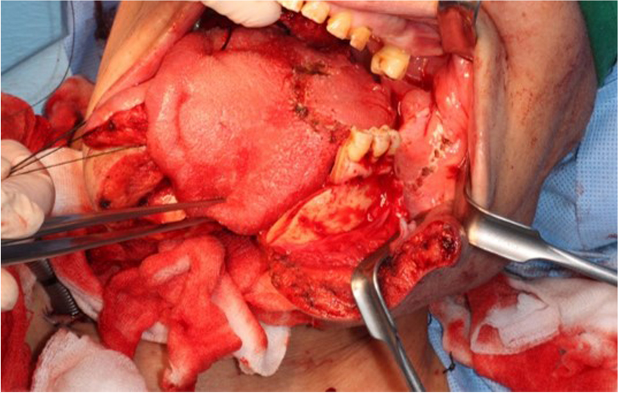
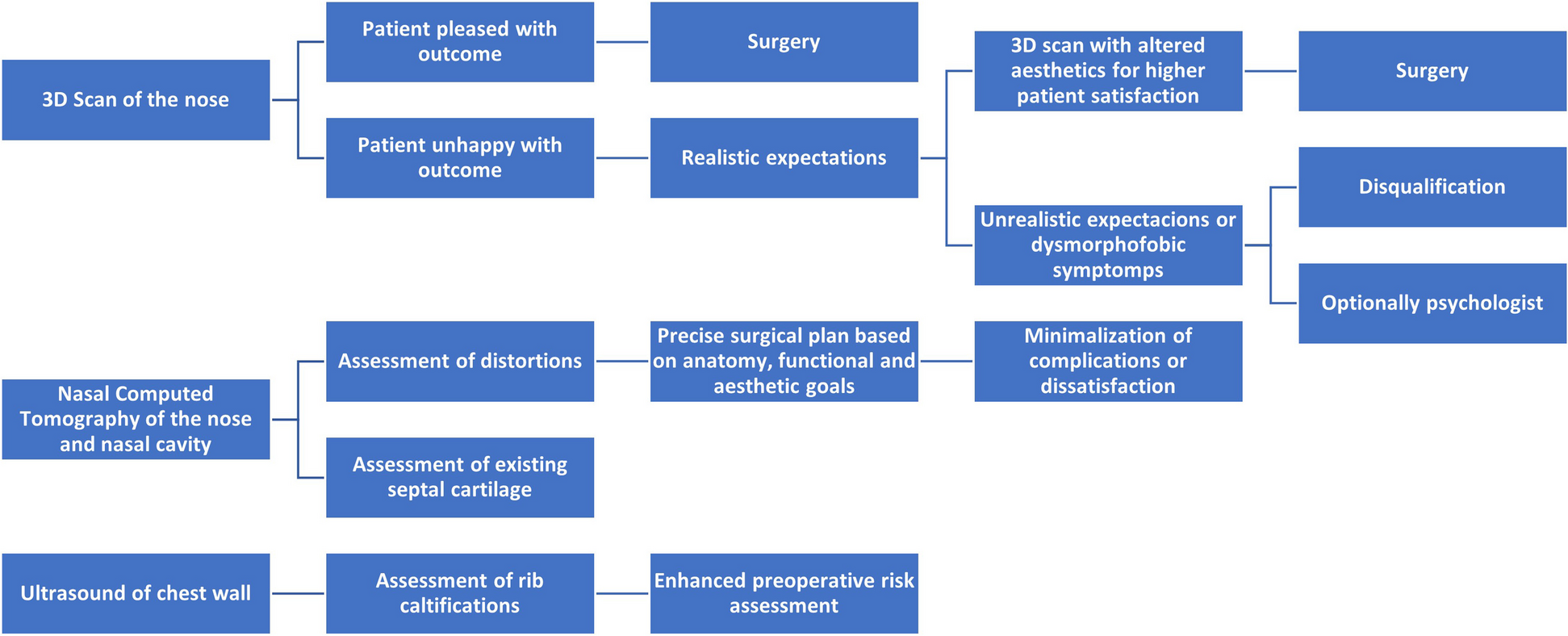
The integration of 3D scanning and CT imaging in preoperative assessment facilitates a nuanced understanding of the nasal architecture, crucial for tailoring the revision surgery to individual needs [8, 9]. The 3D scan, offering a high-resolution external view, is instrumental in assessing aesthetic deformities and asymmetries. It aids in envisioning the expected outcomes and precisely planning the external modifications of the grafts, such as their shape and contour, to harmonize with the patient’s facial features (Table 3). The preoperative nasal CT serves as a crucial tool in meticulously planning and executing this intricate operation [8]. Through a comprehensive evaluation of the nasal anatomy and any prior surgical alterations, the CT scan provides invaluable insights into the patient’s nasal framework, including the presence of septal deviation, nasal bone morphology, and the integrity of existing cartilage structures [4]. This detailed assessment allows for a tailored surgical approach, particularly in the harvesting and sculpting of rib cartilage grafts [5]. By precisely delineating the dimensions and contours of the rib cartilage, the preoperative CT scan facilitates strategic planning to ensure optimal graft size and shape required for structural support and functional restoration, thereby minimizing donor site morbidity and enhancing the aesthetic and functional outcomes of the revision rhinoplasty. Furthermore, the CT evaluation aids in identifying potential challenges such as previous scar tissue, septal perforations, or asymmetric nasal anatomy, enabling the surgeon to anticipate and address these complexities during the surgical procedure. In essence, the integration of preoperative nasal CT imaging into the surgical workflow serves as a fundamental step in achieving successful outcomes in revision rhinoplasty with rib cartilage grafting, allowing for meticulous planning and precise execution tailored to the individual patient’s anatomical nuances and surgical goals.
The utility of preoperative 3D scanning in revision rhinoplasty with costal grafts represents a significant advancement in surgical planning and outcomes [9]. This technology enables precise anatomical assessment, allowing surgeons to visualize the complex structures of the nose with unparalleled clarity. By creating a detailed three-dimensional model of the patient’s nasal anatomy, surgeons can accurately plan the size, shape, and placement of costal grafts before the procedure. This preoperative insight facilitates tailored interventions, minimizes intraoperative guesswork, and enhances the accuracy of graft fitting, leading to improved aesthetic and functional results. The integration of 3D scanning technology into revision rhinoplasty with costal grafts thus marks a pivotal shift towards more predictive and patient-specific surgical outcomes.
In summary, the detailed preliminary evaluation using 3D and CT scans is pivotal in guiding the harvesting and sculpting of rib cartilage for revision rhinoplasty. This approach allows for a highly customized surgical plan, addressing a range of clinical scenarios with appropriately designed chondral grafts, thereby enhancing both the functional and aesthetic outcomes of the procedure.
Our study introduces several advancements and refinements in both the surgical technique and evaluation metrics, distinguishing it from traditional methodologies. Firstly, our approach integrates advanced imaging techniques, specifically high-resolution 3D scanning and CT imaging, not as mere diagnostic tools but as integral components of surgical planning. This goes beyond the conventional use of these imaging methods. The detailed visualization afforded by these scans allows for a more precise and tailored approach to graft harvesting and sculpting. Unlike traditional techniques, which often rely on surgeon’s experience and intraoperative assessment, our method uses these scans to create a detailed map of the nasal architecture, guiding the surgeon in crafting grafts with specific dimensions and shapes suited to each patient’s unique anatomical requirements. This precision in graft preparation, especially in the challenging realm of revision rhinoplasty, potentially reduces the risk of over- or under-correction and enhances both aesthetic and functional outcomes. Additionally, our study offers new insights into the relationship between preoperative imaging findings and postoperative outcomes. By correlating specific anatomical features identified in preoperative scans with postoperative NOSE and ROE scores, we aim to establish a more predictive model of patient outcomes. This model could potentially guide surgeons in making more informed decisions during both the planning and execution phases of the surgery.
The summarized clinical implications drawn from the current experiences serve as a guidepost for plastic surgeons navigating the challenges of revision rhinoplasty. Notably, the study advocates for the strategic integration of costal grafts, refined preoperative planning methodologies, and a comprehensive understanding of patient expectations. These implications offer insights that extend beyond the confines of the study, providing a foundation for fellow plastic surgeons seeking to enhance their practice in revisionary settings. The study presents innovative elements by integrating advanced imaging techniques (3D scanning and CT imaging) for precise preoperative planning in revision rhinoplasty with costal grafts, moving beyond traditional diagnostic uses. This approach allows for detailed mapping of nasal architecture, enabling the creation of grafts tailored to individual anatomical needs. It offers a refined surgical technique that potentially reduces the risk of over- or under-correction, enhancing both aesthetic and functional outcomes. The study also proposes a predictive model correlating preoperative imaging findings with postoperative outcomes, aiming to guide surgeons in making more informed decisions. This novel integration of technology and technique provides new insights into optimizing revision rhinoplasty outcomes.
Despite the valuable insights garnered, this study has inherent limitations. The single-center design and the absence of a control group limit the generalizability of findings. Further, the relatively short follow-up period necessitates cautious interpretation of long-term outcomes. Future research could explore larger multicenter studies with extended follow-up periods to enhance the robustness of findings and address potential confounders.


留言 (0)