Stem cells maintenance and cardiac differentiation
The H1 cell lines of hESCs and hiPSCs were maintained on Matrigel-coated plates with mTeSR1 medium (Stem Cell Technologies, Canada) for 3 days till 80% confluence. To initiate cardiac differentiation, the cells were cultured with a series of cardiac differentiation medium (I, II and III) for 12 days according to the manufacturer’s protocols (CELLAPY, China). Meanwhile, the maintenance and cardiac differentiation of mESCs were performed as previously described [22]. Briefly, the mESCs were seeded onto gelatin-coated plates and cultured in high glucose DMEM medium (GIBCO, USA) supplemented with 10% FBS (ExCell Bio, China) and 1,000 U /ml of leukemia inhibitory factor (LIF) (Millipore, USA). To generate embryoid bodies (EBs), mESCs were developed into EBs (1,250 cells/drop) in a 3D hanging-drop culture for 4 days with LIF-free medium. The early EBs were later transferred to gelatin-coated 12-well culture plates for 8-day adherent differentiation with LIF-free medium.
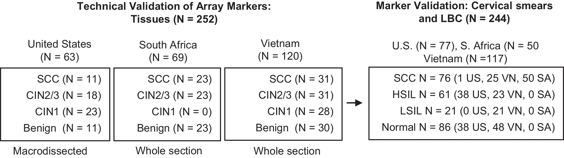
Specimen collection and ethical statement
This study was approved by the Ethics Committee of Guangdong Provincial People's Hospital (KY-Q-2021-277-01) and was performed in accordance with Helsinki declaration. In brief, peripheral blood samples were collected from 10 ASD patients and 9 healthy controls aged from 2 to 8 years, with written informed consent obtained from the parents of each participant. Whole transcriptome sequencing was later performed on plasma extracted from these peripheral blood samples. The comparison of the age and gender between the two groups did not reveal significant difference (Additional file 1: Table S7). The ASD patients were recruited from Guangdong Provincial People's Hospital and confirmed by echocardiography, while healthy controls were from the community. None of the participants had any other underlying health conditions.
MicroRNA expression profiling
The total RNA was extracted from cardiac-differentiated hESCs using TRIzol (Invitrogen, USA) in accordance with the manufacturer's protocol. The quantity and quality of small RNA were evaluated using RiboBio Co., Ltd. The yield and integrity of the RNA were then determined by Qubit®2.0 (Life Technologies, USA) and Agilent 2200 TapeStation (Agilent Technologies, USA), respectively. Subsequently, 1 µg of RNA from each sample was employed for the preparation of small RNA libraries using NEBNext® Multiplex Small RNA Library Prep Set for Illumina (NEB, USA) according to the manufacturer's instructions. The libraries were then sequenced using a HiSeq 2500 (Illumina, USA) platform with a single end 50 bp configuration by RiboBio Co., Ltd. (RiboBio, China). The differential expression of miRNAs between groups was calculated using the edgeR algorithm, with the criteria of | log2(Fold Change) |≥ 1 and P-value < 0.05. Gene ontology (GO) and Kyoto Encyclopedia of Genes and Genomes (KEGG) pathway analysis were further carried out using the KOBAS tool.
Total RNA in plasma samples were extracted from peripheral blood samples collected from ASD patients or healthy individuals using TRIzol® Reagent (Plant RNA Purification Reagent for plant tissue) according to the manufacturer's guidelines (Invitrogen, USA). The quality of extracted RNA was assessed using 2100 Bioanalyzer (Agilent Technologies, USA) and quantified with ND-2000 (NanoDrop Technologies, USA). The RNA purification, reverse transcription, library construction and sequencing procedures were carried out at Shanghai Majorbio Bio-pharm Biotechnology Co., Ltd. (Shanghai, China), in accordance with the manufacturer's instructions (Illumina, San Diego, CA). A total of 3 µg of total RNA per sample was utilized as the input material for the small RNA library. The sequencing libraries were generated using the Truseq TM Small RNA sample prep Kit from Illumina (San Diego, CA) in compliance with the manufacturer's recommendations. The differential expression of miRNAs between the two sets of samples was calculated using the NOIseq algorithm, with the criteria of Prob value > 0.8.
Animal study and ethical statement
The animal experiments were approved by the Animal Research Committee of Guangzhou Medical University (GY2021-054) and the procedures conformed to the National Institutes of Health (NIH) Guide for the Care and Use of Laboratory Animals. C57BL/6 J mice (8 weeks old) were purchased from the Medical Experimental Animal Center of Guangdong Province. All the mice were raised with food and water ad libitum and housed in humidity and temperature-controlled environment with a 12/12-h light/dark cycle. Female and male mice were mated in cages, and vaginal plug was monitored every morning. Once the vaginal plug was observed, it was designated as embryonic day 0.5 (E0.5). Besides, the first postnatal day was designated as postnatal day 0 (P0). Eventually, the mice were euthanized by intraperitoneal injection of 200 mg/kg pentobarbital. The heart samples from either the embryos or neonates were collected on certain embryonic or postnatal day according to experimental requirements.
Knockdown of TET2 in hESCs
A lentivirus vector (LV)-shTET2 with enhanced green fluorescent protein (eGFP) was obtained from Vector Builder (Guangzhou, China), while an empty vector with eGFP was utilized as a negative control (LV-NC). hESCs were cultured in 12-well plates till 50% confluence and then infected with lentivirus particles in the presence of 5 μg/ml polybrene at a multiplicity of infection of 50. The cells were maintained for a minimum of 4 days and then screened for puromycin resistance. The knockdown efficiency of TET2 was evaluated through qRT-PCR and western blotting. The primers used for the shRNA target sequences are listed in Additional file 1: Table S8.
MicroRNA transfection
The agents including negative control of mimic (m-NC), miR-20b-5p mimic, negative control of inhibitor (i-NC) and miR-20b-5p inhibitor were obtained from RiboBio (Guangzhou, China). To investigate the role of miR-20b-5p in hESCs-derived cardiac differentiation, the hESCs were subjected to 12-day cardiac differentiation with transfection of either 100 nM of m-NC or miR-20b-5p mimic, 300 nM of i-NC or miR-20b-5p inhibitor using the miRNA transfection reagent (RiboBio, China) in accordance with the manufacturer's instructions. The transfection was carried out bi-daily throughout the 12-day process of cardiac differentiation.
Western blotting
Total protein was extracted from cell samples or heart tissues using a protein lysis buffer containing protease and phosphatase inhibitors (Beyotime, China). The protein concentration was then determined using a Bicinchoninic Acid protein assay kit (Thermo Fisher Scientific, USA). The proteins were separated using either a 6% or 12% Sodium dodecyl sulfate–polyacrylamide gel electrophoresis (Beyotime, China) and transferred onto polyvinylidene difluoride membranes (Millipore, USA). To block non-specific binding, the membranes were incubated in 5% non-fat milk for 1 h. Subsequently, the membranes were incubated overnight at 4 °C with primary antibodies including rabbit anti-α-tubulin (diluted 1:1000, Abcam, ab52866), rabbit anti-GAPDH (diluted 1:1000, Cell Signaling Technology, #5174), rabbit anti-TET2 (diluted 1:1000, Cell Signaling Technology, #45010), rabbit anti-TBX5 (diluted 1:1000, Abcam, ab137833), mouse anti-NKX2-5 (diluted 1:1000, Abcam, ab91196), rabbit anti-GATA4 (diluted 1:1000, Abcam, ab134057), rabbit anti-cTnT (diluted 1:1000, Abcam, ab209813) and mouse anti-MYH6 (diluted 1:1000, Abcam, ab50967). The membranes were then incubated with horseradish peroxidase-conjugated secondary antibodies (diluted 1:5000, ZSGB-Bio, ZB-2301, ZB-2305) for 1 h at room temperature. Antibody binding was visualized using an ECL detection reagent (Beyotime, China) and captured with Amersham Imager 600 (GE Healthcare, USA). Density analysis of the bands was performed using Image J, and the relative protein levels were normalized to the expression of either GAPDH or α-tubulin.
Quantitative real time-PCR (qRT-PCR)
Total RNA was extracted from cell samples or heart tissues using TRIzol reagent (Accurate Biology, China). Complementary DNA (cDNA) was then synthesized by a reverse transcription kit (Accurate Biology, China) according to the manufacturer's protocol. qRT-PCR was carried out on the Lightcycler480 system (Roche, USA) using SYBR Green PCR Kit (Accurate Biology, China). The level of miR-20b-5p was detected using stem-loop primers obtained from RiboBio (Guangzhou, China), while other gene primers used for qRT-PCR were obtained from Tsingke Biotechnology (Beijing, China). The results were normalized to the expression of either U6 or GAPDH, and relative fold changes were calculated using the 2−△△Ct method. The primers used for qRT-PCR are listed in Additional file 1: Table S9.
Immunofluorescence staining
The cardiac-differentiated hESCs were fixed with 4% paraformaldehyde (Biosharp, China) for 20 min, followed by washing with PBS. The cells were then permeabilized with 0.25% Triton X-100 for 20 min, followed by washing with PBS. The cells were blocked with 10% goat serum for 1 h at room temperature and then incubated with primary antibodies overnight at 4 °C. The primary antibodies included rabbit anti-TET2 (diluted 1:200, Invitrogen, PA5-85488) and rabbit anti-cTnT (diluted 1:200, Abcam, ab209813). After washing with PBS, the cells were incubated with secondary antibodies at room temperature for 1 h. The secondary antibodies included Anti-rabbit IgG (H + L), F(ab’)2 Fragment (Alexa Fluor® 488 Conjugate) (diluted 1:800, Cell Signaling Technology, #4412) and Anti-Rabbit IgG (H + L) Cross-Adsorbed Secondary Antibody (Alexa Fluor™ 647) (diluted 1:800 Invitrogen, A-21244). The nuclei were then stained with DAPI (Beyotime, C1002), and fluorescence images were obtained by a laser scanning confocal microscope (LSM 880, Carl Zeiss).
Flow cytometry analysis
The cardiac-differentiated hESCs were isolated and analyzed by flow cytometry. Approximately 1 × 10⁵ cells per sample were fixed with 4% paraformaldehyde (Biosharp, China) and washed with staining buffer (1% fetal bovine serum in PBS) (Beyotime, China). The cells were then permeabilized with 100% cold methanol and incubated with primary antibodies, followed by the corresponding secondary antibody. The primary antibodies contained rabbit anti-TET2 (diluted 1:500, Abcam, ab94580) and rabbit anti-cTnT (diluted 1:500, Abcam, ab209813). The secondary antibodies used were Anti-rabbit IgG (H + L), F(ab’)2 Fragment (Alexa Fluor® 488 Conjugate) (diluted 1:1000, Cell Signaling Technology, #4412) and Anti-Rabbit IgG (H + L) Cross-Adsorbed Secondary Antibody (Alexa Fluor™ 647) (diluted 1:1000, Invitrogen, A-21244). As shown in Additional file 1: Fig. S6, cell gating was performed by using isotype controls, such as Rabbit (DA1E) mAb IgG XP® Isotype Control (Alexa Fluor® 647 Conjugate) (diluted 1:1000, Cell Signaling Technology, #2985) and Rabbit (DA1E) mAb IgG XP® Isotype Control (Alexa Fluor® 488 Conjugate) (diluted 1:1000, Cell Signaling Technology, #2975). The samples were acquired using Amnis Image StreamX MarkII flow cytometer (Merck & Millipore, Germany) and analyzed with IDEAs.6.2 software.
Dot blot analysis
The detection of 5-hydroxymethylcytosine (5hmC) was performed by dot blot analysis. The genomic DNA was isolated by the DNA Purification Kit (TIANGEN, China) and was then diluted with TE buffer. The DNA samples were subjected to denaturation at 95 °C for 10 min, followed by rapid cooling on ice. The denatured samples were applied onto a nitrocellulose membrane (Biosharp, China) which had been pre-wetted. The membrane was washed for 10 min in 2 × SSC solution, dried for 10 min at room temperature and exposed to ultraviolet (UV) radiation for 15 min. The membrane was then blocked with 3% bovine serum albumin-TBST (BSA-TBST) for 1 h at room temperature (Beyotime, China) and incubated with mouse anti-5hmC antibody (diluted 1:1000, Cell Signaling Technology, #51660) at 4 °C overnight. Subsequently, the membrane was incubated with HRP-conjugated anti-mouse IgG secondary antibody (diluted 1:2000, ZSGB-Bio, ZB-2305) for 1 h at room temperature. The antibody binding was visualized using ECL detection reagent (Beyotime, China), and digital images were captured using Amersham Imager 600 (GE Healthcare, USA). Methylene blue staining of the membranes was used to confirm equal DNA loading. Finally, the densitometry of the bands was quantified by Image J.
Dual luciferase reporter assay
HEK-293 T cells were seeded into a 96-well plate and allowed to reach 50–70% confluence prior to transfection. The pmiR-RB-Report™ luciferase vector (RiboBio, China) was modified by the introduction of either wild-type or mutant TET2 fragments (524 bp) into its restriction sites and designated as LUC-WT-TET2 or LUC-MUT-TET2, respectively. The transfection was performed using lipofectamine 3000 (Invitrogen, USA) with 150 ng of plasmids of LUC-WT-TET2 and LUC-MUT-TET2, and then exposed to 100 nM of m-NC and miR-20b-5p mimic, 300 nM of i-NC and miR-20b-5p inhibitor. The culture medium was replaced 6 h post-transfection. After 48-h incubation, the Dual-Luciferase system (Promega, USA) was utilized to determine the firefly and Renilla luciferase activities. The medium was aspirated, and the cells were treated with 50 μL of PBS and luciferase reagent. The 96-well plate was then incubated on a shaker at room temperature for 10 min before the determination of firefly luciferase activity. The Renilla luciferase activity was determined by adding 30 μL of stop reagent per well and shaking for 10 min. The luciferase activities were then detected using Agilent BioTek Synergy LX. Finally, the differences between firefly and Renilla luciferase activities were calculated to determine the relative luciferase activity.
Statistical analysis
Quantitative data were demonstrated as means ± SEM. A two-tailed unpaired Student’s t test was used for analysis of two-group difference, while a one-way ANOVA followed by Bonferroni multiple comparisons test was introduced for assessment of multiple group variations. The differences were considered to be statistically significant, when *P < 0.05, **P < 0.01 and ***P < 0.001.


留言 (0)