
記住我


The same composition of the enriched 155Gd targets employed by Dellepiane [10] for cross-section measurements (154Gd 0.5%, 155Gd 91.90%, 156Gd 5.87%, 157Gd 0.81%, 158Gd 0.65%, 160Gd 0.27%) was used in the irradiation simulations In Fig. 1 the experimental cross sections for 155Tb production are compared with theoretical calculations. The gray band represents the models variability between Q1 and Q3, the dashed lines are the minimum/maximum values of the cross sections obtained with all models, and the blue solid line is the Talys default option (PE2-LD1), commonly referred to as the standard simulation in the literature. For the 155Tb case all models reproduce equally the cross section up to 10 MeV, and for higher energy the band is relatively thin and this corresponds to a limited model variability, as expected from a typical (p,n) reaction. On account of this, and because the Talys default reproduces the very recent data measured by Dellepiane [10] quite satisfactorily, the analysis will be presented considering the Talys default as the benchmark calculation.
Fig. 1
Experimental 155Tb cross sections from enriched 155Gd target and theoretical curves expressing the variability of nuclear reaction models
Figure 2 shows the contribution of each isotopic component of the target to the 155Tb production. The main contribution to the 155Tb cross section comes, as should be expected, from the 155Gd component of the target. For energies higher than 10 MeV, also the contribution from the 156Gd component becomes significant, and this explains the increase of the experimental data with respect to an ideal target with 100% 155Gd enrichment. The contribution from 157Gd is quite small and that from other Gadolinium components of the targets, such as 154Gd, is negligible. This figure includes cross-section data for 100%-enriched targets assessed by Dellepiane et al., 2023 [30]; they were published after the submission of this study and added in the final revision of the text.
Fig. 2
155Tb cross sections from enriched 155Gd target: Talys calculations and contributions from different Gd isotopes of the target
Next, the 156tTb total cross section, which refers to the cumulative cross sections of 156g,156m1,156m2Tb (ground and the first two metastable states), is presented in Fig. 3A. The curve including all contributions is in agreement with the data measured by Dellepiane et al. It is evident that the main contribution belongs to the 156Gd component of the target, followed by the 157Gd component at slightly higher energies. At even higher energies, about 19 MeV, the contribution from 158Gd also becomes significant. Instead, the contribution to the 156Tb cross section from the main 155Gd component of the target remains quite small in the entire range of considered energies.
Fig. 3
156g, 156m1, 156m2, 156tTb (where t in panel A is the cumulative cross section of 156g, 156m1, 156m2Tb) cross sections from enriched 155Gd target obtained from Talys calculations: contributions from different Gd isotopes of the target
Figure 3B, C and D describe the 156Tb production cross sections for ground and the two metastable states, respectively. For the ground state, the model calculations are in fair agreement with the measurements, although at higher energies there is a tendency to underestimate the data. For the first metastable state there are no measured production data, while for the second one the curves underestimate the measurements. In all cases, by separating the individual isotopic components in the Gd target, it is evident that the main contributions to the production of the contaminants derive from the Gd isotopes heavier than 155.
The remaining contaminants that may have an impact on the quality of the produced 155Tb are 153Tb and 154Tb (separated in ground and the first two metastable states) and their cross sections are given in Fig. 4. The theoretical curves are in clear agreement with the measurements, with the exception of 154m2Tb where slight discrepancies can be observed. In all figures it is evident that the contamination at energies lower that 10 MeV can be entirely ascribed to the small presence of 154Gd in the target, responsible of a characteristic bump seen in the lower energy data. At higher energies, 155Gd becomes the principal contributor to the production of these contaminants.
Fig. 4
153, 154g, 154m1,154m2Tb cross sections from enriched 155Gd target obtained from Talys calculations: contributions from different Gd isotopes of the target
Yields, isotopic, and radionuclidic purityStarting from the cross section analysis of the effects of the isotopic components of the target used for 155Tb production, yields, isotopic purity and RNP were assessed for different target compositions. Four different targets were considered in the simulations to evaluate the minimum enrichment required for a 155Tb production with the purity needed for medical applications: the first target with the exact isotopic composition considered in measurements by Favaretto et al. [9] and Dellepiane et al. [10] (in particular with 91.90% of 155Gd and 5.87% of 156Gd), two highly-enriched targets (99% and 98% of 155Gd and 156Gd the only contaminant), and the ideal target with 100% of 155Gd. The irradiation conditions, for all cases, were set to 1 µA current, 1 h irradiation time and 10.5–8 MeV energy interval, corresponding to the optimal energy selection defined by Dellepiane.
Table 2 reports the yields of the main Tb radionuclides involved in the production. The 156Tb contamination grows proportionally with the fraction of 156Gd in the target. At the energies considered, the production of 153Tb is negligible in all cases. The 154g, 154m1, 154m2Tb contamination remains small and stable when varying the 156Gd component in the target. It increases only if the target contains a fraction of 154Gd, as in the case of the target employed by Dellepiane, with a 0.5% contribution. Table 2 also exhibits the yields 72 h and 96 h after EoB, when the activities of 154gTb and all metastable states are considerably reduced, since their half-lives are about 1 d or less. Figure 5 shows the time evolution of the 155Tb RNP considering the four different target enrichment. The solid green line (156Gd contamination 5.87%) levels at 93.5%, in agreement with the RNP measured [10] after 96 h from EoB. Significantly higher values are reached in the other three cases. In particular, 97.8, 98.8, and 99.8% RNP is obtained, after 96 h, with a target enrichment of 98, 99, and 100%, respectively. It is evident that the contamination of the target with 156Gd directly affects the 155Tb RNP, so it is crucial to limit it as much as possible.
Table 2 xxxTb radioisotopes yields (MBq/µA·h) for different 155Gd-enriched targets at the EoB, 72, and 96 h afterFig. 5
Time-evolution of 155Tb radionuclidic purity for the different target enrichments. The curves start 1 min after EoB to eliminate the transient effects of the rapidly decaying products
Figure 6 shows the fraction of total activity of 154gTb, 156gTb, 156m1Tb, and 156m2Tb. Clearly, the amount of 156Gd in the target proportionally influences the activity of all three states of 156Tb. However, the main problem is represented by 156gTb because its long half-life is comparable to the one of 155Tb. Conversely, the production of 154gTb represents a minor issue, because of its shorter half-life. Moreover, in the selected energy region, any residual amount of 156Gd does not produce 154gTb. This contaminant is mainly produced from 154Gd, which appears, with a residue of 0.5%, only in the less enriched target.
Fig. 6
Fraction of total activity for the main contaminants 154gTb (blue curves), 156gTb (black curves), 156m1Tb (red curves), 156m2Tb (green curves), for different enrichment of the 155Gd targets
In addition, we have evaluated the isotopic purities and they correspond to 93.9, 97.9, 98.9, and 99.9% for the target enrichments of 91.90, 98, 99, and 100%, respectively, 96 h after the EoB.. This implies that the production route is essentially carrier-free, without the presence of contaminants, including stable or long-lived ones.
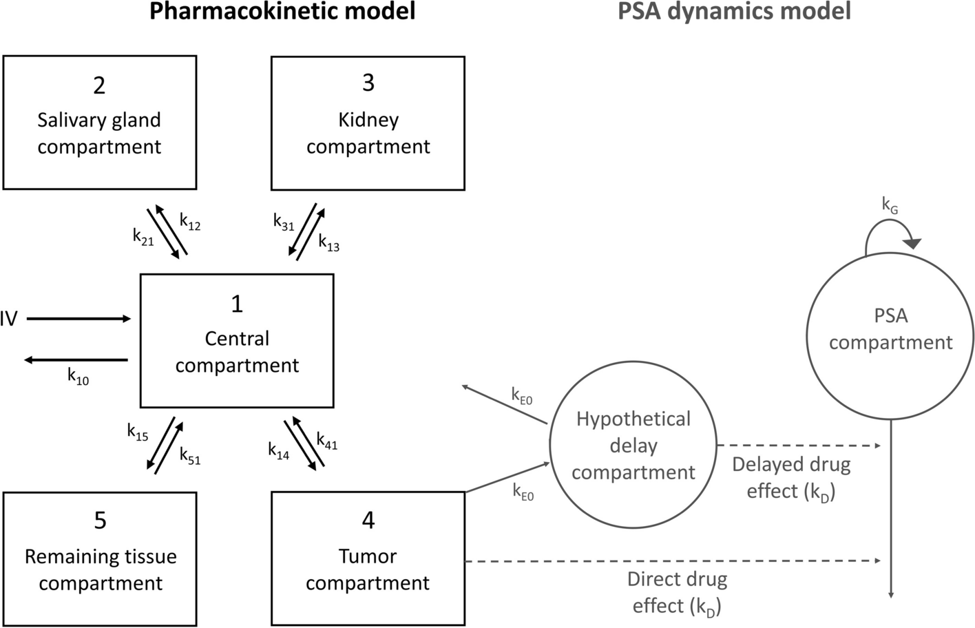
Organ absorbed doses and effective dose due to XXXTb-cm09 injectionBiokinetics curves were obtained plotting the radiopharmaceutical concentration corrected by the radioactive decay vs. time for each source organ of the ICRP 89 male phantom of 73 kg [31]. The total volume of the blood (5110 ml) was obtained using the specific volume value of 70 ml/kg. Figure 7 shows a fast blood clearance followed by a quick radiopharmaceutical uptake by the main organs, with a slow wash-out. Liver and kidneys were the organs with slower clearance.
Fig. 7
Time-activity curves of Tb-cm09 in the main source organs: symbols show experimental data obtained from biodistribution studies; the lines depict fitted time-activity curves
The number of disintegrations in the source organs, calculated by assuming that the injected radiopharmaceutical was labelled with only one of the radioisotopes 154gTb, 155Tb, 156gTb, 156m1Tb and 156m2Tb, are reported in Table 3. These are the main radionuclides expected to be produced by proton irradiation of 155Gd-enriched targets. The dosimetric properties of 154m1Tb and 154m2Tb were not assessed because these two metastable states are not included in the OLINDA software. However, at an irradiation energy of 10.5 MeV, 154m1Tb and 154m2Tb production is essentially due to the presence of 154Gd in the target, not considered in the case of 155Gd target enrichment ≥ 98%. Besides, since the energy of these two metastable states is very close to that of the ground state, their contribution to the absorbed dose occurs mainly through the decay of the ground state, properly taken into account through the application of the Bateman equations.
Table 3 Number of nuclear transitions (MBq × h/MBq) in source organs per unit administered activity of xxxTb-cm09 for male ICRP 89 human phantomFor the dosimetric assessment, the total activity in the intestines was distributed in left colon, small intestine, right colon and rectum, according to their mass ratio with respect to the total intestine mass. The mean maximum volume of blood that can be contained in the four chambers of the heart, two atria and two ventricles, of an adult man is about 505 ml [32], therefore, just the 10% of the blood activity was assigned to the “heart contents” and the rest to “Remaining”. The activity in mouse muscle was extrapolated to humans considering that the human muscle is 40% of total body weight. Muscle activity was also assigned to the”Remaining” organs when performing calculations with OLINDA 2.2.3, because this tissue is not included in the source organs of the ICRP 89 phantom model.
For each xxxTb-radiopharmaceutical, the organs with the highest number of disintegrations were the kidneys, followed by the liver and the cortical bone. Comparing the different radioisotopes, it was found that the radionuclides with the highest number of disintegrations were 155Tb and 156gTb, due to their long half-life. 156m2Tb showed the lowest number of disintegrations because of its relatively short half-life.
The absorbed doses per unit of administered activity in the main human male organs, calculated for different xxxTb-cm09, are reported in Table 4. For all the radioisotopes, the organs receiving the highest absorbed dose are the kidneys, followed by the osteogenic cells for 155Tb, 156m1Tb and 156m2Tb, and by the adrenals for 154gTb and 156gTb. 156gTb is the radioisotope giving the highest values of absorbed doses, due to its long half-life and the large amount of energy emitted per decay (see Table 1). The absorbed doses due to 154gTb administration are also higher than those of 155Tb, because, even if the 154gTb half-life is much shorter than that of 155Tb, the energy emitted per decay is about ten times higher. The absorbed doses due to 156m1Tb and 156m2Tb are about one order of magnitude lower than those of 155Tb. The ED after administration of 156m1Tb-cm09 and 156m2Tb-cm09 are lower than the one of 155Tb-cm09, consequently their presence as contaminants in the produced 155Tb will not increase the dose to the patient. In contrast, when the radiopharmaceutical is labelled with 156gTb or 154gTb, the ED is 5.9 and 2.4 times the ED values of 155Tb-cm09. Therefore, to guarantee a dose increment per unit of activity administered lower than 10%, the presence of 156gTb or 154gTb as contaminants must be lower than about 2% or 7%, respectively.
Table 4 Organ absorbed doses (mGy/MBq) and ED values (mSv/MBq) per unit administered activity calculated for xxxTb-cm09 for male ICRP 89 phantomsThe dose increase due to 155Tb contaminants obtained with different levels of target enrichmentThe DI due to 155Tb-cm09 obtained with different levels of target enrichment is plotted in Fig. 8 vs. time t post-production. The DI is well above 25% for the 91.9% target enrichment and this confirms that the isotopic contamination of that target is inadequate for medical purposes. The ideal case of a 155Gd target with 100% enrichment is described by the solid red line. Here, the RNP is very close to 100% and the DI is negligible. Quite interesting are the dashed and dot-dashed curves obtained with 99% and 98% enrichment. The former reaches a 5% DI, while the latter remains within the 10% limit.
Fig. 8
Dose increase for 155Tb-cm09 radiopharmaceutical labelled at different times after the end of irradiation of different 155Gd-enriched targets
Impact on the image qualityThe imaging quality of 155Tb produced with the different 155Gd-enriched targets was assessed at the EoB and 96 h later by considering the 156gTb and 154gTb yields reported in Table 2. The metastable states 156m1Tb, 156m2Tb and 154m1Tb were disregarded in the simulations because of their lower energy γ-ray emission.
Using the data in Table 2, a γ-ray spectrum produced at the EoB from the irradiation of 100% 155Gd-enriched target was simulated, as shown in Fig. 9. The γ rays with the energies of 86.55 keV and 105.318 keV emitted from 155Tb and the low intensity 88.97 keV γ ray emitted by 156gTb are situated close with respect to the energy resolution of the imaging system and form a compound peak with a barycenter of 88.5 keV. Other four peaks from 155Tb with energies of 148.64 keV, 161.29 keV, 163.28 keV and 180.08 keV form a less intense compound peak with a barycenter of 167 keV. A smaller 155Tb peak at 262 keV (5.3%) is exposed to the higher energy γ rays from the isotopes of 154gTb and 156gTb only. For imaging purposes, the compound peak with the energy of 88.5 keV is most convenient, for its higher intensity, although the other two peaks at 167 keV and 262 keV can also be used for imaging.
Fig. 9
Simulated spectrum for 155Tb, obtained immediately after the proton bombardment of 100% 155Gd-enriched target. The predicted observable spectrum is presented with a thicker black line
Table 5 shows the calculated Compton-to-peak ratio for the three principal peaks of 155Tb with respect to the γ-ray background, generated by the contaminant isotopes 156gTb and 154gTb. The analysis of the data reveals that in all cases the level of noise in the γ-ray images reconstructed using a 10% energy window around the barycenter of the peaks does not exceed 27%. For the intense peak at 88.5 keV the Compton-to-peak ratio remains within the interval of 19%– 23% for all enriched targets immediately after the EoB and no significant improvement could be achieved 96 h later. Lower Compton-to-peak values, slightly below 20%, are obtained for the compound peak at 167 keV. This suggests its use as a single peak for image reconstruction in some particular cases. The lower intensity of the peak at 262 keV makes the ratio more affected by the presence of the contaminant nuclides 156gTb and 154gTb, although its maximum value still remains below 27%. However, it is worth noting that the present image quality estimation is made for point-like γ-ray sources in air, hence the values quoted refer to the imaging system only and do not take into account Compton scattering inside the tissues. To estimate this contribution for the case of small animals imaging, a water phantom was used in our laboratory, obtaining a noise increase level smaller than 5%.
Table 5 Compton-to-peak ratio calculated from the simulation of 155Tb γ-ray spectra, obtained for 100%, 99% and 98% enrichment of 155Gd targets, at the EoB and 96 h later
留言 (0)