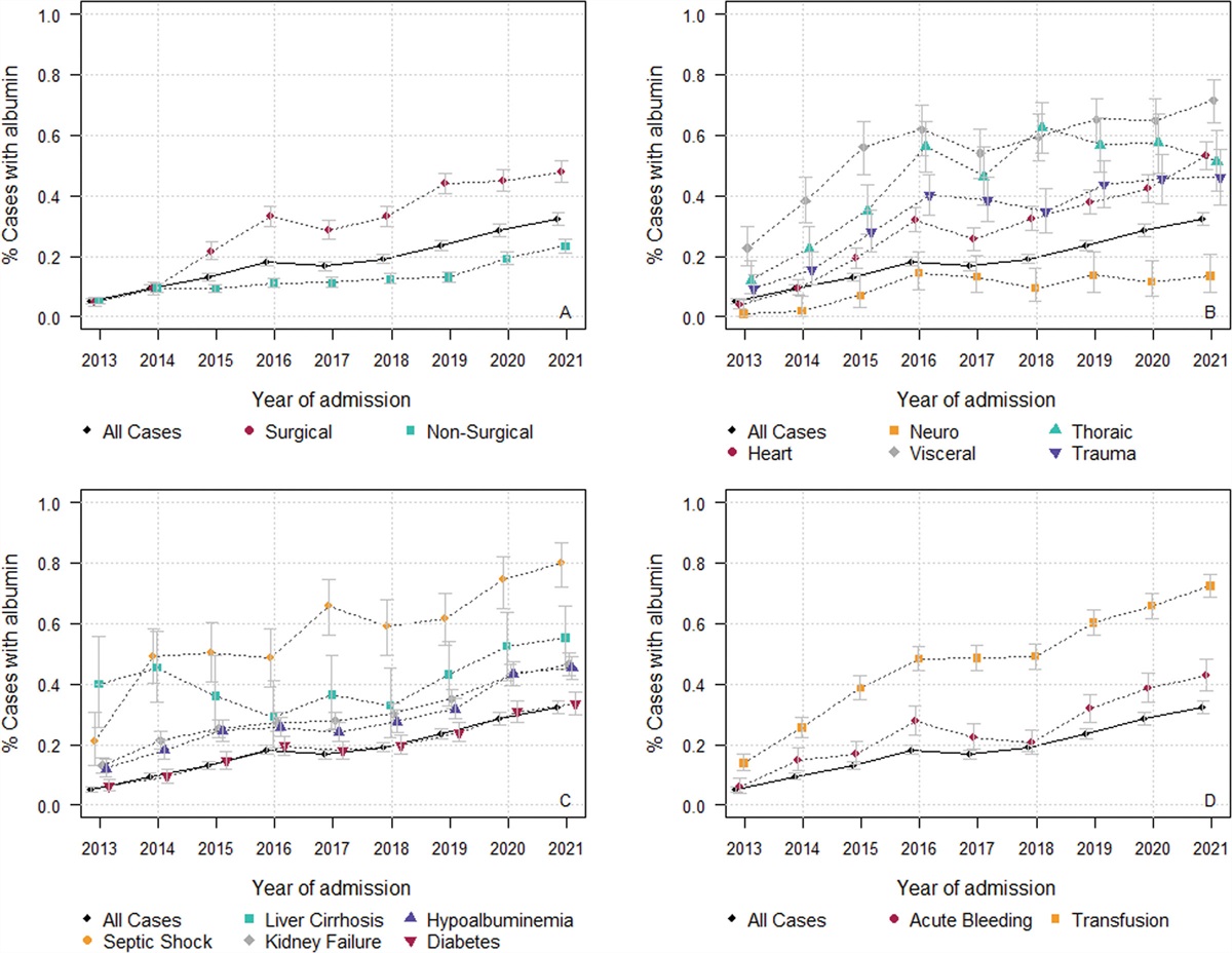
Extracorporeal membrane oxygen (ECMO) is well established as a resuscitative adjunct or treatment modality in severe respiratory failure (1), refractory cardiogenic shock (2,3), and during sudden cardiac arrest (4,5)—where it can be applied during active chest compressions and is known as extracorporeal cardiopulmonary resuscitation (ECPR). Acknowledging some controversy over optimal use, we can observe that survival on ECMO is better among patients who are alive and have a pulse at the time of cannulation than among patients who are not and do not. Further, among patients who receive ECMO, be it for respiratory failure, cardiogenic shock, or cardiac arrest, observational data suggest earlier initiation (6–8) and initiation among lower severity of illness (9) is generally associated with better outcomes. What is relatively unknown is exactly how early.
It is against this brief summary of adult ECMO that we discuss the significance of signs of life (SOL) among patients who have cardiac arrest. SOL are physical manifestations of cerebral activity, and include gasping, movement, and pupillary reflexes. It has been previously shown that +SOL during cardiac arrest resuscitation are associated with improved outcomes, although data are sparse (10,11). Furthermore, research into the prognostic value of these SOL has been limited given the rarity of the outcome of interest (survival), and the fact that a given prognostic factor (e.g., SOL) has limited utility in situations where we cannot control the outcome. In the situation of out-of-hospital cardiac arrest (OHCA), by 20 minutes of refractory arrest, only 5% of patients survive (12); prognostic factors are not needed when the outcome is nonmodifiable death.
Enter ECPR, and patients can be kept hemodynamically alive on ECMO for days or more, when their condition was otherwise nonsurvivable. Now the utility of a given prearrest or intra-arrest prognostic factor for the outcome of survival becomes much more useful.
In this issue of Critical Care Medicine, Bunya et al (13) analyzed 1395 adults from the Study of Advanced Cardiac Life Support for Ventricular Fibrillation with Extracorporeal Circulation in Japan (SAVE-J) cohort, examining the prognostic value of SOL upon hospital arrival for patients with OHCA who then received ECPR. The authors examined the occurrence rate of SOL upon hospital arrival, and the value of these SOL as independent predictors of neurologically intact survival. Their findings are thematically consistent with previous ECPR studies that demonstrated a survival association with gasping (14), and transient return of spontaneous circulation (15); Bunya et al (13) found that +SOL upon hospital arrival were individually and cumulatively associated with dramatically higher survival for each added SOL. The adjusted odds ratio (aOR) of survival increased from a baseline aOR of 1 (0 SOL) to 4.7 (1 SOL) to 18.66 (2+ SOL). Furthermore, the authors demonstrated that survival outcomes were stratified by initial rhythm when patients had at least 1 SOL, with 12.5% survival for asystole, 27.3% for pulseless electrical activity (PEA), and 42.2% for ventricular fibrillation (VF).
It should be noted that these survival statistics are artificially inflated compared with an otherwise unrestricted ECPR cohort, that is, as has been done with other studies out of the SAVE-J cohort (16,17), the analysis a priori removed patients whom the authors identified as having noncardiac etiologies, diagnosed after the fact. Many of these etiologies (aortic dissection, aneurysm, primary cerebral disorder) are likely nonsurvivable if presenting with sudden cardiac arrest. Removing these “nonrecoverable” patients does bias the population, functionally enriching it for potential survivors. Although it inflates the survival numbers, it also cleans up the analysis, tightening the scientific relationship between pre-ECPR prognostic features such as SOL, and eventual outcome. This is not bad, as long as it is acknowledged and considered. It leads to a better understanding of the science, but lower generalizability of the prognostic factors.
This functional enrichment for survival might partially explain the higher survival seen in this study compared with a 2021 study by Debaty et al (18) from Paris which demonstrated that SOL at any point of the resuscitation were associated with an adjusted odds 7.35 for neurologically intact survival, with 12% and 23% survival (nonshockable/shockable) if +SOL, compared with 0% and 4% without.
Bunya et al (13) make a point of the significance of restricting their analysis to patients who had SOL after hospital arrival, rather than at any point of the arrest. It means that patients had to have had consistently sufficient perfusion to not only avoid irreversible ischemia throughout the resuscitation, but then were able to achieve sufficient cerebral blood flow upon hospital arrival to elicit neurologic activity and corresponding motor movement.
This last point may be the most important conclusion—the suggestion that patients who exhibit SOL during resuscitation are not quite dead yet. The theoretical construct goes something like this: SOL occur in response to brain activity. Brain activity requires some cerebral blood flow and that that particular portion of the brain has thus far escaped death, which indicates that the perfusion up to that point was at least sufficient to prevent irreversible neurologic death.
Stepping back, the hypothesis that these patients still have at least partial preserved cerebral blood flow and brain activity is more radical than one might initially think. Most cardiac arrest (and ECPR patients) either do not have preserved cerebral blood flow, or already sustained irreversible neurologic injury. Many patients already demonstrate signs of brainstem injury (dilated pupils, agonal breaths) and myocardial injury (asystole and PEA) early on in the resuscitation—and never normalize—despite receiving perfusion through CPR, or even full ECMO. This irreversibly injured population may constitute ~60% of ECPR patients, which is still less than the typical proportion of adult ECPR patients who die.
Another demonstration of the value of the SOL in prognosticating survival is that the survival numbers in the no-SOL group are low, even comparable to the historical outcomes in the SAVE-J dataset (16,19). By removing patients who had +SOL, it is easy to see how the no-SOL group had survival of 5% (PEA and asystole) and 9% (VF). Further, the patients with +SOL had much higher survival by rhythm than typically observed (42% VF, 27% PEA) for ECPR.
Coming back to the initial rhythm, let us consider the finding that survival was stratified by initial rhythm among patients with +SOL. Initial rhythm has been a reliable predictor of outcome in just about all previous studies of ECPR, although in this analysis, the initial rhythm fell out of the multivariate model after adding SOL. A potential reason for this could be due to the knowledge that initial rhythm not only correlates with etiology (20,21), but also correlates with duration of no-flow (22,23), with VF becoming less common over time. As patients with SOL upon hospital arrival have had cerebral blood flow since arrest sufficient to avoid neurologic death of the region generating the movement, this temporally late prognostic factor (+SOL) may subsume the prognostic value of the initial rhythm. Although the demonstration that survival was still stratified by initial rhythm shows that initial rhythm retains some value. As a take away for the clinician, patients with initial VF and at least 1 SOL upon hospital arrival had a nearly 50% probability of neurologically intact survival when treated with ECPR.
The demonstration by Bunya et al (13) of the value of SOL upon hospital survival in prognosticating neurologic intact survival, and our discussion of the physiologic implications of +SOL, taken together beg the question of why we are not initiating ECPR—or ECMO writ large—earlier. I suggest that the answer is a combination of the aspirational “we certainly should be” and the pragmatic “it’s both logistically difficult and will lead to harm if done too early.” Earlier ECPR implementation requires patients get to the hospital faster, or that ECPR goes to them. Both have been demonstrated (24,25), but neither is easy. Even if we take the cardiac arrest out of the picture and look at respiratory and cardiac ECMO, there are legitimate concerns around implementing ECMO too early. ECMO has a known rate of complications, including structural (26,27) and hemorrhagic (28), and despite examples to the contrary (29), the majority of patients do not have high levels of mobilization (30,31), which is associated with, and may lead to, better outcomes.
In summary, this study by Bunya et al (13) demonstrates that +SOL upon hospital arrival, in a multicenter cohort of adult patients who did eventually receive ECPR and had nonsurvivable etiologies excluded, are strongly and cumulatively associated with neurologically intact survival. As we can surmise that these patients had intact cerebral perfusion throughout the resuscitation, we now have more data that getting ECMO if you are not quite fully dead is better than if you are already fully dead. It sounds elementary, but as clinicians, we still struggle with it. There is an optimal time of initiation, and that time is early in the course of cardiac arrest, or critical illness—just not too early, where the complications and side effects from ECMO add unnecessary risk. Identifying this optimal balance, for various patients and conditions, should drive prospective research and trials. The closer we get to it, the better patients will do.
1. Combes A, Hajage D, Capellier G, et al.; EOLIA Trial Group, REVA, and ECMONet: Extracorporeal membrane oxygenation for severe acute respiratory distress syndrome. N Engl J Med 2018; 378:1965–1975
2. Ostadal P, Rokyta R, Karasek J, et al.; ECMO-CS Investigators: Extracorporeal membrane oxygenation in the therapy of cardiogenic shock: Results of the ECMO-CS randomized clinical trial. Circulation 2023; 147:454–464
3. Thiele H, Zeymer U, Akin I, et al.; ECLS-SHOCK Investigators: Extracorporeal life support in infarct-related cardiogenic shock. N Engl J Med 2023; 389:1286–1297
4. Belohlavek J, Yannopoulos D, Smalcova J, et al.: Intraarrest transport, extracorporeal cardiopulmonary resuscitation, and early invasive management in refractory out-of-hospital cardiac arrest: An individual patient data pooled analysis of two randomised trials. EClinicalMedicine 2023; 59:101988
5. Suverein MM, Delnoij TSR, Lorusso R, et al.: Early extracorporeal cpr for refractory out-of-hospital cardiac arrest. N Engl J Med 2023; 388:299–309
6. Urner M, Barnett AG, Bassi GL, et al.; COVID-19 Critical Care Consortium Investigators: Venovenous extracorporeal membrane oxygenation in patients with acute COVID-19 associated respiratory failure: Comparative effectiveness study. BMJ 2022; 377:e068723
7. Lee HH, Kim HC, Ahn CM, et al.: association between timing of extracorporeal membrane oxygenation and clinical outcomes in refractory cardiogenic shock. JACC Cardiovasc Interv 2021; 14:1109–1119
8. Bartos JA, Grunau B, Carlson C, et al.: Improved survival with extracorporeal cardiopulmonary resuscitation despite progressive metabolic derangement associated with prolonged resuscitation. Circulation 2020; 141:877–886
9. Jentzer JC, van Diepen S, Barsness GW, et al.: Cardiogenic shock classification to predict mortality in the cardiac intensive care unit. J Am Coll Cardiol 2019; 74:2117–2128
10. Bobrow BJ, Zuercher M, Ewy GA, et al.: Gasping during cardiac arrest in humans is frequent and associated with improved survival. Circulation 2008; 118:2550–2554
11. Steen-Hansen JE, Hansen NN, Vaagenes P, et al.: Pupil size and light reactivity during cardiopulmonary resuscitation: A clinical study. Crit Care Med 1988; 16:69–70
12. Goto Y, Funada A, Goto Y: Relationship between the duration of cardiopulmonary resuscitation and favorable neurological outcomes after out-of-hospital cardiac arrest: A prospective, nationwide, population-based cohort study. J Am Heart Assoc 2016; 5:e002819
13. Bunya N, Ohnishi H, Kasai T, et al.; Study of Advanced life support for Ventricular fibrillation with Extracorporeal circulation in Japan II (SAVE-J II) Study Group: Prognostic Significance of Signs of Life in Out-of-Hospital Cardiac Arrest Patients Undergoing Extracorporeal Cardiopulmonary Resuscitation. Crit Care Med 2024; 52:542–550
14. Nara S, Bunya N, Ohnishi H, et al.; SAVE-J Study Group: Long-term prognostic significance of gasping in out-of-hospital cardiac arrest patients undergoing extracorporeal cardiopulmonary resuscitation: A post hoc analysis of a multi-center prospective cohort study. J Intensive Care 2023; 11:43
15. Otani T, Hifumi T, Inoue A, et al.; SAVE-J II study group: Transient return of spontaneous circulation related to favourable outcomes in out-of-hospital cardiac arrest patients resuscitated with extracorporeal cardiopulmonary resuscitation: A secondary analysis of the SAVE-J II study. Resusc Plus 2022; 12:100300
16. Inoue A, Hifumi T, Sakamoto T, et al.; SAVE-J II study group: Extracorporeal cardiopulmonary resuscitation in adult patients with out-of-hospital cardiac arrest: A retrospective large cohort multicenter study in Japan. Crit Care 2022; 26:129
17. Shibahashi K, Hifumi T, Sugiyama K, et al.; SAVE-J II study group: Comparison of sedation using propofol vs midazolam in patients admitted to the intensive care unit after extracorporeal cardiopulmonary resuscitation for out-of-hospital cardiac arrest: A multicentre observational study. Eur Heart J Acute Cardiovasc Care 2023; 12:246–256
18. Debaty G, Lamhaut L, Aubert R, et al.: Prognostic value of signs of life throughout cardiopulmonary resuscitation for refractory out-of-hospital cardiac arrest. Resuscitation 2021; 162:163–170
19. Sakamoto T, Morimura N, Nagao K, et al.; SAVE-J Study Group: Extracorporeal cardiopulmonary resuscitation versus conventional cardiopulmonary resuscitation in adults with out-of-hospital cardiac arrest: A prospective observational study. Resuscitation 2014; 85:762–768
20. Youngquist ST, Hartsell S, McLaren D, et al.: The use of prehospital variables to predict acute coronary artery disease in failed resuscitation attempts for out-of-hospital cardiac arrest. Resuscitation 2015; 92:82–87
21. Yannopoulos D, Bartos JA, Raveendran G, et al.: Coronary artery disease in patients with out-of-hospital refractory ventricular fibrillation cardiac arrest. J Am Coll Cardiol 2017; 70:1109–1117
22. Tanguay-Rioux X, Grunau B, Neumar R, et al.: Is initial rhythm in OHCA a predictor of preceding no flow time? Implications for bystander response and ECPR candidacy evaluation. Resuscitation 2018; 128:88–92
23. Hara M, Hayashi K, Kitamura T: Outcomes differ by first documented rhythm after witnessed out-of-hospital cardiac arrest in children: An observational study with prospective nationwide population-based cohort database in Japan. Eur Heart J Qual Care Clin Outcomes 2017; 3:83–92
24. Bartos JA, Frascone RJ, Conterato M, et al.: The Minnesota mobile extracorporeal cardiopulmonary resuscitation consortium for treatment of out-of-hospital refractory ventricular fibrillation: Program description, performance, and outcomes. EClinicalMedicine 2020; 29-30:100632–100630
25. Lamhaut L, Hutin A, Puymirat E, et al.: A pre-hospital extracorporeal cardio pulmonary resuscitation (ECPR) strategy for treatment of refractory out hospital cardiac arrest: An observational study and propensity analysis. Resuscitation 2017; 117:109–117
26. Wang L, Yang F, Zhang S, et al.: Percutaneous versus surgical cannulation for femoro-femoral VA-ECMO in patients with cardiogenic shock: Results from the extracorporeal life support organization registry. J Heart Lung Transplant 2022; 41:470–481
27. Griffee MJ, Zimmerman JM, McKellar SH, et al.: Echocardiography-guided dual-lumen venovenous extracorporeal membrane oxygenation cannula placement in the ICU-A retrospective review. J Cardiothorac Vasc Anesth 2020; 34:698–705
28. Willers A, Swol J, Buscher H, et al.: Longitudinal trends in bleeding complications on extracorporeal life support over the past two decades-extracorporeal life support organization registry analysis. Crit Care Med 2022; 50:e569–e580
29. Pasrija C, Mackowick KM, Raithel M, et al.: Ambulation with femoral arterial cannulation can be safely performed on veno-arterial extracorporeal membrane oxygenation. Ann Thorac Surg 2018; 107:1389–1394
30. Marhong JD, DeBacker J, Viau-Lapointe J, et al.: Sedation and mobilization during venovenous extracorporeal membrane oxygenation for acute respiratory failure: An international survey. Crit Care Med 2017; 45:1893–1899
31. Tonna JE, Bailey M, Abrams D, et al.: Predictors of early mobilization in patients requiring VV ECMO for greater than 7 days: An international cohort study. Heart Lung 2023; 62:57–63


留言 (0)