
記住我


The present study is a case–control study that was conducted from January 2021 to January 2022 on 240 Egyptian subjects; 120 clinically defined MS sufferers recruited from MS unit, Neurology department in our university hospitals. 120 healthy control subjects (they did not suffer from hypertension, liver or renal diseases) matched for age, and sex with MS sufferers’ group were recruited from the outpatient clinics during the same period of the study and included in this study. MS sufferers and control subjects assigned an informed written consent.
All clinically defined multiple sclerosis sufferers according to the McDonald criteria of 2017 [6] were eligible for the current study. We omitted MS sufferers with a history of autoimmune disorders, vascular disease, intracranial or intraspinal tumor, antihormonal therapy, exacerbations in the month before the study, steroids for one month prior to study enrollment, active acute or chronic infections, use of antibiotics in the last month, participants refused to give an informed written consent.
Participants of this study underwent complete medical history taking (with special emphasis on the date of onset of multiple sclerosis, duration of the disease, and type of MS), full general and neurological examinations. We assessed the clinical disability of multiple sclerosis group by using the Kurtzke Expanded Disability Status Scale (EDSS) score. It provides a total score on a scale ranging from zero to ten (from normal examination to death from multiple sclerosis, respectively) [7]. Magnetic resonance imaging of the brain and spine was conducted by using one and half Tesla Philips superconducting magnetic resonance imager with a standard head coil.
Fresh blood samples were collected from all included subjects under complete aseptic condition, divided into three parts: the first part of fresh blood was collected in heparin containing vacutainer used for isolation of peripheral blood mononuclear cells (PBMCs). We collected the second part into EDTA containing vacutainer for DNA extraction. And the third part was collected into plain vacutainer for serum separation and estimation of SIRT 1 serum level and lipid profile.
Quantitative measurement of SIRT1 level was determined using a commercially available ELISA assay purchased from thermofisher.com (Catalog NO. EH427RB), following the instructions of manufacturer.
Peripheral blood mononuclear cells were separated from peripheral blood by standard density-gradient centrifugation using lymphocyte separation medium (Ficoll). In brief, blood was diluted one: three with sterile phosphate buffered saline then became layered over the separating medium then centrifuged at 2000 rpm for 20 minutes. PBMCs layer were carefully aspirated after centrifugation. Extraction of RNA and reverse transcription was done in the same day.
Extraction of genomic DNA from whole blood: using the commercially available G-spin TM Total DNA Extraction Kit (iNtRON Biotechnology, Seongnam, Korea). The extracted DNA was detected by submarine agarose gel electrophoresis and visualized on ultraviolet transilluminator (Fig. 1). We determined purity and concentration of DNA spectrophotometrically at 260 and 280 nm. The purified genomic DNA was stored at − 20 °C until use. Genotyping of rs7895833 A > G, rs7069102 C > G and rs2273773 C > T polymorphism in SIRT1 gene polymorphism was achieved using TaqMan SNP Genotyping Assays (Applied Biosystems, Foster City, CA; for rs7895833 A > G SNP Assay ID C_29163689_10 was used, for rs7069102 SNP Assay ID C_1340389_10, for rs2273773 C > T SNP Assay ID C_16179813_10 Thermo Scientific, Waltham, MA, USA).
Fig. 1
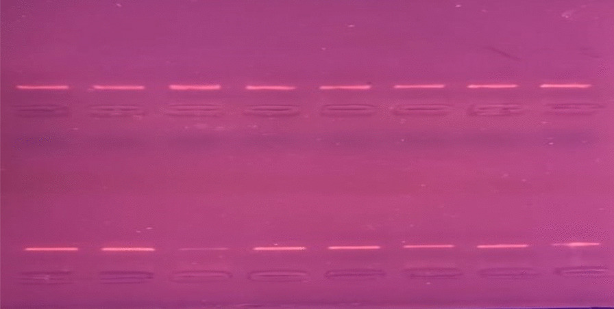
Total RNA extraction from PBMC, and complementary DNA (cDNA) synthesis: total RNA was isolated according to RNA isolation kit (Gentra, Minneapolis, MN 55441 USA) following the manufacturer's protocol. We monitored total RNA purity and integrity by the absorbance of ultraviolet light spectrophotometrically at 260/280 nm. For the synthesis of cDNA, reverse transcription of the extracted RNA was performed by QuantiTect SYBR Green reverse transcription (RT–PCR) kit (Qiagen; catalog no.204243) as recommended by the manufacturer. Complementary DNA was detected by agarose gel electrophoresis (Fig. 2) and stored at − 20 °C until time of analysis.
Fig. 2
Level of SIRT1 mRNA expression were estimated by real time polymerase chain reaction using Step One TM System (Applied Biosystems). We used glyceraldehyde-3-phosphate dehydrogenase gene (G3PDH) as an internal control. Primers sequence for SIRT1 gene were (For: 5′-TGGCAAAGGAGCAGATTAGTAG-3′, Rev: 5′-GGCATGTCCCACTATCACTGT-3′). The PCR was performed in 25 µL containing 12.5 µL QuantiFast SYBR Green (Cat.No.204141), PCR Master Mix, 1 µL of each primer (Invitrogen, USA) and 2 µL cDNA.
The thermal cycling profile was an initial denaturation at 95 °C for two minutes followed by 40 cycles of denaturation at 94 °C for 30 s, annealing at 52 °C for 60 s and the elongation at 72 °C for 2 s for 40 cycles with a final incubation at 72 °C for seven min. All genes expression was reported as the D cycle threshold (DCt) value. Expression levels of mRNAs for target genes were compared with endogenous control β-actin using the 2 − ∆∆CT method. All kits were supplied by QIAGEN (Valencia, CA).
Statistical analysisVariables of this study are expressed as means ± SD for continuous data, and as numbers and percentages for categorical data. Student’s “t” tests, Chi-square test, one-way analysis of variance, Mann–Whitney tests, Kruskal–Wallis, Spearman’s correlation coefficient (r), multiple logistic regression analysis, and linear regression analysis were done when appropriate in the current study. Values of p less than 0.001, 0.05 in this study were considered statistically highly significant and significant, respectively. We carried out statistical computations using the statistical package of social science program for Windows version 24 that is released 2016 by International Business Machines Corporation, USA.
留言 (0)