Animals
Animal experiments were carried out in accordance with the National Institutes of Health Guidelines for the Use of Laboratory Animals, and all procedures were approved by the Animal Care Committee of Shandong Provincial Third Hospital (Shandong, China). The animals were housed in normal cages at a temperature of 24 ± 2°C and relative humidity of 60% on a 12:12 h light and dark cycle and given free access to water. To model MI, 8 ~ 10-week-old male C57BL/6 mice were subjected to MI surgery. The mice were randomly assigned to the sham and MI groups. Cardiomyocytes were isolated from neonatal Sprague–Dawley rats (1 ~ 3 d old).
Myocardial infarction surgery
Eight-week-old male C57BL/6 mice in the MI group were anesthetized via 2% isoflurane inhalation and fixed on a heating pad in the left supine position. Left thoracotomy was performed at the 4th left intercostal space. The heart was subsequently exposed, and the pericardium was opened. The left anterior descending (LAD) artery was ligated with a 7–0 suture immediately below the tip of the left auricle. When the left ventricle became pale, LAD ligation was considered successful. The sham mice underwent the same procedure except occlusion of the LAD. Ensuring a hermetic closure of the surgical site, air was evacuated, and the chest was closed with 6–0 sutures. The mice were placed on another heating pad until recovery. At different time points after the operation, the hearts were analyzed.
Cardiomyocyte isolation, culture and treatments
NRVMs were isolated from Sprague–Dawley rats (1 ~ 3 d old) using a differential adhesion method as described previously (Chen et al. 2019). Briefly, the heart was minced into 1 mm3 fragments and digested with 0.1% collagenase type II (Worthington, LS004177, Lakewood, NJ) and 1% BSA in PBS. The cells were then separated into cardiomyocytes and cardiac fibroblasts via differential adhesion. NRVMs were used for subsequent experiments. The cells were cultured in high-glucose Dulbecco’s Modified Eagle Medium (DMEM, GIBCO, c11995500bt, Waltham, MA) containing 20% M199 medium (Basal Media, L649KJ, Shanghai, China), 10% fetal bovine serum (FBS, EXCELL, FND500, Taicang, China), 100 μM 5-bromo-2'-deoxyuridine (Brdu, Sigma, B5002-250MG, St. Louis, MO) and 1 × penicillin–streptomycin (P/S, Gibco, 15,070,063, Waltham, MA). Afterward, the cells were cultured in high-glucose DMEM containing 20% M199 medium and 2% FBS.
Transfection was performed with Lipofectamine™ RNAimax transfection reagent (Invitrogen, 13,778,150, Carlsbad, CA) and Lipofectamine® 3000 transfection reagent (Invitrogen, L3000015, Carlsbad, CA) according to the manufacturer’s instructions. Briefly, for the knockdown assay, small interfering RNA (siRNA) against Clk3 (40 nmol/L) and negative control siRNA were transfected into NRVMs using Lipofectamine™ RNAimax transfection reagent. For the overexpression assay, 4 μg Flag-CLK3 plasmid (Sino Biological, HG10716-CF, Beijing, China), 4 μg Flag-vector plasmid (Sino Biological, CV012, Beijing, China), and 5 μL P3000 were mixed with 125 μL of Opti-MEM medium (GIBCO, 11,058–021, Waltham, MA). Then, 5 μL Lipofectamine™ 3000 was diluted in 125 μL Opti-MEM. After incubation for 5 min at RT, diluted DNA and diluted Lipofectamine™ 3000 were combined and incubated for another 15 min at RT. Afterward, DNA-lipid complex was added to each well. Forty-eight hours post-transfection, the medium was replaced with fresh complete medium containing 2% FBS and CoCl2 or PBS for another 24 h. Finally, the NRVMs were collected for analysis. The siRNA sequences are listed in Table 1.
Table 1. The specific siRNA sequences
Cell culture
Human embryonic kidney 293 T (HEK293T) cells were obtained from Shanghai Zhong Qiao Xin Zhou Biotechnology (ZQ0033, Shanghai, China). The cell line used in this study was tested and authenticated. We confirmed that all experiments used mycoplasma-free cells. HEK293T cells were cultured in DMEM supplemented with 10% FBS and 1% penicillin streptomycin (P/S), and incubated at 37°C with 5% CO2.
Quantitative real-time polymerase chain reaction (qRT-PCR)
Total RNA was extracted from NRVMs and heart tissues using RNAiso Plus (Takara, 9109, Shiga, Japan) according to the manufacturer's instructions. The concentration and purity of the RNA samples were determined using a Nanodrop 2000 spectrophotometer (Thermo Fisher Scientific). cDNA was synthesized using PrimeScript™ RT Master Mix (Takara, RR036A, Shiga, Japan). Real-time PCR was conducted using SYBR Green Real-Time PCR Master Mix (TOYOBO, QPK-201, Osaka, Japan). β-Actin was used as an internal reference. The sequences of the primers used for PCR in this study are listed in Table 2.
Table 2. The primer sequences
Western blotting analysis
Total protein was extracted from cells or tissues by RIPA lysis buffer (Beyotime, P0013B, Shanghai, China) containing protease inhibitor (Roche, 4,906,845,001, 1 ml ddH2O per tablet, 1:50, Mannheim, Germany) and phosphatase inhibitor (Roche, 4,693,116,001, 400 μL ddH2O per tablet, 1:25, Mannheim, Germany) on ice for 30 min. Subsequently, the lysates were centrifuged at 12,000 × g and 4°C for 15 min. The concentration of the protein was measured using a BCA kit (Beyotime, P0009). The protein lysates (35 μg protein per sample) were separated on a 10% NuPAGE Bis–tris gel (Invitrogen, NP0316BOX, Carlsbad, CA) and transferred to PVDF membranes (Millipore, IPVH00010, Burlington, MA). The membranes were then blocked with 5% nonfat milk at RT for 1 h and subsequently incubated with primary antibodies (β-Actin, 1:1000, Santa Cruz, sc-47778, Dallas, TX; CLK3, 1:200, Cell Signaling Technology, 3256, Danvers, MA; HIF-1α, 1:1000, Cell Signaling Technology, 36,169; PARP, 1:1000, Cell Signaling Technology, 9532; Bcl-2, 1:500, Santa Cruz, sc-7382; Bax, 1:1000, Cell Signaling Technology, 2772; Caspase-3, 1:200, Cell Signaling Technology, 9662; Akt, 1:1000, Abcam, ab8805, Cambridge, UK; Phospho-Akt (Ser473), 1:1000, Cell Signaling Technology, 4060 T; Anti-Phospho-(Ser/Thr), 1:500, Abcam, ab300625; p53 (DO-1), 1:200, Santa Cruz, sc-126; p53 (DO-2), 1:200, Santa Cruz, sc-53394) at 4°C overnight. The next day, TBS containing 0.1% Tween 20 (TBST) was used to rinse the membranes. Subsequently, the membranes were incubated with near infrared dyes-conjugated secondary antibodies (Invitrogen, A32735; Invitrogen, A21036, Carlsbad, CA) at RT for 1 h. Images of the protein bands were captured using an Odyssey imager (LI-COR, Biosciences). Then, the band intensity was quantified using ImageJ software (version 1.46r). β-Actin was used as a loading control.
Immunofluorescence and TUNEL staining
NRVMs were fixed in 4% paraformaldehyde for 15 min and permeabilized with 0.5% Triton X-100 in PBS for 15 min. After washing, the cells were blocked with 5% normal goat serum at RT for 1 h. The cells were then incubated with primary antibody against cardiac troponin T (cTnT, 1:500, Abcam, ab8295, Cambridge, UK) at RT for 1 h or overnight at 4°C. The cells were washed three times with PBS with 0.1% Tween 20 (PBST) and incubated for 1 h with Alexa Fluor® 488-conjugated goat anti-mouse IgG secondary antibody (1:500, Abcam, ab150113, Cambridge, UK). Apoptotic cells were stained using reagents from an In Situ Cell Death Detection Kit, TMR Red (Roche, 12,156,792,910, Mannheim, Germany) for 1 h. The cells were washed three times with PBS, and nuclei were stained with DAPI (1:1000, Sigma, D9542, St. Louis, MO) for 15 min. Images were captured using a fluorescence microscope (Leica, DMi8). At least 6 random images from each group were analyzed.
Cell counting kit-8 (CCK-8) assay
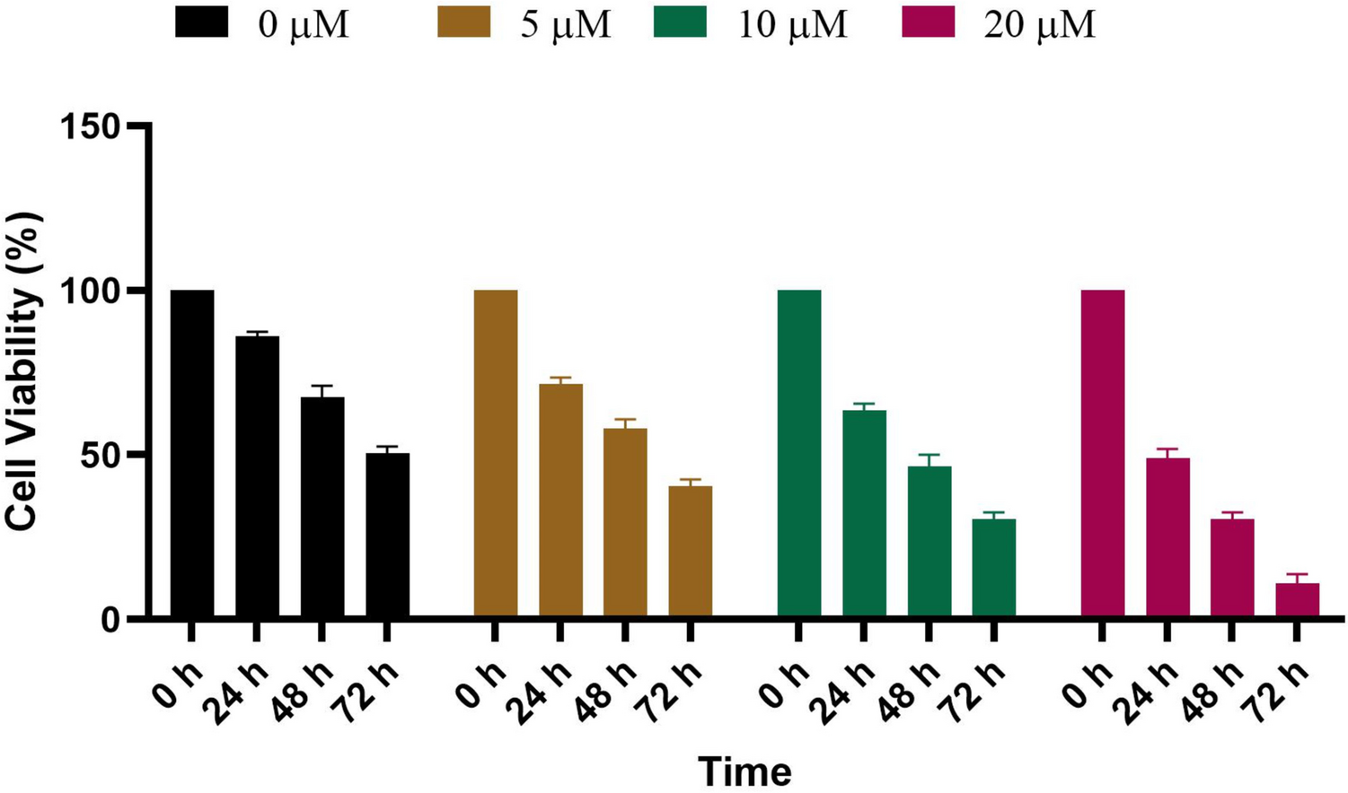
Cell viability was measured using the CCK-8 assay according to the manufacturer’s protocol (Beyotime, C0038, Shanghai, China). Briefly, NRVMs were seeded in 96-well plates and cultured in high-glucose DMEM containing 20% M199 medium and 2% FBS in a humidified 5% CO2 incubator at 37°C. Forty-eight hours post-transfection, the cells were treated with CoCl2 at a final concentration of 400 μM. Ten microliters of CCK-8 solution mixed with 100 μL of medium was added to each well after 24 h of treatment with CoCl2, and the cells were incubated for another 4 h. The absorbance of each well was measured at 450 nm using an M5 Molecular Devices microplate reader.
Immunoprecipitation (IP)
HEK293T cells were transfected with either AKT plasmid alone or co-transfected with Flag and CLK3-Flag plasmids. 48 h post-transfection, the cells were washed three times with pre-cooled PBS and then lysed with 800 µL lysis buffer (RIPA buffer with protease inhibitor and phosphatase inhibitor). The samples were incubated on ice for 30 min and then centrifuged at 12,000 g at 4°C for 15 min. 30–50 µL of the extracted protein was kept as the input sample. Concurrently, magnetic beads (Thermo Fisher Scientific, 88,803, Waltham, MA) were gently mixed, and 30 µL per sample was used and mixed on a rotator. Beads were allowed to settle on a magnetic rack for over 30 s before supernatant removal. After washing, 500 µL of PBST-mixed beads were added to each tube, and the appropriate volume of Flag antibody (1:500, Sigma, F1804-200UG, St. Louis, MO) was added. The mixture was incubated at room temperature on a rotator for 1.5 to 2 h. The beads, now bound to the antibody, were gently washed with PBS and mixed on a rotator three times. After magnetic separation and supernatant removal, the rest of the protein was added to the bead tubes and incubated at 4°C overnight on a rotator. The following day, samples were washed three times with PBS, boiled at 100°C for 10 min in the loading buffer and used for subsequent experiments.
Statistical analysis
The data are presented as the mean ± SEM, and all data were at least three different experiments. A P value < 0.05 was considered significant difference. Statistical analyses were carried out using GraphPad Prism software (version 6.01). Two-tailed Student t-test, and one-way analysis of variance (ANOVA) and two-way ANOVA were carried out as appropriate.


留言 (0)