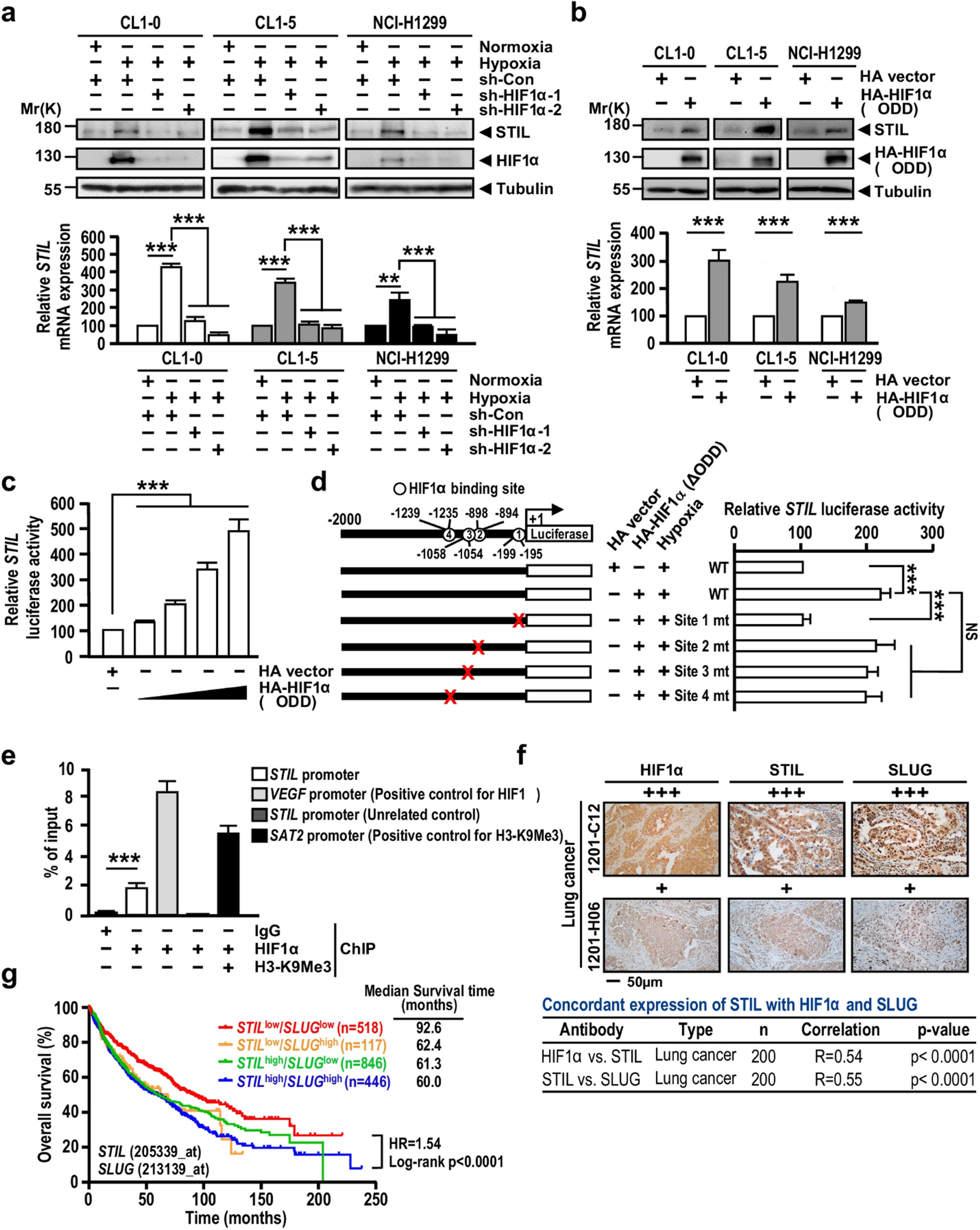
Samples collection
Blood samples were collected from 40 patients with tick-borne encephalitis (TBE), including 14 women and 26 men, with an average age of 40 years (from 23 to 58 years) that were admitted to the Department of Infectious Diseases and Neuroinfections of the Medical University of Bialystok. In addition, blood was collected from 6 patients (including 4 females and 2 males) who had co-infections with other tick-borne pathogens such as Borrelia burgdorferi sensu lato (Bb) causing Lyme borreliosis (LB) and Anaplasma phagocytophilum (Ap) causing human granulocytic anaplasmosis (HGA). According to the European Academy of Neurology (EAN) guidelines [15], TBE was diagnosed based on clinical symptoms, positive serology and lymphocytic pleocytosis in the cerebrospinal fluid (CSF). LB was determined by the presence of erythema migrans (1 patient) or possible neuroborreliosis (1 patient). In addition, in two patients anti-Borrelia burgdorferi sensu lato IgM and IgG antibodies were detected and, consequently, the patients were treated according to the standard. However, HGA was diagnosed based on CDC criteria when all 3 criteria were satisfied (https://wwwn.cdc.gov/nndss/conditions/ehrlichiosis-and-anaplasmosis/case-definition). Human granulocytic anaplasmosis was diagnosed in four cases on the basis of clinical picture, laboratory tests and PCR, of which Lyme disease was additionally diagnosed among two people. In summary, triple co-infection (TBE + borreliosis + anaplasmosis) occurred in two patients, double co-infection: TBE + borreliosis was diagnosed among two patients and TBE + anaplasmosis was detected in two patients. Patients with TBE were treated symptomatically (antioedematous drugs, anti-inflammatory drugs, analgesics and glucocorticosteroids), patients with anaplasmosis received doxycycline, while those with Lyme disease received doxycycline or ceftriaxone. Before compiling the results of the manuscript, we analyzed the parameters of patients who were given glucocorticosteroids during treatment and patients who did not receive glucocorticosteroids during treatment. These parameters did not differ significantly between the two groups.
The study was conducted in accordance with the Declaration of Helsinki, and the protocol for the collection of all blood samples was approved by the Local Bioethics Committee in Medical University of Bialystok (Poland), No. R-I-002/169/2018. Written informed consent was obtained from all participants.
The control group consisted of 20 healthy people (7 women and 13 men) with a mean age of 41 (28–55).
Blood from patients was collected twice, once on admission to the hospital and again after treatment. Blood from control group was obtained once. The mean time between these two examinations in patients with TBE was 29.4 ± 10.99 days, while in patients with co-infection it was 32.8 ± 4.21 days. Blood was collected into EDTA tubes and then centrifuged at 3000×g (4 °C) to separate the plasma as well as buffy coat, and erythrocytes. To obtain clear fraction of granulocytes, obtained buffy coat was layered on Gradisol G (Aqua-Med ZPAM–KOLASA, Łódź, Poland) and subjected to 25 min centrifugation at 300×g at room temperature. The purity of the obtained cell fraction was examined microscopically (Nikon Eclipse Ti, Nikon Instruments Inc., New York, NY, USA). Obtained plasma and granulocytes were stored at − 80 °C and used for analysis.
Tables 1 and 2 contain demographic and clinical data of patients and controls, as well as their laboratory data.
Table 1 Demographic and clinical data of patients with TBEV infection (TBE) and patients with TBEV + Bb/Ap co-infection (co-infection) compared to healthy subjects (control)Table 2 Laboratory data of patients with TBEV infection (TBE) and patients with TBEV + Bb/Ap co-infection (co-infection) compared to healthy subjects (control)MethodsAntioxidant status (TAS) determination
The total antioxidant status (TAS) of the blood serum of patients and healthy subjects was determined using 2,20-azino-bis-3-ethylbenzothiazolin-6-sulfonic acid (ABTS) [16]. For this purpose, a solution ABTS cationic radical was prepared in phosphate buffer (PBS, pH 7.4). 3 times diluted blood serum samples (5 μL) were incubated (37 °C) with ABTS radical working solution (245 μL) for 10 min. Absorbance was measured using an Infinite 200 plate reader (TEKAN, Switzerland) at 734 nm.
The test results are reported with reference to the equivalent antioxidant capacity of Trolox (TEAC) as a standard. Trolox (Hoffman-LaRoche, Basel, Switzerland) is a water-soluble derivative of vitamin E with antioxidant properties and does not interact with other cellular components. The TAS level is therefore expressed in mmol Trolox/l.
Phospholipid metabolism estimationFree and phospholipid fatty acids level
The phospholipid and free fatty acids were analyzed by gas chromatography [17]. Fatty acids were isolated by Folch extraction using chloroform/methanol mixture (2:1, v/v) in the presence of 0.01% butylated hydroxytoluene. Using TLC analytes were separated with the mobile phase as follows: heptane-diisopropyl ether–acetic acid (60:40:3, v/v/v). All lipid fractions were transmetylated to fatty acid methyl esters (FAMEs) with boron trifluoride in methanol. FAMEs were analyzed by gas chromatography with a flame ionization detector (FID) on Clarus 500 Gas Chromatograph (Perkin Elmer). Separation of FAMEs was carried out on capillary column coated with Varian CP-Sil88 stationary phase (50 m × 0.25 mm, ID 0.2 μm, Varian). Identification of FAMEs was made by comparison of their retention time with standards and quantitation was achieved using an internal standard method (nonadecanoic acid (19:0) and 1,2-dinonadecanoyl-sn-glycero-3-phosphocholine (19:0 PC) were used as internal standards). Plasma levels of PL-AA, PL-DHA, free AA, and free DHA, were expressed in μg/mL.
Lipid peroxidation products level
Lipid peroxidation in plasma was estimated by measuring small molecular weight reactive aldehyde, malondialdehyde (MDA) as well as neuroprostanes (NPs). The reactive aldehyde was determined using gas chromatography coupled with mass spectrometry 7890A GC–7000 (Agilent Technologies, Palo Alto, CA, USA) as the O-pentafluorobenzyl-oxime (O-PFB-oxime) or O-pentafluorobenzyl-oxime-trimethyl silane (O-PFB-oxime-TMS) derivatives, based on Luo’s method [18]. Benzaldehyde-d6 was added to plasma as an internal standard. Aldehyde derivatives were separated using an HP- 5 ms capillary column (0.25-mm internal diameter, 0.25-μm film thickness, 30-m length) and analyzed in selected ion monitoring mode (SIM). Samples were deproteinized by the addition of 1 mL of methanol. MDA derivatives were extracted with hexane. The hexane layer was evaporated and N,o-bis (trimethylsilyl) trifluoroacetamide in 1% trimethylchlorosilane was added. The following ions were monitored: m/z 204.0 and 178.0 for MDA-PFB and m/z 307.0 for IS (benzaldehyde-D6) derivatives. Plasma level of MDA was expressed in nmol/mL.Total NPs were quantified using modified LC–MS methods of Coolen and Fam respectively [19]. NPs were isolated using SPE method, after an alkaline hydrolysis step. All analyses were performed using an Agilent 1290 UPLC system interfaced with an Agilent 6460 triple quadrupole mass spectrometer with electrospray ionization source (ESI). The separation was performed using a reverse phase C18 column and linear gradient water (pH 5.7) and acetonitrile. NPs were analyzed by selected ion monitoring (SIM) in the m/z 357, as a series of peaks that have molecular masses and retention times expected for NPs generated from the oxidation of DHA in vitro.
PLA2, COX1/2 and LOX5 activity
The activity of enzymes involved in the metabolism of phospholipids and fatty acids was examined spectrophotometrically using commercially available assay kits, phospholipase A2 (PLA2–EC.3.1.1.4; Cayman Chemical Company, Ann Arbor, MI, USA), cyclooxygenases 1 and 2 (COX-1/2–EC.1.14.99.1; Cayman Chemical Company, Ann Arbor, MI, USA), and lipoxygenase-5 (LOX; Sigma-Aldrich, St. Louis, MO, USA), following the manufacturer’s instructions. The samples were incubated with arachidonoyl thiophosphatidylcholine, a synthetic substrate of cPLA2. The hydrolysis of arachidonoylthio-phosphatidylcholine by PLA2 released the free thiol, which was converted to NTB via a reaction with DTNB. The NTB concentration was determined with a spectrophotometric analysis at 405 nm.
PLA2 activity was expressed in nmol of arachidonoyl thio-phosphatidylcholine/min/mL Cayman’s COX colorimetric inhibitor screening assay measures the peroxidase components of COXs. The peroxidase activity was assessed colorimetrically by monitoring the appearance of oxidized N,N,N′- and n′-tetramethyl-phenylenediamine (TMPD) at 590 nm. One unit of COX’s enzyme activity is defined as the amount of enzyme that will oxidize 1.0 nmol of TMPD per minute at 25 ℃. The specific COX1-inhibitor SC-560 was applied to measure only COX-2 activity. Cyclooxygenases’ activities were expressed in U/mL.
In the Sigma-Aldrich lipoxygenase-5 (LOX-5) activity assay, lipoxygenase converts the LOX substrate to an intermediate that reacts with the probe generating a fluorescent product. The increase in the fluorescent signal can be recorded at Ex/Em 500/536 nm and is directly proportional to LOX-5 activity. One unit (U) of LOX-5 was determined as the amount of enzyme that causes oxidation of 1 μmol of the LOX probe per minute at pH 7.4 and at room temperature. LOX-5 activity was expressed in U/mL.
Endocannabinoids level
An analysis of endocannabinoids [anandamide (AEA) and 2-arachidonylglycerol (2-AG)] and related compounds [palmitoylethanolamide (PEA)] was performed using a Shimadzu UPLC system (Nexera X2) coupled with an electrospray ionization source (ESI) to a Shimadzu 8060 Triple Quadrupole system (Shimadzu, Kyoto, Japan) operating in the positive ion mode [20]. Analyte separation was performed using a 120 EC-C18 analytical column (3.0 × 150 mm; a 2.7 μm particle size) with a 5 μL injection volume. The mobile phase consisted of (A) 0.1% formic acid in MilliQ water and (B) 0.1% formic acid in acetonitrile. The following gradient was employed: 0.0–5.0 min, 70–80% B; 5.0–10.0 min, 80–88% B; 10.0–16.0 min, 78–100% B; 16.0–20.0 min, 100% B; 20.0–21.0 min, 100–70% B; and 21.0–25.0 min, 70% B. Briefly, cell culture samples were thawed on ice and spiked with 10 μL of an internal standard solution (100 ng/mL of AEA-d8, 2-AG-d8, and OEA-d4) and then applied to pre-washed and conditioned solid phase extraction SPE cartridges. After loading the sample, the cartridges were washed, dried under a high vacuum, and eluted. Eluates were concentrated and reconstituted in 30 μL of ACN/H2O (7:3) with 0.1% formic acid and vortexed (if necessary, they were centrifuged to remove any residuals). Solutions were then transferred to LC vials with low-volume inserts and an LC–MS/MS analysis was performed immediately. Transitions of the precursors to the product ions were as follows: m/z 348.3 → 62.15 for AEA, m/z 379.3 → 287.25 for 2-AG, m/z 300.3 → 62.0 for PEA, m/z 356.2 → 63.05 for AEA-d8, m/z 387.3 → 294.0 for 2-AG-d8, and m/z 330.20 → 66.15 for OEA-d4. The level of endocannabinoids was determined using a calibration curve range of 1–100 pmol/mL for AEA (r2−0.9992); of 10–1000 pmol/mL for 2-AG (r2−0.9995); of 10–500 pmol/mL for PEA (r2−0.9993). The levels of endocannabinoids were expressed in pmol/mL.
Eicosanoids level
Eicosanoids analysis was performed using an Shimadzu UPLC system (Nexera X2) coupled with an electrospray ionization source (ESI) to a Shimadzu 8060 Triple Quadrupole mass spectrometer (Shimadzu, Kyoto, Japan) operating in negative mode [21]. Analyte separation was performed using an Eclipse Plus C18 analytical column (2.1 × 100 mm, 1.8 µm particle size) with 3 µL injection volume. The mobile phase consisted of 0.1% acetic acid in MilliQ water (A) and acetonitrile (B). The following gradient was employed: 0.0–1.0 min 25–40% B, 1.0–2.5 min 40–42% B, 2.5–4.5 min 42–50% B, 4.5–10.5 min 50–65% B; 10.5–12.5 min 65–75% B; 12.5–14.0 min 75–85% B; 14.0–14.5 min 85–95% B; 14.5–15 min 95–25% B; 15.0–16.0 min 25% B. Briefly, plasma samples were thawed on ice and spiked with 10 µL internal standard solution (100 ng/mL TXB2-d4, PGD2-d4, 15-d-PGJ2-d4 and 15-HETE-d8) and then applied to pre-washed and conditioned solid phase extraction SPE cartridges. After loading the sample the cartridges were washed, dried under high vacuum and eluted. Eluates were concentrated and reconstituted in ACN/H2O (8:2) with 0.1% acetic acid and vortexed (if necessary centrifuged to remove any residuals). Solutions were then transferred to LC vials with low-volume inserts and LC–MS/MS analysis was performed immediately. The precursor to the product ion transitions were as follows: m/z 351.3 → 271.2 for PGE2, m/z 315.2 → 271.2 for 15-d-PGJ2, m/z 369.3 → 169.1 for TXB2, m/z 355.0 → 275.3 for PGD2-d4, m/z 373.0 → 173.1 for TXB2-d4 m/z 319.3 → 275.2 for 15-d-PGJ2-d4. Levels of eicosanoids were expressed as pmol/mL.
Protein expression determination
Receptor expression was measured using an enzyme-linked immunosorbent assay (ELISA) [22] Cell lysates (granulocytes and lymphocytes) were applied to the wells of an ELISA plate (Nunc Immuno MaxiSorp, Thermo Scientific, Waltham, MA, USA). The plates with attached proteins were incubated at 4 °C for 3 h with blocking solution (5% skimmed milk powder in carbonate binding buffer). After washing with PBS supplemented with 0.1% Tween 20, samples were incubated at 4 °C overnight with appropriate primary antibodies against TRPV1 (host: mouse) (Sigma-Aldrich, St. Louis, MO, USA); CB1, CB2, (host: mouse) (Santa Cruz Biotechnology, CA, USA); PPARγ (host: rabbit) (Invitrogen, Waltham, MA, USA). All antibodies were used at a concentration of 1:1000. Then, after washing (PBS supplemented with 0.1% Tween 20), the plates were incubated for 30 min with peroxidase blocking solution (3% H2O2, 3% skimmed milk powder in PBS) at room temperature. Goat anti-rabbit/mouse antibody EnVision + Dual Link/HRP solution (1:100) (Agilent Technologies, Santa Clara, California, USA) was used as secondary antibody. After 1 h of incubation at room temperature, secondary antibodies were removed and plates were incubated with chromogen substrate solution (0.1 mg/mL TMB, 0.012% H2O2) for 40 min. The reaction was stopped by adding 2 M sulfuric acid and the absorbance was read after 10 min at 450 nm and automatically recalculated from standard curves for each protein CB1: Abcam, Cambridge, UK; CB2: Abnova, Taipei, Taiwan; TRPV1: Lifespan Biosciences, Seattle, WA, USA; PPARγ: Fine, Wuhan Test, Hubei, China;). The values obtained were converted to mg of protein in the samples.
Statistical analysis
Data were expressed as mean ± SD, and were analyzed by one-way analysis of variance (ANOVA) followed by a post hoc Tukey testing using Statistica software (Statistica 13.3, StatSoft, Poland). Results were compared using Mann–Whitney U test and Wilcoxon signed-rank test. Values of p ≤ 0.05 were considered significant, and only these results were discussed in detail.


留言 (0)