Cell culturing
hAMSCs were obtained from the cells bank of the Tissue Engineering and Cell Therapy laboratory and were characterized by flow cytometry (detailed in the Supplemental Material). Tumor cells lines were provided by the Immunobiology and Cell Biology group of Pontificia Universidad Javeriana. Cells were kept in standard conditions as described in the Supplemental Material.
Natural products
Natural products were produced using Caesalpinia spinosa and Petiveria alliacea plant materials from Colombia; the Colombian Environmental Ministry authorized the research by the agreement for Access to Genetic Resources and Derived Products no. 220/2018 (RGE 0287-6).
To obtain P2Et, pods and fruits of Caesalpinia spinosa (Molina) Kuntze (divi-divi or tara) were collected in the wild, in the municipality of Villa de Leyva—Province of Ricaurte (Department of Boyacá, Colombia) in the polygon delimited by the following geographic coordinates: between 5°37 and #39;95 and #39; and #39; north and 5°39 and #39;17 and #39; and #39; north; and between 73°32 and #39;19 and #39; and #39; west and 73°34 and #39;63 and #39; and #39. Annual average temperatures were recorded between 14.7 and 27.5 °C in the month of March 2013 and, identified by Luis Carlos Jimenez, from the Colombian National Herbarium (voucher specimen L 523714). P2Et, was standardized and chemically characterized as previously described (Sandoval et al. 2016).
On the other hand, the leaves of Petiveria alliacea were collected in Quipile, Cundinamarca, Colombia, at a temperature of 24 °C; the plant material was identified by Antonio Luis Mejía from the National Herbarium of Colombia (voucher number COL 333406). The Anamu-SC extract was obtained through supercritical fluids at Corporación Universitaria Lasallista at 60 °C, 400 bar, flow 30 kg/h, and with 15% ethyl acetate as a cosolvent; the procedure was previously described (Ballesteros-Ramírez et al. 2020).
For each assay, both extracts were diluted in 95% ethanol (Merck, Darmstadt, Germany) obtaining a 25 mg/ml fresh solution and homogenized by means of vortex. Working solutions were stored for a maximum of 1 month at 4 °C.
hAMSC-CM preparation
hAMSCs were plated on 75 cm2 culture flask until 80–90% confluency was reached. Then, four washes with Ringer’s lactate, and a final one with MEM (without phenol red and without supplementation, Corning, Corning, USA), were done to remove any traces of platelet lysate. Cells were then cultured in 5 ml Dulbecco’s modified Eagle medium (DMEM)/F12 supplemented with 1% penicillin/streptomycin and 1% l-glutamine for 24 h. hAMSC-CM was collected, centrifugated at 85 RCF for 10 min, and filtered in 0.22 μm syringe filters; then, it was aliquoted and stored at –20 °C until it was used.
MTT assay
hAMSCs, A375, and MCF7 cell viability was determined using the MTT assay (Alfa Aesar, Ward Hill, USA) after treatment with Anamu-SC, P2Et, or hAMSC-CM, alone or in combination with natural products. 3 × 10³ cells were seeded in 96-well plates and allowed to attach overnight. They were treated with serial dilutions of the natural products ranging from 500 to 7.81 μg/ml for 48 h; afterwards, the medium was discarded, and cells were washed once with Ringer’s lactate and MTT was added (final concentration 0.33 mg/ml) and incubated for 4 h. MTT was discarded, and formazan crystals were solubilized in dimethyl sulfoxide (DMSO, Fisher Scientific, Pittsburgh, USA) to measure the absorbance at 540 nm (Liu et al. 1997) (EPOCH microplate reader, BioTek, Winooski, USA). Absorbance values were normalized to negative controls and Doxorubicin 10 nM served as positive control.
Indirect coculture and viability by Alamar Blue
hAMSCs and tumor cells were cocultured in a transwell system (Corning, Corning, USA), and tumor cell viability was determined by Alamar Blue (InvitrogenTM, Waltham, USA) to model their interaction within the tumor microenvironment. A total of 19 × 103 hAMSCs were seeded on 0.4 μm pore-size transwell inserts directly on the membrane or embedded in 100 μl of fibrin gel (Apical side), and the same amount of A375 or MCF7 (1:1) was seeded on 24-well plate dishes (Basal side) and allowed to attach overnight. Then, cells were treated (both sides equally) with Anamu-SC or P2Et for 48 h using the corresponding IC50 for tumor cells; 10 nM doxorrubicin was used as a positive control and fresh medium as a negative control. Fibrin gels were prepared following the protocol described in (Gaviria et al. 2020). Cell viability was determined by Alamar Blue as instructed by the manufacturer. Briefly, wells were washed once with Ringer’s lactate, and then 500 μl of fresh medium and 50 μl of Alamar Blue were added to each well and left in incubation for 3 h. After that, 100 μl of each well was transferred to a new 96-well plate to measure the absorbance at 570 and 600 nm (EPOCH microplate reader, BioTek, Winooski, USA).
Colony forming units assay
The clonogenicity of tumor cells treated with natural products was determined using the corresponding IC50, conditioned media, or a combination. For this purpose, 250 × 103 cells per well were seeded in 6-well plates. The following day, treatments or serum-free medium were added as a positive control for 48 h. Cells were then detached and reseed (1 × 10³ cells per well) in triplicate in 6-well plates; they were maintained for 10 days, allowing colonies to form in the control group. Colonies were fixed and stained with 0.5% crystal violet (Merck, Darmstadt, Germany) diluted in 80% methanol (Franken et al. 2006). Colonies were segmented and counted using Fiji.
Migration assay
Tumor cell migration was assessed by the wound healing assay (Grada et al. 2017). A total of 3 × 10⁵ cells were plated on 24-well plates, forming a confluent monolayer using 2.5% fetal bovine serum (FBS) medium. The next day a scratch was done using a 200-μl micropipette tip, the medium was removed, and all wells were washed once with Ringer’s lactate. Serum-free DMEM/F12 medium with 1% l-glutamine or hAMSC-CM was then added with the respective treatments using sublethal doses of the extracts (IC50/10); doxorubicin 1 nM was used as a control. Two pictures per well were taken at 0, 24, and 48 h. The images were then analyzed using Fiji software to determine the percentage of migration inhibition.
$$} = \frac}}}} \times 100\%$$
(1)
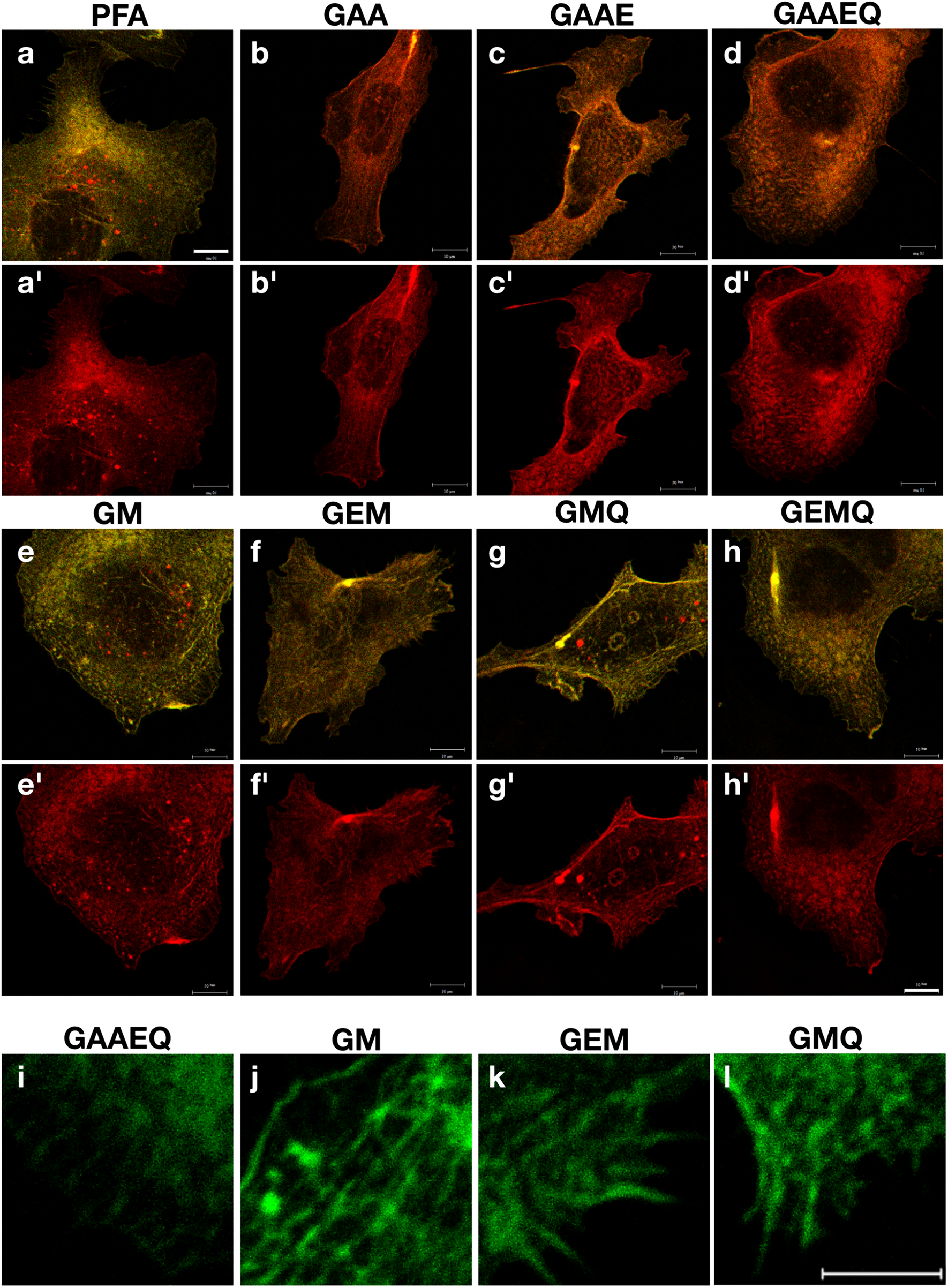
Confocal microscopy
MCF7 cytoskeleton arrangement was observed by confocal microscopy using the same treatments as in the wound healing assay. A total of 20 × 103 cells were seeded on fibronectin-pretreated glass slides (Gibco, Grand Island, USA). The next day, the respective treatments were added in duplicate for 24 h. The cells were washed with blocking solution [phosphate-buffered saline (PBS) and 2% SBF], fixed with 4% paraformaldehyde (Sigma, Burlington, USA) for 10 min and permeabilized with triton X 100 0.1% (ICN Biomedicals, Santa Ana, USA) for 5 min; then, they were labeled with Alexa Fluor™ 594 Phalloidin (2 μl/ml in PBS, InvitrogenTM, Waltham, USA) for actin filaments, and DAPI 600 nM (InvitrogenTM, Waltham, US) as nuclear staining. After each labeling they were washed twice with blocking solution. Finally, they were placed on slides with a drop of Prolong (Molecular Probes, Eugene, USA) and allowed to dry overnight to be observed under the microscope using a FV1000 laser scanning microscope from Olympus (Tokyo, Japan). Cells were visualized using an UPLSAPO 60× NA 1.35 objective and fluorescence emission was obtained using 50 mW diode laser line 405 and multiargon FV5-LAMAR 30 mW 543 nm laser line. A total of 640 × 640 images were obtained using X, Y, Z laser scanning and a z-projection was constructed for each field. The experiment was performed in duplicate, and from each condition, at least three different fields were analyzed; on this occasion, only hAMSC-CM from sample 4 was used.
RT-qPCR for secretome cytokines expression
Total RNA of hAMSC was extracted using TRIzol LS reagent according to the manufacturer’s instructions (Life Technologies Corporation, InvitrogenTM, Waltham, USA) after 48 h treatment with both natural products at a concentration of 60 μg/ml, and RNA quality and quantity were assessed by NanoDrop spectrophotometry (NanoDrop Technologies, Wilmington, USA). Then, complementary DNA (cDNA) was synthesized with the SuperScript III Reverse Transcriptase (InvitrogenTM, Waltham, USA) following the manufacturer’s protocol. Afterwards, the real time polymerase chain reaction (RT-PCR) was carried out using 600 ng of cDNA, iTaq Universal SYBR Green Supermix (BIORAD, Hercules, USA), and 250 nM forward and reverse primers in a total volume of 20 μl (Supplemental Material Table S2). Reactions were done in duplicates using the QuantStudioTM 3 Real-Time PCR system (InvitrogenTM, Waltham, USA).


留言 (0)