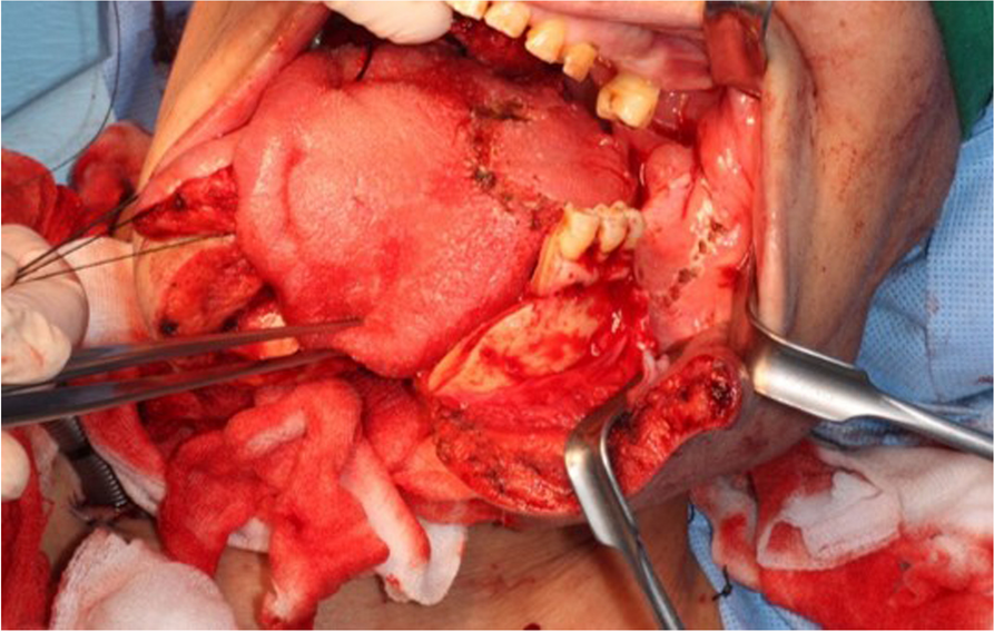
In oral cancer surgery, various reconstructive approaches are used in surgically removed defect, depending on the extent and size of the soft and hard tissues involved. Flaps are categorized based on anatomical location and blood supply. The fibular flap was introduced by Taylor et al. in 1975 for tibial fractures [4] and reported its superiority [5]. Starting with Hidalgo’s report on the reconstruction of mandibular defects in 1989 [6, 7], it was used for defects in the oral and maxillofacial region. The fibular flap can be transplanted to a defect of up to 20 cm, providing a sufficient length of bone flap, and the blood supply of the flap is well maintained even when the size of the flap is large [8]. Additionally, the use of a tourniquet during dissection confers several advantages, including reduced blood loss and enhanced visibility within the surgical field, and allows direct suturing of the donor site even when the size of the flap is large.
In addition, the donor site complications are minimal and stable, and the large diameter of the peroneal artery makes anastomosis with cervical vessels relatively easy during microsurgery.
Additionally, since the donor site is located far away from the head and neck area, there is an advantage that two teams can operate simultaneously on both the donor and recipient sites. Due to these advantages, fibular flap is considered primarily during oral and maxillofacial reconstruction when reconstruction of a large defect in the mandible is required.
In 1987, Taylor anatomically described the perforator of the human body, and in 1989, Koshima introduced the perforator flap based on the musculocutaneous type. In the 2000s, with the development of microsurgical techniques, flaps based on perforator began to be introduced for the reconstruction of oral and maxillofacial defects. Reconstruction with a perforator flap requires microsurgical techniques and has the disadvantage of prolonging the surgery time due to microvascular anastomosis. However, perforator flaps have a lower thickness, allowing for more free tissue placement, and the perforator flaps are easier to handle, resulting in satisfactory results after reconstruction.
The average number of perforators originating from the peroneal artery is 4.8 [9], and most perforators range in size from 0.5 to 1 mm. In addition, since the majority of the perforators distributed in the fibula are located in the seventh or eighth position from the top when the tibia is divided, the primarily harvested site for fibular free flap during the harvest process is 8 to 12 cm above the ankle, which has a good blood supply [10].
The peroneal artery mainly supplies blood to the distal third of the fibula, and most perforators branch from the peroneal artery. However, it has been reported that the perforator of the fibula branches from sources other than the peroneal artery [11], rarely branching separately from the anterior tibial artery or popliteal artery [12,13,14]. There are also cases where the perforator is absent [15,16,17].
The skin paddle of fibula free flap receives blood supply from septocutaneous or musculocutaneous perforators originating from peroneal artery. The distal perforator is mostly a septocutaneous flap, whereas the proximal perforator is a musculocutaneous flap. Several cadaveric and radiographic studies concluded critical vascular anomalies in 10% of population, which can lead to failure of flap survival with ischemia on donor site [12].
For these reasons, it is essential to check the distribution of blood vessels in the lower extremities through angiography and Doppler ultrasonography before surgery [18]. Most (70–96%) of the perforators in the distal third of the fibula are septocutaneous type, branching from the peroneal artery and comes out along the intermuscular septum to supply blood on flap [19, 20]. On the other hand, perforators mainly distributed in the proximal third of the fibula are in the form of musculocutaneous type [21, 22], which penetrate the soleus muscle and travel to the skin to supply blood circulation [23,24,25].
The perforators branching from the peroneal artery are mainly concentrated in the distal third of the fibula with better blood supply. Fibula free flap harvest is based on the distal perforator in this reason [6]. In particular, an average of one to three perforators were observed in the distal third of the fibula. On the other hand, fibula flap with proximal perforators is not considered as the primarily method, due to disadvantages that come with its proximal location and low usefulness [26]. When forming a fibular flap, preoperative angiography of the lower extremities is performed to identify the branches of the peroneal artery [27], and the location or distribution of the subcutaneous perforators can be confirmed through Doppler examination. Therefore, preparing surgery with preoperative identification of the location of the perforator before the incision is critical [28, 29]. The distal perforator, which mainly distributes in the distal third of the fibular flap, is responsible for the blood supply to the flap and is known to be relatively stable and has little anatomical variation [30].
However, as in this case, there may be cases where the distal perforator is not observed due to aberrant anatomical defect, and such vascular abnormalities sometimes show contradictory results to the preoperative Doppler examination [12]. This is an error that may occur in Doppler examination when a perforator overlaps with other arteries in the surrounding area, making it difficult to rely solely on Doppler examination results. For this reason, clinicians should consider the possibility of identifying a defect in the perforator after incision of the skin, as in this case, and understand alternative approaches to flap formation [9].
In this case, by using a proximal flap design, the reconstruction was proceed as planned. The conventional fibula flap is connected with bone by the posterior intermuscular septum, and it is difficult to inset due to limitation of movement between the bone and skin paddle if defect is complex. This can also lead to failure to offer sufficient soft tissue volume. In contrast, the proximal perforator skin paddle is more flexible and provides extended soft tissue and offers advantages such as better visualization and possibility of chimeric flap elevation. The fibula free flap with proximal perforator is a reliable approach method in the reconstruction of extensive mandibular defects [8].
While reports exist regarding alterations in blood vessel dynamics during the formation of fibular free flaps and subsequent modifications in the harvesting process employing alternative vessels, documentation of perforator abnormalities remains limited [19, 25]. Nevertheless, it is imperative for clinicians to possess precise anatomical knowledge pertaining to blood vessel distribution in the microsurgical field and to be adept in responding to encounters with aberrant anatomical variations [26]. Though anatomic variations in perforator flaps are infrequent and distinctive, it is crucial to recognize the potential for deficiencies in the distal perforator. Moreover, preserving the proximal perforator identified during skin incision and comprehending flap formation with the proximal perforator, when necessary, are deemed pivotal factors in ensuring successful flap formation and a favorable prognosis in the presence of vascular abnormalities during the flap harvest process [31,32,33].


留言 (0)