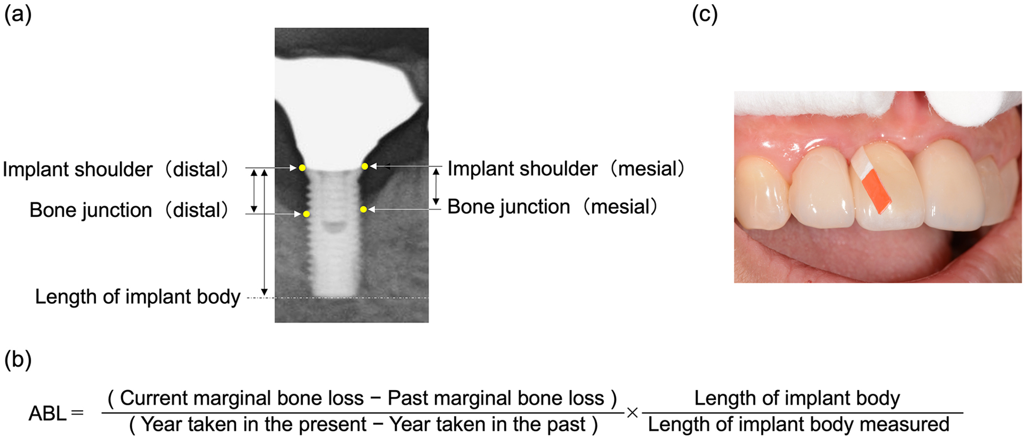
The analysis of the deviations between planned and placed implant positions showed that there were deviations both in the group of postgraduate dentists and in the group of undergraduate students. However, no significant differences were found between the two groups, thus, the null hypothesis was accepted.
Apical deviations of 0.97 ± 0.35 mm, coronal deviations of 0.63 ± 0.25 mm, angulation deviations of 2.92 ± 1.16°, and a vertical deviation of 0.47 ± 0.24 mm were found in the postgraduate dentists’ group. In the undergraduate students’ group, apical deviations of 1.17 ± 0.58 mm, coronal deviations of 0.80 ± 0.56 mm, angulation deviations of 3.75 ± 1.86°, and vertical discrepancies of 0.56 ± 0.60 mm were determined. Thus, the range of these discrepancies between the planned and placed implant positions is in line with the results in the literature. Rungcharassaeng et al. and Cassetta and Bellardini presented comparable deviations in inexperienced practitioners (coronal deviations of 0.64 ± 0.21 mm, apical deviations of 1.22 ± 0.63 mm, vertical deviations of − 0.51 ± 0.21 mm and angulation deviations of 3.21 ± 1.99° vs. coronal deviations of 0.75 ± 0.18 mm, apical deviations of 1.02 ± 0.44 mm, and angulation deviations of 3.07 ± 2.70°, respectively) [31, 32]. Interestingly, both studies also found deviations between planned and placed implant positions in experienced practitioners, although they did not significantly differ from those of inexperienced practitioners (Rungcharassaeng et al.: coronal 0.47 ± 0.15, apical 1.32 ± 0.25, vertical − 0.26 ± 0.23, angulation 4.11 ± 0.76°; Cassetta and Bellardini: coronal 0.60 ± 0.25 mm, apical 0.67 ± 0.34 mm, angulation 3.21 ± 1.57°). This phenomenon can be explained by the fact that inherent errors in computer-guided implant surgery can lead to imprecise implant placement, regardless of the experience of the operator [33, 34]. In addition to intraoperative errors, which could be due to the lack of expertise of the surgeon, incorrect matching of CBCT and scan data, errors in the preparation of the surgical template, and fixation of the guiding sleeve [31, 35]. In a systematic review, Schneider et al. reported mean coronal, apical, and vertical deviations of approximately 1.07 mm, 1.63 mm, and 0.43 mm, respectively, and a mean angular deviation of 5.26 degrees [33].
Interestingly, in the present study and other previous studies, coronal deviations were found to be smaller than apical deviations. D'Haese et al. explained this by pointing out that minimal coronal drilling defects can result in axial deviation in bone depth, leading to larger apical deviations [36], especially for longer implants. In the current study, the analysis of the influence of implant length did not reveal relevant differences between shorter and longer implants. Nevertheless, the significance of these results should be regarded as very weak. This is attributed to the considerable variation in group sizes for implants with lengths of 8, 9, 10 and 11 mm, which were combined into two groups (8 to 9 mm and 10 to 11 mm) for the statistical analysis.
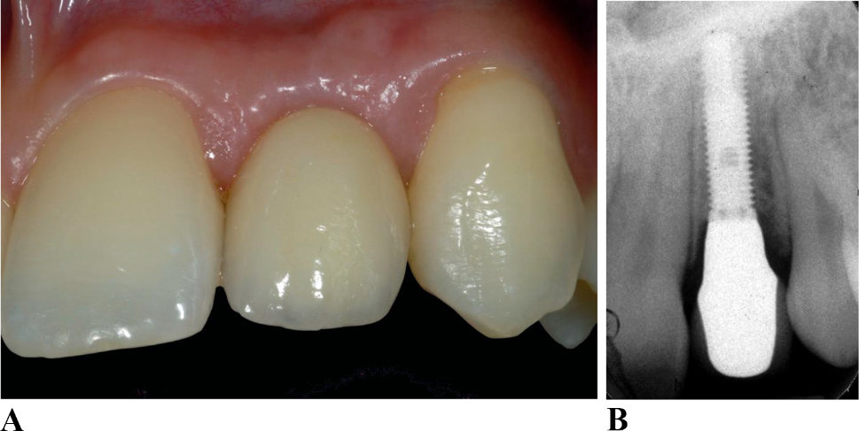
However, the question of whether the deviations found are clinically relevant remains unanswered. Di Giacomo et al. divided the apical and coronal deviations into slight deviations (≤ 1 mm), moderate deviations (> 1 to ≤ 2 mm), and relevant deviations (≥ 2 mm) [37]. Considering this classification, the mean deviations found in this study should be considered as slight to moderate in both groups. However, this did not apply to the two implants that had to be inserted without the use of a surgical template by the supervising assistant because of the lack of primary stability after fully guided preparation of the implant bed. In these cases, the greatest deviation between planned and placed implant positions were measured.
The lack of primary stability has been highlighted in the literature as one of the most common problems in fully guided implant surgery [25, 37, 38]. In the present study, the deviation from the surgical protocol led to significantly higher deviations than in implants placed according to the fully guided protocol (apical 3.41 ± 0.06, coronal 1.81 ± 0.12, vertical 1.31 ± 0.61, and angulation deviations of 9.96 ± 1.14°). Due to the minimal sample size (n = 2), these findings have to be critically evaluated. However, a meta-analysis by Putra et al. showed that freehand insertion after pilot drilling led to greater deviations than fully guided implantation [23]. Even under ideal in vitro conditions, higher deviations that were comparable to those in the present study were observed with freehand insertion [39]. The apical deviations found in this study support the recommendations of Mistry et al., who proposed that, in the context of freehand implantations, safety distances of 3 mm should be taken into account to prevent severe consequential damage [22].
The literature has discussed the significance of the surgeon’s experience as instrumental in achieving precision in implantation [1, 9]. The present data indicate that postgraduate dentists tended to achieve smaller deviations from the planned implant position than students, but this difference was not found to be statistically significant or clinically relevant. However, Choi et al. and Cushen et al. demonstrated that the precision of implant placement improved with the increasing surgical experience of the surgeon [20, 40]. In the present study, postgraduate dentists already had more surgical experience than the undergraduate students based on at least two years of clinical experience. It is possible that the training resulted in the dentists achieving an overall higher level of accuracy than the students. However, it is important to note that there was still no significant and clinically relevant difference, which is an additional indication of the safety of the fully guided method.
Regarding the implant survival rate, no differences were found between the two groups as no implant loss or postoperative complications were observed during the observation period. However, it is important to note that the significance of this result is limited by the relatively short follow-up period of 598.33 ± 195 days (postgraduate dentists) and 1641 ± 302.1 days (undergraduate students), indicating that long-term survival cannot be evaluated in this context (see Table 3). It was also observed that the implant healing time (88 ± 110.4 days for postgraduate dentists vs. 142.9 ± 66.9 days for undergraduate students), as well as the time until integration of the prosthetic restoration (243.5 ± 138.4 days for postgraduate dentists and 218.3 ± 103.9 days for undergraduate students), differed between both groups. This can be attributed to logistic challenges in student training (e.g., semester breaks) as well as the fact that, in the undergraduate students’ group, solely implant-supported fixed prosthodontics were more frequently fabricated, whereas in the postgraduate dentists’ group, prosthetic treatment included more comprehensive prosthetic restorations.
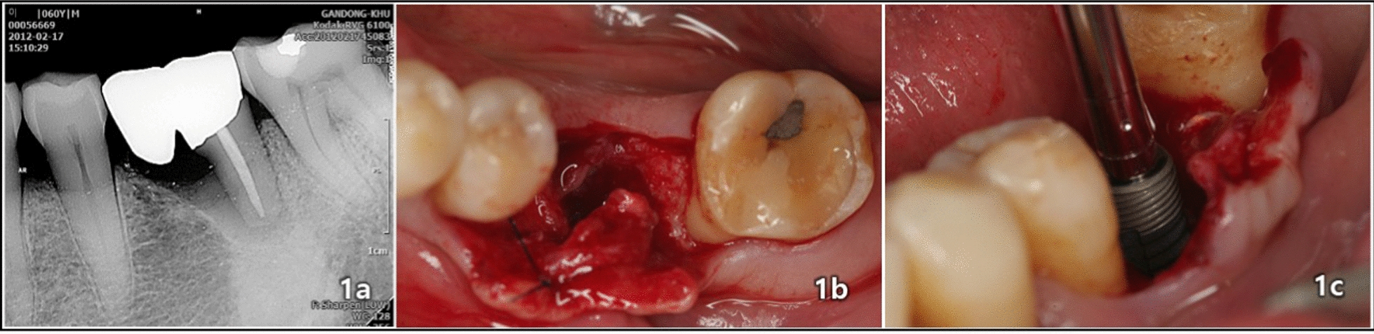
Besides the surgeon’s experience, various other factors that influence the precision of fully guided implant placement have been discussed in the literature. For example, whether the use of a flapless protocol can lead to less deviation in the implant position has been discussed. Although some studies showed better outcomes with the flapless protocol [23, 41], this was not confirmed in other studies [42]. In the present study, a flap was created to improve the surgical overview. The extent to which flapless surgery would have reduced the discrepancies between the planned and inserted implants can only be speculated. However, in the two cases in which freehand implant placement was required due to a lack of primary stability, the flap that had already formed proved to be extremely helpful in retrospect.
In addition, an assessment was conducted to determine whether affiliation to the jaw led to varying deviations between the planned and placed implant positions. In the present study, smaller deviations were observed for implants placed in the mandible than those placed in the maxilla. This tendency was also reported by Ersoy et al. [42]. However, clinical relevance has not yet been reported [23, 42].
The edentulous area has also been discussed as a factor influencing the precision of fully guided procedures [23]. In the present study, implants were placed only in patients with minor tooth loss and the templates were well supported by the available teeth. Similar to the results of the present study, Raico Gallardo et al. found smaller deviations between the planned and placed implant positions in implant placement in interdental gaps than in free-end situations [43]. This is because of the better fixation of the template without the risk of lateral thrust in the interdental gaps. Furthermore, there is a risk of displacement of the surgical template due to differently resilient mucosa, mucosa thickness, and possibly swelling due to local anesthesia in free-end situations [23, 43].
Several other limitations should be considered when evaluating the results of this study. Overall, although our results were comparable with those reported in existing literature, the number of implant cases and participants both in the undergraduate students’ and postgraduate dentists’ groups were comparatively small. Moreover, the postgraduate dentists used two different implant systems and planning software, whereas the undergraduate students used only one. Nevertheless, because the evaluation of implant accuracy depending on the implant system showed no significant difference in the postgraduate dentist group, all implants were included in the analysis. This decision was based on results of the literature that described comparable implant accuracy between different planning and implant systems [29].
The informative value of the study is further limited by the fact that only straightforward cases were selected, based on the SAC classification [44]. The extent to which novice practitioners achieve better results than undergraduate students in more difficult cases (e.g., partially edentulous or edentulous patients) should be the subject of future investigation.
Finally, it must be noted that in two cases within the group of undergraduate students, the operation had to be performed by the supervising assistant. As a result, the final implant bed preparation and implant placement took place without the previously constructed template, which resulted in significantly higher deviations from the planned and achieved implant position. This raises the question of whether dynamic implant placement might not have provided significant advantages in such cases. This technique is supported by a computed-navigation system, in which planned implant position and real-time position of the drill tip could be visualized. Thus, the preoperatively intended implant location as well as implant configuration can be changed in real-time in a controlled way when needed [45, 46]. Future investigations should focus on the potential benefits of dynamic implantation in undergraduate dental education.


留言 (0)