Two theories of aging, not mutually exclusive, have been projected. The first is a concept whereby a cell is programmed to fulfill the cell cycle a finite number of times before undergoing senescence and this is supported by the cellular senescence-telomere theory suggesting the presence of a biological clock (Bernadotte, Mikhelson, & Spivak, 2016) (Goldsmith, 2016). While the alternative idea is one where an accumulation of damage ensues at multiple sites and by various sources (Liguori et al., 2018).
Cells under stress can undergo senescence potentially caused by mutations in signaling, DNA damage from free radicals, and/or replication (Kumari & Jat, 2021). By activating the enzyme telomerase, which aids in telomere regeneration, telomere deterioration, which occurs after each cell division and causes replicative senescence, can be halted. According to the theory underlying stress-induced senescence, external stressors cause mutations in repair enzymes altering DNA further resulting in dysfunction and accelerated aging (Polsky, Rentscher, & Carroll, 2022). The gene regulation theory of aging is consistent with the concept whereby genes govern life and death as some genes promote lifespan by diminishing insulin-like signaling and gene expression may influence lifespan similarly to the sirtuin family of antiaging genes (Moskalev, Aliper, Smit-McBride, Buzdin, & Zhavoronkov, 2014). Other hypotheses, such as the immunological theory, support the premise that as people age, their immune systems begin to function less typically, rendering them more susceptible to infectious diseases and, ultimately, death (De la Fuente, 2008).
The hormonal theory, defined by the stress theory of aging, asserts that protracted stress and stressful situations damage cells and disrupt normal cellular function, resulting in system dysfunction and aging (Maldonado, Morales-Pison, Urbina, & Solari, 2023). The prevalence of stress-related illnesses and conditions, such as resistance to insulin, anxiety disorders, depression, hypertension, coronary heart disease, and cerebrovascular disease, increases significantly across the lifespan. The heterogeneity of the aging process may also contribute to individual differences in susceptibility and resistance to stress and stress-related diseases (Maldonado et al., 2023). The principal stress-related signaling pathways are the insulin/IGF, TOR, and sirtuin networks. Variations in dietary quality or the number of stressors affect these signaling pathways, thereby altering mitochondrial function and metabolic activity via genome proteostasis and maintenance circuits. During early development, the network integration and activity of the stress response system and the maintenance circuitry, which are both designed to increase endurance, develop (Hughes, Ellefsen, & Baar, 2018). The inability to respond to stress has been linked to age and age-related disorders while further research supports that this decline may be reversed (Childs, Durik, Baker, & Van Deursen, 2015). Instead, it appears that increasing stress tolerance via conserved signaling pathways can increase longevity and delay the aging process (Haigis & Yankner, 2010).
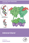
Every human body naturally has a pair of adrenal glands, each nesting above a kidney, and are each composed of the innermost medulla and outer cortex. Once the structure and function of the cells within these two regions are clear then the effects aging has on them may be explored.
The medulla is overall composed of granular chromaffin cells responsible for preparing the body for action through production and secretion of epinephrine, norepinephrine, dopamine along with other catecholamines. This stimulation is under the control of the sympathetic nervous system, hence the presence of sympathetic ganglion cells in the region. Activity of the medulla affects fight-or-flight response, motivation, arousal, and additional physiological responses. The cortex on the other hand is histologically distinguished by three concentric zones known as the innermost zona reticularis (ZR), zona fasciculata (ZF) and outermost zona glomerulosa (ZG). The ZG region of the organ is involved in the production of steroids, such as aldosterone that is critical in maintaining salt-water homeostasis in the body and is regulated by the renin-angiotensin system. Cortisol, a glucocorticoid, is primarily produced by the ZF and is implicated in multiple physiological functions among which are regulation of stress, blood pressure and immune response (Fig. 1). Finally, the yield of dehydroepiandrosterone (DHEA), essential for regulating sexual development and function, is from the ZR (De Silva & Wijesiriwardene, 2007).
Both the adrenal cortex and medulla have distinct embryonic origins as they develop from the mesoderm and ectoderm, respectively. The adrenal gland appears around one month post conception and its development is concluded postnatally (Kim and Choi, 2020, Pignatti et al., 2023, Xing et al., 2015). The cortex is the first to make an appearance as mesothelial cells differentiate into acidophilic cells, followed by the invasion of the cortex by neural crest cells a couple of weeks later which give shape to the medulla (Costanzo Physiology, n.d.). Subsequently, a second group of mesothelial cells surrounds the acidophilic cells to form the permanent cortex. By week eight of development, the fetal and permanent cortexes surround the adrenal medulla. The permanent cortex differentiates into the zona glomerulosa, zona fasciculata, and zona reticularis, which secrete glucocorticoids, mineralocorticoids, and adrenal androgens (Kim and Choi, 2020, Pignatti et al., 2017).
The adrenal cortex is responsible for producing steroid hormones, including glucocorticoids, adrenal androgens, and mineralocorticoids. Mineralocorticoids, such as aldosterone, are synthesized by the zona glomerulosa, while the zona fasciculata and zona reticularis are responsible for synthesizing glucocorticoids, including cortisol, corticosterone, and cortisone, and adrenal androgens such as dehydroepiandrosterone (DHEA) and androstenedione (Costanzo Physiology, n.d.). The biosynthesis of these hormones starts with the conversion of cholesterol to pregnenolone, a reaction catalyzed by cholesterol desmolase, which is stimulated by ACTH (Miller & Bose, 2011).
Cholesterol is a crucial precursor for steroid hormone synthesis and can be synthesized de novo or obtained from lipid droplets within the cells of the adrenal cortex through hydrolysis by cholesteryl ester hydrolase. High-density lipoprotein also transports cholesterol to the adrenal cortex, ensuring a continuous supply of cholesterol to the adrenal glands (Hu, Zhang, Shen, & Azhar, 2010).
Glucocorticoids are 21-carbon steroids that have a ketone group attached to carbon 3 and hydroxyl groups attached to carbon 11 and carbon 21 (Costanzo Physiology, n.d.). The production of glucocorticoids occurs mainly in the zona reticularis and the zona fasciculata, with cortisol being the most potent and responsible for 95% of all glucocorticoid activity. Corticosterone is another glucocorticoid produced from pregnenolone via similar reactions involving the same enzymes but accounts for only 4% of all glucocorticoid activity (Khonsary, 2017).
The zona reticularis and the zona fasciculata also produce adrenal androgens such as DHEA and androstenedione (Burger, 2002). The enzyme 17,20-lyase is essential for the biosynthesis of adrenal androgens (O’Donnell et al., 2004). A small amount of testosterone and estrogens are also produced by the zona reticularis and the zona fasciculata, although the main source for these hormones is the testes and ovaries, respectively (Burger, 2002).
The zona glomerulosa produces mineralocorticoids, the main one being aldosterone, which is essential for electrolyte homeostasis. The enzyme aldosterone synthase, expressed exclusively in the zona glomerulosa, is essential for the synthesis of aldosterone and is regulated by angiotensin II.
The adrenal glands are essential components of the hypothalamic-pituitary-adrenal (HPA) axis, which plays a critical role in the stress response and homeostasis maintenance (Costanzo Physiology - 7th Edition, n.d, Khonsary, 2017) In response to stress, the hypothalamus secretes corticotropin-releasing hormone (CRH), which enters the hypothalamic-hypophysial portal circulation and stimulates the anterior pituitary gland to release adrenocorticotropic hormone (ACTH) (Bollag, 2014). ACTH then triggers the adrenal gland’s secretion of adrenocortical hormones. The zona reticularis and the zona fasciculata’s endocrine secretions are regulated exclusively by the HPA axis, while the zona glomerulosa’s secretion is initially controlled by the HPA axis and then regulated by the renin-angiotensin-aldosterone system (Gao et al., 2021).
Glucocorticoids, particularly cortisol, exhibit pulsatile and diurnal secretion patterns, with ten secretory bursts observed over a 24-hour period, and the highest concentration secreted in the morning and the lowest in the evening (Costanzo Physiology - 7th Edition, n.d, Khonsary, 2017). Cortisol secretion is also regulated by negative feedback mechanisms, where cortisol can inhibit hypothalamic CRH or pituitary gland ACTH secretion ACTH is required for the initial secretion of the mineralocorticoid aldosterone, but it does not significantly regulate its rate of secretion. Aldosterone secretion is mainly regulated by the renin-angiotensin-aldosterone system and the serum potassium concentration, playing a crucial role in electrolyte homeostasis. Renin secretion increases in response to a decrease in extracellular fluid volume, leading to the conversion of angiotensinogen to angiotensin I. Angiotensin I is then converted to angiotensin II by the angiotensin-converting enzyme, controlling the final step of aldosterone synthesis. Additionally, an increase in extracellular potassium levels leads to cell membrane depolarization, opening of voltage-sensitive calcium channels, and subsequent aldosterone secretion.
The adrenocortical hormones act as steroid hormones, binding intracellular steroid hormone receptors, causing conformational changes that allow the receptor to translocate into the cell nucleus. The receptor then dimerizes, binds to the steroid response elements in the DNA, and activates transcription as a transcription factor (Griekspoor, Zwart, Neefjes, & Michalides, 2007). The newly synthesized mRNA exits the nucleus and is translated into proteins by ribosomes in the rough endoplasmic reticulum. The newly synthesized proteins exert their physiological actions on the target tissue.
Glucocorticoids exhibit catabolic and diabetogenic effects, stimulating gluconeogenesis and glycogen storage and inducing lipolysis and proteolysis, resulting in an increase in free amino acids and glycerol, which serve as the primary substrates for gluconeogenesis (Mir, Chin, Riddell, & Beaudry, 2021). In addition to these metabolic effects, the proteins synthesized following glucocorticoid receptor activation have anti-inflammatory, immunosuppressive, and anti-osteoblastic effects (Cain and Cidlowski, 2017, Hardy et al., 2020).
The adrenal medulla is responsible for the synthesis and secretion of the catecholamines epinephrine and norepinephrine, which are amines derived from the amino acid tyrosine. Interestingly, glucocorticoids produced by the nearby adrenal cortex stimulate the expression of phenylethanolamine-N-methyl transferase (PNMT), the enzyme required for the conversion of norepinephrine to epinephrine. Following synthesis, epinephrine and norepinephrine are stored in the large chromaffin granules in the medullary chromaffin cells (Coupland & Tomlinson, 1989).
The adrenal medulla is considered a specialized ganglion and is part of the sympathetic autonomous nervous system. Neural mechanisms regulate the secretion of the medullary hormones. Specifically, the neurotransmitter acetylcholine is released by preganglionic neurons and activates the nicotinic receptors on the chromaffin cells. The nicotinic receptors are transmembrane receptors that consist of five subunits and act as potassium/sodium ion channels (Ho, Abraham, & Lewis, 2020). Upon acetylcholine binding on the α subunit, a conformational change of the receptor occurs, leading to opening of the ion channel (Liu et al., 2008). An influx of sodium and outflux of potassium leads to membrane depolarization. As a result, the voltage-sensitive calcium channels open, leading to an increase in intracellular calcium. Consequently, the secretory chromaffin vesicles fuse with the plasma membrane of the chromaffin cells, releasing the catecholamines into the bloodstream (Feher, 2012).
Around 80% of catecholamines are secreted in the circulation as epinephrine, while the remaining 20% are secreted as norepinephrine (Feher, 2012). The catecholamines then travel to their target organs, where they bind to adrenoreceptors and exert their physiological actions. Activation of the α1 adrenoreceptors leads to activation of phospholipase C and generation of IP3, resulting in vasoconstriction, intestinal relaxation, and contraction of the uterine smooth muscle and the intestinal and bladder sphincters. In contrast, activation of the β1 and β2 adrenoreceptors leads to activation of adenylyl cyclase and generation of cAMP, resulting in vasodilation, uterus relaxation, glycogenolysis, lipolysis, and increased heart rate.


留言 (0)