
記住我


The Paecilomyces genus includes species of fungus not frequently associated with human infection.1 Thirty pediatric cases of Paecilomyces spp. infections have been reported, with most in patients with indwelling devices and immunocompromise; very few involved central nervous system (CNS) infections and all were fatal.2–4 We present a case of a pediatric Paecilomyces spp. ventriculoperitoneal (VP) shunt infection treated with shunt replacement, liposomal amphotericin B and posaconazole.
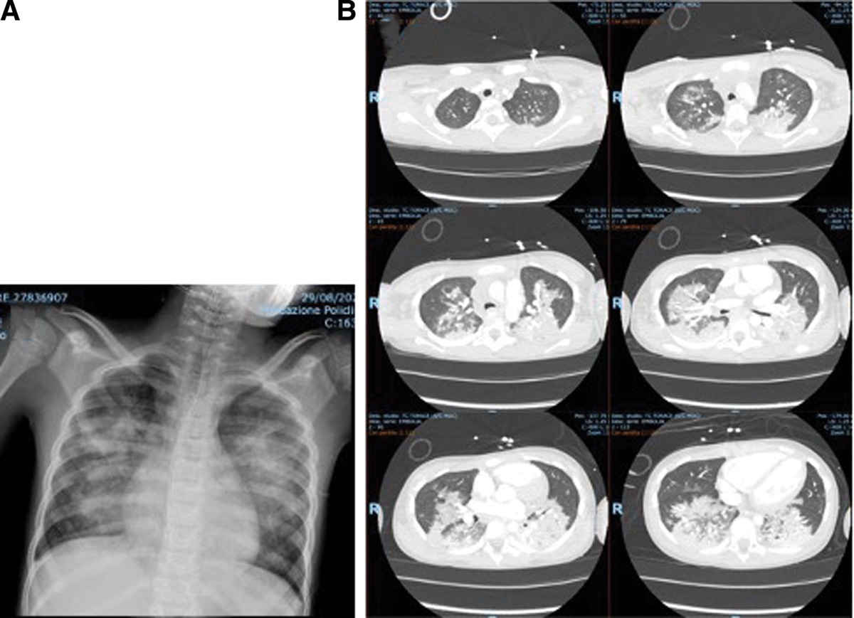
A 14-year-old girl with a history of myelomeningocele and VP shunt, tethered cord release and procedures for neurogenic bowel and bladder (appendicostomy, appendicovesicostomy), presented with 2 weeks of increasing headache without fever or other symptoms and underwent VP shunt externalization. The illness was preceded by a shunt revision 2 months earlier, at which time the cerebrospinal fluid (CSF) laboratory profile was normal and cultures were sterile. During the externalization, catheter tip fenestrations were noted to be filled with proteinaceous debris, and the distal abdominal catheter appeared obstructed. An abdominal computed tomography scan showed a small rim-enhancing fluid collection with surrounding inflammation, possibly representing an abscess. Broad-spectrum antibacterial therapy was begun.
Examination of the CSF showed pseudohyphae and septate hyphae on Gram stain, prompting the addition of fluconazole and liposomal amphotericin B (L-AmB) to maximize antifungal activity. Within 3 days, cultures were growing mold from intraoperative peritoneal fluid and CSF. Repeated samples of CSF yielded mold through the first week of antifungal therapy; during the second hospital week, the mold was identified by colonial morphology and matrix-assisted laser desorption ionization–time of flight mass spectrometry as Paecilomyces spp. (Fig. 1). The antifungal regimen was revised to L-AmB and posaconazole, and a left ventriculopleural shunt was placed. After 18 days of L-AmB and 6 months of posaconazole, the child has remained well-appearing. However, she has required several shunt revisions due to malfunctions of uncertain etiology, each with sterile CSF. Due to possible shunt plugging from undetected infection, her current therapy (8 months after the initial diagnosis) has empirically been changed to oral isavuconazole.
 FIGURE 1.:
 Photomicographs and photographs of isolate. A: Gram stain (B) calcofluor-white (C) lactophenol cotton blue and (D) colony morphology of the Paecilomyces isolate.
The Paecilomyces genus includes many species of fungus (P. varioti, P. marquandii and P. javanicus, etc.) that are commonly plant and insect pathogens in nature and are not frequently associated with human infection.1 One clinically important species recently was reclassified as Purpureocillium lilacinum.2 Most human infections have been associated with indwelling devices or peritonitis among immunocompromised hosts or patients with underlying chronic diseases, including diabetes.2–4 These molds are hyaline hyphomycetes that grow as filamentous molds on numerous media, forming a thin powdery yellow-brown colony (P. lilacinum is notable for pink/lilac colonies) (Fig. 1). Mycelia have broad, branching and septate hyphae, and phialides have broad bases ending in a long slender neck. Speciation is by molecular targeting of the internal transcribed spacer regions within ribosomal DNA.1–5
Our case represents the first documented pediatric CNS Paecilomyces spp. VP shunt infection. Whether peritonitis seeded the VP shunt, or whether the VP shunt infection seeded the abdomen, is unclear. Previous reports have described Paeciliomyces peritonitis in association with peritoneal dialysis catheters; our patient’s prior abdominal procedures for neurogenic bowel and bladder may have caused the primary abdominal infection.2
One prior report of Paeciliomyces spp. VP shunt infection involved an immunocompetent adult with shunted hydrocephalus following mesh placement for a basilar artery aneurysm.5 Several subsequent shunt revisions were required in this adult for catheter occlusions with “fibrous and proteinaceous material.”5 The authors suspected a possible undiagnosed fungal infection may have existed for months to years, with repeated shunt failures due to “recurrent mycelial plugging of the device.”5 This adult ultimately succumbed to a cerebral aneurysm said to be unrelated to infection.
Our patient’s recovery and CSF sterilization indicate that amphotericin B and posaconazole, along with source control, is a possible treatment for CNS Paecilomyces spp. infection. With the increasing complexity of abdominal and CSF diversion surgeries, clinicians should be aware of rare environmental mold infections in children and adolescents.
REFERENCES 1. Houbraken J, Verweij PE, Rijs AJ, et al. Identification of Paecilomyces variotii in clinical samples and settings. J Clin Microbiol. 2010;48:2754–2761. 2. Sprute R, Salmanton-García J, Sal E, et al. Characterization and outcome of invasive infections due to Paecilomyces variotii: analysis of patients from the FungiScope® registry and literature reports. J Antimicrob Chemother. 2020;76:765–774. 3. Sprute R, Salmanton-García J, Sal E, et al.; FungiScope® ECMM/ISHAM Working Group. Invasive infections with Purpureocillium lilacinum: clinical characteristics and outcome of 101 cases from FungiScope® and the literature. J Antimicrob Chemother. 2021;76:1593–1603. 4. Alharbi M, Alruqaie N, Alzahrani A, et al. Paecilomyces/Purpureocillium infection in children, case report, and review of the literature. J Fungi (Basel). 2022;8:930. 5. Fagerburg R, Suh B, Buckley HR, et al. Cerebrospinal fluid shunt colonization and obstruction by Paecilomyces variotii. case report. J Neurosurg. 1981;54:257–260.
留言 (0)