2.1 Animals
Male naive Rhesus (Macaca mulatta) monkeys (four to eight years old and six to nine kg, Sichuan Green-house Biotech) and naive Wuzhishan Inbred’ Miniature Pig (WZSP) (10 to 24 months old, 35 to 50 kg for males and 30 to 45 kg for females, Grand Life Science & Technology Ltd) were acclimated to the laboratory conditions for 21 days at the animal facility of Sichuan Huashen Veterinary Biological Products Co., Ltd.
The animals were treated in strict accordance with good animal practice under the supervision of veterinarians. Monkeys were housed in individual stainless steel cages in a controlled environment under a 12-h light–dark cycle (lights on at 8:00 and off at 20:00), fed completely formulated monkey feed (twice per day, 100–150 g each time), and had free access to drinking water produced by a reverse osmosis system. In addition, seasonal fresh fruits were provided three times weekly.
Environment enrichment includes a metal mirror attached to each cage, toy playing (plastic balls and swing ring) and video watching twice a week. Pigs were group-housed in the pigpen during the acclimation period, and postgraft implantation animals were housed individually in stainless steel cages in a facility. The facility had a permit for laboratory animal use. The animals were fed twice daily with certified experimental minipig maintenance feed supplied by Beijing Keao Xieli Feed Co., Ltd.
The diet is routinely analyzed by the manufacturer for nutritional components and environmental contamination. Qualified drinking water was supplied via an automatic drinking tap or water bowl ad libitum. Water is routinely analyzed for contaminants and specific microbes, and available information indicates that no contaminants present in the drinking water at a concentration likely influence the outcome of this study. All animal procedures described here were approved by the Institutional Animal Care and Use Committee at Sichuan University West China Hospital.
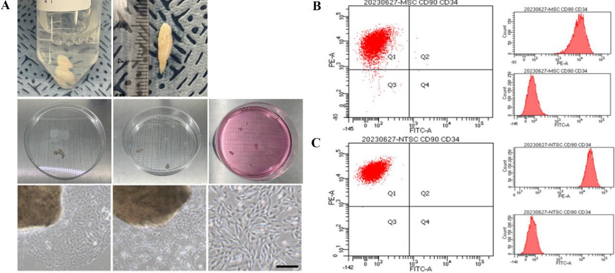
2.2 Adipose-derived mesenchymal stromal cells (ADSCs)
ADSCs were isolated from rhesus monkeys or miniature pigs as follows: Approximately five g (1 mm3) adipose tissue was harvested from the lateral hypogastric region (monkey) or the chest region (swine) after sterilization from anesthetized animals by aseptic surgery. ADSCs were isolated following a procedure described previously [15, 16]. The isolated ADSCs were propagated for three generations within 14 days, and the propagated ADSCs were cryopreserved until the use of biosynsphere bioinks.
2.3 Bioink preparation and 3D bioprinting
Cell-laden microgels were prepared according to a Biosynsphere® (Revotek Co., Ltd) technology [17] and used to prepare the stromal cell-based bioink for the fabrication of the ADSC vascular grafts. The bioink was a mixture of sterilized collagen Type I (17 mg/mL) and biosynspheres in a volumetric ratio of 2:1. An in-house 3D bioprinter was used to “print” the bioink into the lumen structure, and then the bioink was adhered to the surface of the endovascular lumen of ePTFE prosthetic grafts by tissue adhesive. The parameters of 3D bioprinter are described in the Table 1. The ePTFE prosthetic grafts used in this study were obtained from W.L. Gore & Associates, Inc. The main component of tissue adhesive is α-N-octyl cyanoacrylate, and the tissue adhesive was obtained from Guangzhou Baiyun Medical Glue Co. LTD, China.
Table 1 The parameters of in house 3D bioprinter for the construction of the 3D bioprinted ADSC vascular graft2.4 Determination of the stemness of ADSCs
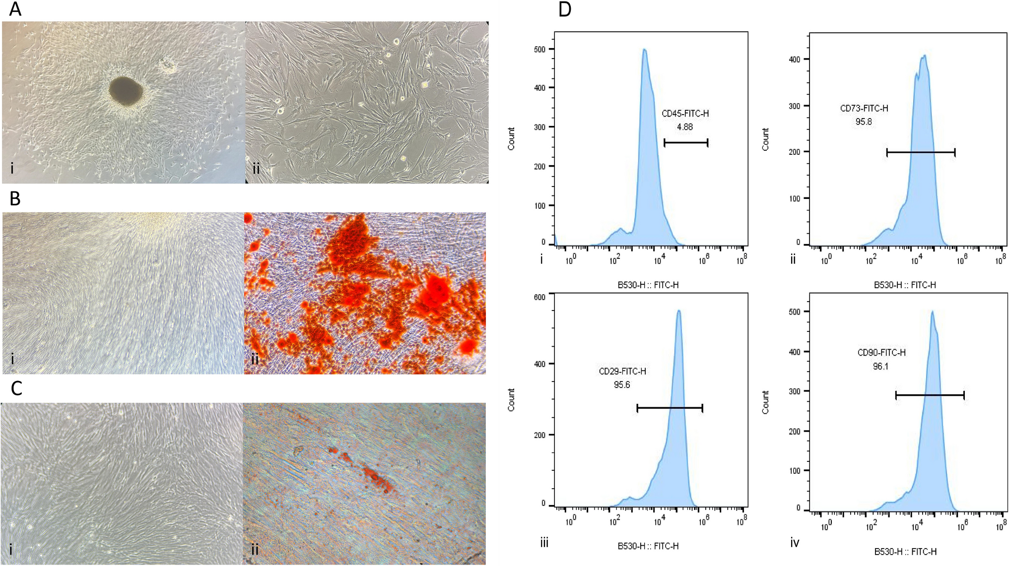
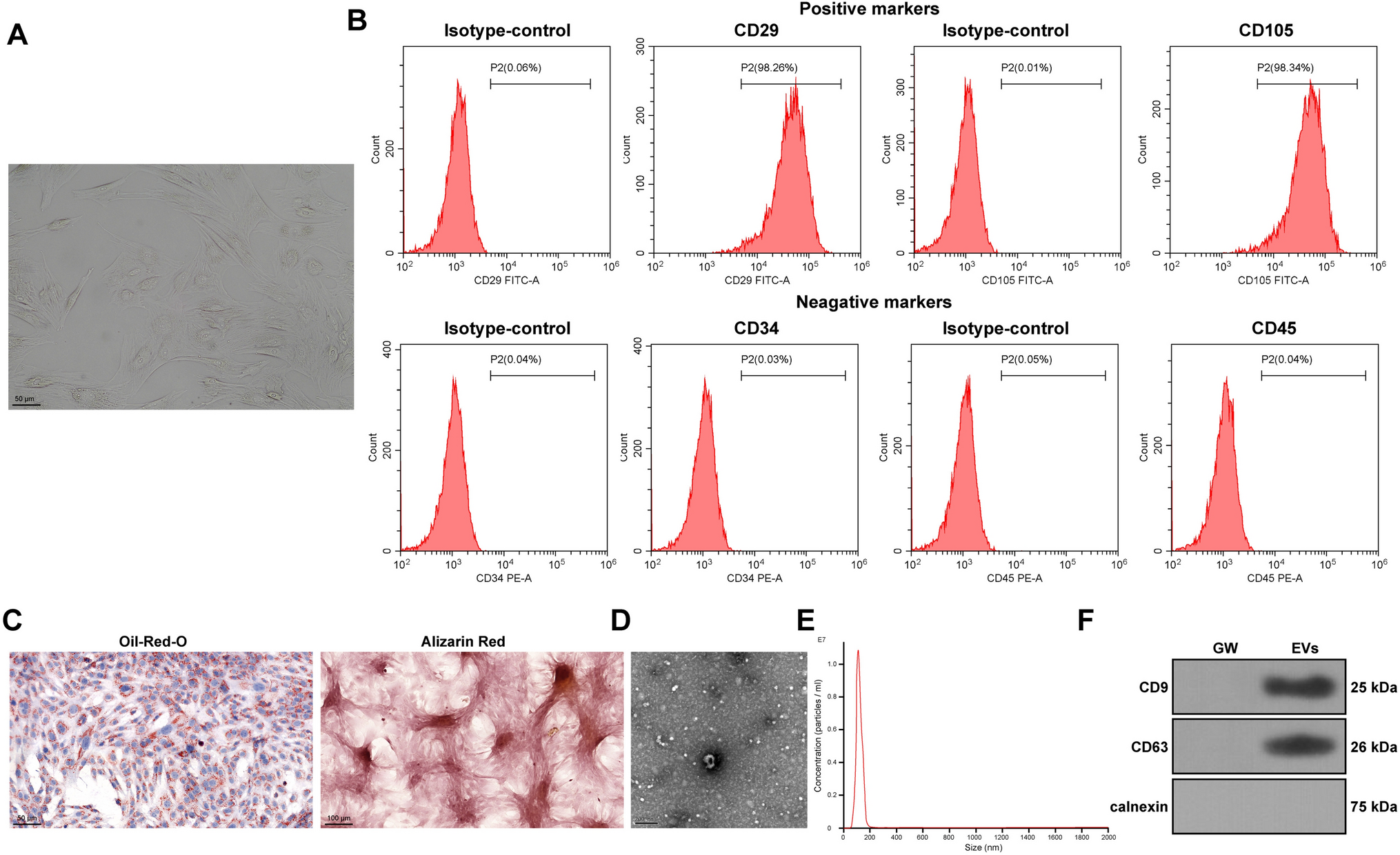
The surface marker of ADSCs was analyzed by flow cytometry with the following antibodies used: (1) CD90/CD44-PE (BD Pharmingen, US, 561970, 561858), (2) CD73/HLA-DR-PerCP-CyTM5.5 (BD Pharmingen, US, 561260, 552764), (3) CD29-APC (BD Pharmingen, US, 561794), (4) CD45/CD31-FITC (BD Pharmingen, US, 557803, 557508), and (5) CD105-PE-Cy7 (eBioscience, US, 25-1057). The induced differentiation of ADSCs was performed by culturing the ADSCs in conditioned induction medium (Cyagen, US). Alizarin red staining was used to detect the osteoblast differentiation of ADSCs. Oil red O staining was used to detect the adipogenic differentiation of ADSCs. Alcian blue staining was used to detect the chondrogenic differentiation of ADSCs.
2.5 Vascular graft implantation
A total of 30 male Rhesus monkeys four to eight years old (approximately six to nine kg body weight) were subjected to routine aseptic surgical procedures as described previously [18,19,20]. A 3-cm-long segment of the infrarenal abdominal aorta was extracted through a midline laparotomy incision. The bioprinted ADSC vascular graft was implanted by End-to-end anastomosis to replace the 3-cm-long segment. Anticoagulant (heparin, i.v., 0.5 mg kg−1) was given immediately after the operation and every day thereafter for the first and maximum five days post-operation.
They were given an anticoagulant, low molecular weight heparin (LMWH), only for the first 5 days (unless sampling less than five days) immediately post-implantation. Histological and immunohistochemical analyses were performed on the graft samples harvested after implantation at 4 and 24 h and on 5, 7, 14, 21, 28, and 70 days. There were three monkeys allowed to survive for an indefinite time after implantation and subjected to long-term noninvasive monitoring for blood vessel function unless otherwise specified.
A total of 34 miniature pigs 10 to 24 months old (approximately 35–50 kg body weight for males, approximately 30–45 kg for females) were subjected to routine aseptic surgical procedures and three pigs remained untreated as controls. The segment of the infrarenal abdominal aorta and left external iliac aorta were exposed and extracted through a midline laparotomy incision. Approximately 10 cm long 3D bioprinted ADSC vascular grafts (26 pigs) or ePTFE grafts (eight pigs) were bypassed from the infrarenal abdominal aorta to the left external iliac artery. The 3D bioprinted ADSC vascular grafts or ePTFE grafts were anastomosed end-to-side to the infrarenal aorta with 6-0 Prolene sutures, and the 3D bioprinted ADSC vascular grafts or ePTFE grafts were anastomosed end-to-side to the left iliac artery with 7-0 Prolene sutures. The starting section of the left iliac artery was ligated to simulate the clinical setting of artery occlusion, and the left iliac artery was replaced by 3D bioprinted ADSC vascular grafts or ePTFE grafts.
2.6 Immunohistochemistry/immunofluorescence staining
The vascular grafts or regenerated vessels explanted from monkeys and pigs were processed for histological and immunohistochemical analyses as described previously (21). The following antibodies were used: rabbit anti-Oct4 polyclonal antibody (Abcam, US, ab19857), mouse anti-CD31 monoclonal antibody (Maixin, China, MAB-0031), goat anti-CD31 (R&D, US, AF3628), rabbit anti-α-SMA polyclonal antibody (Abcam, US, ab5694), rabbit anti-calponin monoclonal antibody (Abcam, US, ab46794), rabbit anti-SMM-HC (Myh11) monoclonal antibody (Abcam, US, ab133567), rabbit anti-collagen I polyclonal antibody (Abcam, US, ab34710), rabbit anti-PGP9.5 monoclonal antibody (Abcam, US, ab108986), donkey anti-goat IgG (H+L) cross-adsorbed secondary antibody, Alexa Fluor 488 (Thermo Fisher, US, A11055), goat anti-rabbit IgG (H+L) cross-adsorbed secondary antibody, Alexa Fluor 568 (Thermo Fisher, US, A11011), and SignalStain Boost IHC Detection Reagent (HRP, Rabbit/Mouse) (Cell Signaling Tech., Tech., US, 8114/8125). All the tissues prepared for staining were harvested from the middle part of the implants.
2.7 Ultrasound examination
For monkeys, after fasting for eight hours (water provided ad libitum), sedation was induced by intramuscular injection of ketamine (10 mg kg−1) and midazolam (1 mg kg−1). Monkeys were subjected to ultrasound evaluation with a 3.5-MHz ultrasonic probe (L9-3, iU22, Philips Medical Systems) in the supine position. B-mode, color Doppler and pulsed Doppler ultrasound were performed to examine abdominal aorta hemodynamics. Longitudinal and transverse views of the abdominal aorta (AO) were examined.
For pigs, the animal was sedated by intramuscular injection of Zoletil® 50 (50 mg/mL) with 3–3.5 mL (approximately three to five mg/kg). Color Doppler ultrasound examination was performed on the engraftment area (ePTFE or 3D bioprinted ADSC vascular grafts) or sham area to evaluate blood flow through the implanted vessel graft, including vascular stenosis or occlusion and thrombosis detection, at or near the graft section.
2.8 Computed tomography angiography (CTA) examination
For monkeys, before the test, sedation was induced by intramuscular injection of ketamine (10 mg·kg−1) and midazolam (one mg·kg−1), and then vein catheter access was established through a 22-gauge venous indwelling needle via the small vein on the right forearm for contrast delivery. X-ray images of the abdominal aorta were captured using a CT scanner (Lightspeed, GE). An iodine-containing contrast dye was injected intravenously to improve the quality of the images. Then, axial images were reconstructed and used to generate high-quality multiplanar reformatted images.
For pigs, the animal was sedated by intramuscular injection of Zoletil® 50 (50 mg/mL) with 3–3.5 mL (approximately three to five mg/kg). In addition, before the CTA test, IV-line access was established through a 20-gauge venous indwelling needle via the marginal ear vessels for CTA contrast agent delivery. CTA examination was conducted on the engraftment area using intravenous injection of iodine-rich contrast agent (iopamidol) to evaluate blood flow through the implanted vessel graft, detect malformation of the implanted graft, and identify abnormalities of vascular stenosis or occlusion and thrombosis at or near the graft section.
2.9 Statistical analysis
Data were obtained from three separate experiments and expressed as the means ± standard errors of the means (SEM). A single factor design was applied to this study. After a significant interaction was detected by analysis of variance (SPSS), the significance of the main effects was further determined by T test. The level of significance was considered when P < 0.05.


留言 (0)