
記住我


A 12-year-old previously healthy boy presented to our hospital with headache and vomiting for 1 month and fever, drowsiness, and behavioral changes for 1 week. He lived in a rural area in Kyrgyzstan, where his family was engaged in livestock. On admission, he was lethargic and confused. Neurologic examination was significant for neck stiffness and right-sided hemiparesis. There was no skin lesion. Laboratory studies revealed a white blood cell count of 7.8 × 10³/uL with 52% neutrophils, 42% lymphocytes, 5% monocytes and 0.3% eosinophils; hemoglobin of 13.6 g/dL; platelets of 298 × 103/uL; C-reactive protein of 3.5 mg/L; and erythrocyte sedimentation rate of 11 mm/h. The blood chemistry panel was normal. Serologic tests for human immunodeficiency virus, Toxoplasma gondii and Bartonella henselae were negative. Brucella tube agglutination and interferon-gamma release assay tests were negative. Cranial magnetic resonance imaging (MRI) revealed an invasive-appearing, calcified herniating mass with ill-defined borders (Fig. 1). Chest radiography, abdominal ultrasonography and echocardiogram were all normal.
 FIGURE 1.:
 Magnetic resonance imaging of the brain: axial (A) and coronal (B) T1-weighted images showing invasive-appearing, calcified mass with unclear borders. T2-diffusion-weighted image (C) shows hyperintensity of the giant lesion in midline.
With the preliminary diagnosis of a brain tumor, the patient underwent diagnostic and therapeutic surgery with the removal of a 5-cm necrotic mass. Stains of the tissue biopsy did not reveal any bacteria, but hyphae-like structures were detected. There was no evidence of malignancy.
Empiric antimicrobial therapy with vancomycin (60 mg/kg/day), ceftriaxone (100 mg/kg/day), metronidazole (40 mg/kg/day) and liposomal amphotericin B (5 mg/kg) was initiated for presumed central nervous system (CNS) infection. The patient was given steroids (prednisone 1 mg/kg/day) for cerebral edema. External ventricular drainage (EVD) was placed due to the development of hydrocephalus. Cerebrospinal fluid (CSF) obtained from EVD showed no leukocytes, erythrocytes of 130/mm3, glucose of 61mg/dl (blood glucose was 80 mg/dl) and protein of 118 mg/dl. Multiple bacterial and fungal cultures from blood and CSF were negative. Serum and CSF galactomannan were negative. The results of fungal and bacterial cultures of the brain biopsy specimen were negative, as was the result of the Mycobacterium tuberculosis polymerase chain reaction (PCR) test. Histopathologic examination of the brain biopsy revealed the diagnosis.
DENOUEMENTHistopathologic examination of the brain tissue showed diffuse necrosis, inflammation, and the presence of microorganisms compatible with amebic trophozoites (Fig. 2). Therefore, while vancomycin and ceftriaxone were discontinued, liposomal amphotericin B and metronidazole were continued. Since the species of ameba had not yet been identified, treatment with intravenous azithromycin (500 mg once daily) and trimethoprim-sulfamethoxazole (12 mg TMP/kg/day) was initiated for coverage of amebic encephalitis.1 Miltefosine or pentamidine treatment was also considered, but neither drug was available in our country. The amebas were eventually identified as Balamuthia mandrillaris by real-time PCR of the biopsy specimen. The patient was diagnosed with granulomatous amebic encephalitis (GAE) caused by B. mandrillaris.
 FIGURE 2.:
 Ameba trophozoites (arrows) in brain tissue (hematoxylin and eosin stain, original magnification x600).
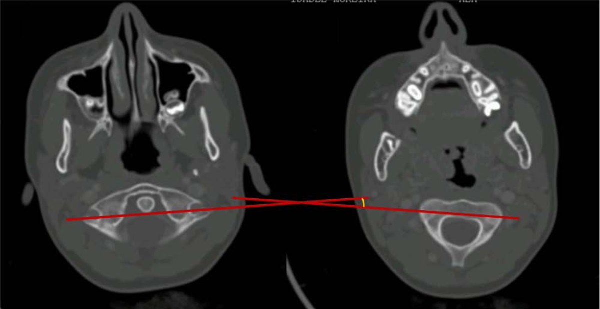
The patient’s fever did not recur in the postoperative period, but his clinical status progressively deteriorated. On postoperative day 6, he became unconscious with a Glasgow coma score of 7 (eye-opening to pain 2, verbal response none and motor response 4). On postoperative day 10, he developed status epilepticus, requiring intubation and mechanical ventilation. A noncontrast computed tomography (CT) of the brain on postoperative day 14 revealed a partially removed calcific mass located in the midline and slightly to the left. Diffuse gyral edema was evident in both cerebral hemispheres. Brain death occurred 11 weeks after admission to the hospital. The patient died 3 days later.
B. mandrillaris is a free-living ameba naturally found in the environment. It can cause rare cases of human disease, including cutaneous and CNS disease called GAE.1,2 The true incidence of this infection is unknown and data about the epidemiology of Balamuthia infections have been derived from small case series and individual case reports.2,3 More than 200 cases of Balamuthia disease have been reported worldwide since 1989.4 Infection can occur in both immunocompetent and immunocompromised patients and has been associated with agricultural exposure and activities such as swimming and gardening.2,4 Balamuthia enter the human body through a lesion in the skin or via the lungs by inhalation of cysts. The incubation period of Balamuthia GAE is unknown but is believed to range from several weeks to months.1
Balamuthia infection can present with a local skin lesion, isolated brain involvement or disseminated disease affecting the brain, skin and other organs.2,4 Although not common, some patients with Balamuthia GAE may develop an initial skin lesion 1 month to 2 years before the development of neurologic symptoms.1,4Balamuthia encephalitis has an insidious onset and develops as a subacute or chronic disease. Initial symptoms may include fever, headache, vomiting, stiff neck, personality changes, confusion, lethargy, seizures, hemiparesis, aphasia, cranial nerve palsies, diplopia and/or dizziness.1–4 Fever often is low grade and intermittent.1 CNS symptoms are generally related to bleeding, infarction and necrosis.5 Signs of increased intracranial pressure may be present, followed by meningeal symptoms and progressive loss of consciousness. The clinical course may resemble that of a brain tumor or a brain abscess.3
CSF indices may demonstrate lymphocytic pleocytosis and increased protein concentration, with normal to low glucose concentration, but Balamuthia organisms are rarely seen in the CSF.1,2,4 In our patient, lumbar puncture was not performed due to the risk of cerebral herniation. CSF samples were obtained from EVD during the postoperative period, and therefore, CSF cell count and chemistries could not be interpreted clearly. CT or MRI of the brain may show single or multiple space-occupying or ring-enhancing lesions. Hydrocephalus or edema may also be present.1,4,6 These findings may cause patients to be initially misdiagnosed as having a brain tumor, as in our patient.
Confirmation of Balamuthia species can be made by direct immunofluorescence, immunoperoxidase staining or real-time PCR in a reference laboratory.4 Brain biopsy typically shows granulomas with foamy macrophages, multinucleated giant cells accompanied by lymphocytes. Amebae tend to settle in the vascular walls of capillaries and venules, and vascular damage may be present as tissue bleeding. Neutrophils may predominate, and amebae may be seen.7
The optimal treatment regimen and duration for B. mandrillaris infection have not been established;1,2,4 experience is based on a limited number of case reports2,8–11 and in vitro drug activity.12 The Centers for Disease Control and Prevention currently recommends the following multidrug regimen: pentamidine, sulfadiazine, flucytosine, fluconazole plus either azithromycin or clarithromycin.4 Additionally, miltefosine has shown good in vitro activity against B.mandrillaris,12 and has been used successfully in combination therapy of Balamuthia infection.2,11 Unfortunately, our patient died shortly after the diagnosis of Balamuthia GAE was confirmed, so the treatment regimen recommended by the Centers for Disease Control and Prevention could not be applied. Surgical resection of the CNS lesions can also play a critical role in the rapid diagnosis and treatment of Balamuthia GAE.13 Despite treatment, mortality is high for Balamuthia infections and patients generally die several weeks to months after the onset of symptoms.1,14
In conclusion, Balamuthia GAE should be kept in mind in patients presenting with subacute or chronic meningoencephalitis and intracerebral lesions resembling brain tumors. However, even with early diagnosis, the prognosis remains quite poor.
REFERENCES 1. Tan TQ. Naegleria, Acanthamoeba, and Balamuthia Infections. In: Cherry JD, Harrison GJ, Kaplan SL, Steinbach WJ, Hotez PJ, eds. Feigin and Cherry’s Textbook of Pediatric Infectious Diseases. 8th ed. Philadelphia: Elsevier; 2019:2199–2208. 2. Cope JR, Landa J, Nethercut H, et al. The epidemiology and clinical features of Balamuthia mandrillaris disease in the United States, 1974-2016. Clin Infect Dis. 2019;68:1815–1822. 3. Schuster FL, Yagi S, Gavali S, et al. Under the radar: balamuthia amebic encephalitis. Clin Infect Dis. 2009;48:879–887. 4. Centers for Disease Control and Prevention. Parasites - Balamuthia mandrillaris - Granulomatous Amebic Encephalitis (GAE). Available at: http://www.cdc.gov/parasites/balamuthia/health_professionals/index.html. Accessed on July 1, 2023. 5. Recavarren-Arce S, Velarde C, Gotuzzo E, et al. Amoeba angeitic lesions of the central nervous system in Balamuthia mandrilaris amoebiasis. Hum Pathol. 1999;30:269–273. 6. Healy JF. Balamuthia amebic encephalitis: radiographic and pathologic findings. AJNR Am J Neuroradiol. 2002;23:486–489. 7. Guarner J, Bartlett J, Shieh WJ, et al. Histopathologic spectrum and immunohistochemical diagnosis of amebic meningoencephalitis. Mod Pathol. 2007;20:1230–1237. 8. Cary LC, Maul E, Potter C, et al. Balamuthia mandrillaris meningoencephalitis: survival of a pediatric patient. Pediatrics. 2010;125:e699–e703. 9. Yi Z, Zhong J, Wu H, et al. Balamuthia mandrillaris encephalitis in a child: case report and literature review. Diagn Microbiol Infect Dis. 2021;100:115180. 10. Deetz TR, Sawyer MH, Billman G, et al. Successful treatment of Balamuthia amoebic encephalitis: presentation of 2 cases. Clin Infect Dis. 2003;37:1304–1312. 11. Martínez DY, Seas C, Bravo F, et al. Successful treatment of Balamuthia mandrillaris amoebic infection with extensive neurological and cutaneous involvement. Clin Infect Dis. 2010;51:e7–11. 12. Schuster FL, Guglielmo BJ, Visvesvara GS. In-vitro activity of miltefosine and voriconazole on clinical isolates of free-living amebas: Balamuthia mandrillaris, Acanthamoeba spp., and Naegleria fowleri. J Eukaryot Microbiol. 2006;53:121–126. 13. Orozco L, Hanigan W, Khan M, et al. Neurosurgical intervention in the diagnosis and treatment of Balamuthia mandrillaris encephalitis. J Neurosurg. 2011;115:636–640. 14. Bakardjiev A, Azimi PH, Ashouri N, et al. Amebic encephalitis caused by Balamuthia mandrillaris: report of four cases. Pediatr Infect Dis J. 2003;22:447–453.
留言 (0)