
記住我


Distal femoral fractures are rare, accounting for <1% of all fractures, and ~3% to 6% of all femur fractures.1,2 Distal femur fractures can be difficult fractures to treat owing to challenges relating to poor bone quality, the need for sufficient bone stock for distal fixation, and pre-existing hardware in the case of periprosthetic fractures. These fractures are typically treated with lateral plating—both locked and unlocked (rarely used), retrograde intramedullary nails, a combined nail and plate fixation, or in cases where fixation is not possible—a distal femoral replacement.3 The lateral locked plate remains the workhorse for the fixation of these complex injuries.4,5
The development of locked lateral plates has been beneficial in preserving periosteal blood supply and providing more secure, angle-stable fixation in an osteoporotic setting. However, the rigidity of the fixation may increase the risk of nonunion.6,7–12 Bicondylar fractures may require dual plates to support both columns. A retrograde nail has the advantage of being a load-sharing device, which can allow for earlier weight-bearing. The disadvantage of these plating and retrograde nailing techniques is that there is often inadequate bone stock for fixation. This is particularly true in very distal femur fractures, periprosthetic fractures, and when the skeleton is small, either because of dysplasia or immaturity. The presence of osteoporosis further compromises the quality of fixation. In addition, in very distal fractures, femoral condylar locked plates may only allow for 2 points of distal fixation. Lateral-locked plates also cannot be used to treat medial-sided fractures as they do not fit. Lastly, these plates are not malleable, and thus cannot be contoured to fit the specific anatomy and fracture pattern of each individual patient.
In our center, we have developed a technique for treating distal femur fractures with a periarticular tibial plate. This technique is used infrequently in carefully indicated patients, in fewer than 5% of patients. We select these plates when the distal fragment appears <2 to 3 cm and planning suggests we will potentially only get 2 locked screws from a standard lateral supracondylar plate. In addition, those distal fractures that are primarily medially based, such as those with a medial spike, and are best treated with medial plating, are ideal candidates for these plates. The included plates are a plateau plate fixed upside down or a pilon or distal medial tibial plate contoured to the distal femur and we choose our plates based on which one fits the patient’s anatomy best. For a predominately laterally based fracture, this plate is placed laterally in a similar manner to other fixed angle devices alone or in combination with a medial plate. The advantages of this technique are its versatility across a wide range of ages and fracture patterns, the ability to get multiple points of fixation in very distal fractures, and the low profile of the plates, which can be useful in periprosthetic fractures and skeletally small individuals. The objective of this report is to introduce to the reader that there are other options available from routine orthopedic inventory for plate fixation for these fractures when a conventional lateral locked plate is not feasible.
TECHNIQUEOnce a distal femur fracture is identified on orthogonal radiographs, we routinely obtain computed tomography scans with three-dimensional reconstruction to further assess the fracture pattern, extent of articular involvement, and the available bone stock.
Patients are placed supine on a radiolucent operating room table with a bump under the hip to avoid external rotation of the lower extremity. A radiolucent triangle is often used as an aide for positioning. The periarticular tibial plates we utilize are Synthes proximal tibia locking, plates, Zimmer pilon plates, and Synthes pilon plates.
For Medial Plating ApproachA standard medial parapatellar incision is made from the level of the joint extending proximally for 15 to 18 cm. A formal medial arthrotomy is made and the vastus medialis muscle belly is retracted anteriorly. This retraction and dissection are extended proximally, carefully separating the muscular layers from the medial longitudinal fascia until an adequate femoral shaft is visualized to allow a minimum of 6 holes through the plate on the intact proximal diaphysis. We use 6 holes above the fracture to improve the working length and to span at least 2.5 times the diameter of the bone above the fracture site. Typically, the screws used in the diaphysis are 3.5 mm screws. Proximal dissection is limited by the adductor hiatus, where the femoral vessels cross posteriorly. By definition, this means that only the distal 12 to 15 cm of the femur are easily accessible using this approach. Reduction and fixation proceed as on the lateral side. On the medial side, periarticular plates usually need slight rotational contouring only.
For Lateral PlatingA lateral incision is made over the distal thigh, curved anteriorly towards the tibial tubercle. The iliotibial band is incised in line with the skin incision. A subvastus approach is made to the distal femur and perforating vessels are ligated as needed. Lateral parapatellar arthrotomy is made based on the fracture pattern. Fracture is reduced, and temporary reduction is held with K-wires, Schantz pins, or pointed reduction clamps as dictated by the fracture anatomy. The articular block is now fixed with 3.5 mm or 4.0 mm screws planned in a way to keep the fixation away from the proposed plate. For the lateral condyle, typically the proximal tibial plateau plate will need to be contoured to fit the patient’s anatomy. Typically locking screws are used distally and cortical screws proximally to finish the construction. After intraoperative films confirm the appropriate position of the implant and anatomic reduction, the wound is closed in a standard manner.
Typically, patients are non–weight-bearing in a knee immobilizer from surgery until week 2 or 3. Graduated increase of motion begins at week 3 until full range of motion by week 8 depending on the soft tissue condition and wound healing. Patients are partial weight-bearing from week 8 to week 12 and full weight-bearing by 3 months postoperatively.
EXPECTED OUTCOMESAfter Institutional Review Board approval, all patients with distal femur fractures treated with periarticular tibial plates were reviewed. Records were searched from 2010 to 2020 and patients were included if they had achieved radiographic healing or if they had at least 6 months of follow-up. Data were pulled from a trauma registry, which includes the type of implant used for the fixation of a particular fracture. Data collected included: patient demographics, medical comorbidities, history of prior ipsilateral limb surgeries, Orthopedic Trauma Association (OTA) fracture classification, implant information, medial versus lateral plating, time to healing, complications, and final ambulatory status.
Periarticular tibial plates were used in 19 distal femur fractures in 18 patients. Seven patients with <6 months of follow-up who had not achieved full radiographic healing were excluded. Twelve fractures in 11 patients who had achieved radiographic healing or who had at least 6 months follow-up were included.
Six patients were males and 5 patients were females. Patient characteristics are summarized in Table 1. Five fractures occurred in patients younger than 18 years old (Fig. 1) and 3 fractures occurred in patients over age 65. The average age was 33.2 years (range: 10 to 81). There were 2 Salter-Harris II fractures, and 3 extra-articular fractures consistent with OTA type 33A fractures. There were 2 partial articular fractures consistent with OTA type 33B fractures. There were 3 complete articular fractures consistent with OTA type 33C fractures (Fig. 2). There were 2 periprosthetic fractures associated with total knee arthroplasties (Fig. 3). Three patients had prior surgery relating to the distal femur, including 2 total knee arthroplasties and 1 intramedullary nail. Three patients were staged with an external fixator before definitive plating.
TABLE 1 - Summary of Distal Femur Fracture Cases Treated with Periarticular Tibal Locking Plates Sex Age Implant Implant location Fracture description Prior surgery Weeks to healing Months of follow-up Complications Ambulatory status at final follow-up Male 10 Distal medial tibial plate Lateral 33A No 18.9 25.1 Planned removal of implants Full Male 14 Tibial plateau plate Lateral Salter-Harris II No 24.1 6.9 Planned removal of implants Full Female 67 Tibial plateau plate Medial Periprosthetic fracture Total knee arthroplasty 14.1 41.9 None Full Male 12 Tibial plateau plate Lateral 33A External fixator 16.1 13.2 Multiple irrigation and debridement, grafting with a cell, skin grafting Full Male 12 Tibial plateau plate Lateral 33A External fixator 16.1 13.2 Full Female 73 Tibial plateau plate Lateral 33C None 18.9 7.3 None Cane (per baseline) Female 64 Tibial plateau plate Lateral 33B None 13.1 3 None Walker Female 56 Tibial plateau plate/pilon plate Dual Periprosthetic fracture Total knee arthroplasty 33.9 7.8 None Full Male 14 Posteromedial distal tibia plate Lateral Salter-Harris II None 16.1 16 Mild valgus deformity Full Male 58 Tibial plateau plate Lateral 33B Intramedullary nail 13.1 3.0 None Full Female 81 Pilon plate/distal medial tibial plate Dual 33C None 26.1 6.0 None Full Male 26 Pilon plate/pilon plate Dual 33C External fixator 28.7 14.9 None Full FIGURE 1:
 Twelve-year-old boy, 6 months postoperatively demonstrates a healed Salter-Harris II fracture treated with an upside-down lateral tibial plateau plate.
 FIGURE 2:
 Eighty-one–year-old with a T-type supracondylar femur fracture ~6 months postoperatively, demonstrating healed fracture treated with a pilon plate (lateral side) and an upside-down distal medial tibial plate used on the medial distal femur.
 FIGURE 3:
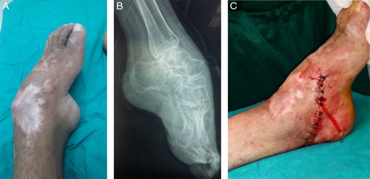
 Fifty-six–year-old with a periprosthetic distal femur fracture ~8 months postoperatively, demonstrating healed fracture with treated with a pilon plate (lateral side) and an upside-down tibial plateau plate (medial side).
Eight fractures were treated with tibial plateau plates. Four patients were treated with pilon plates and 3 patients were treated with distal tibia plates. The distal tibia plates were 2 distal medial tibial plates and 1 posteromedial tibial plate. Three patients were treated with dual plates, including a tibial plateau plate and a pilon plate, 2 pilon plates, and a distal medial tibial plate and a pilon plate.
The average follow-up was 12.2 months (range: 3.0 to 41.9 mo). All patients achieved radiographic healing by final follow-up. The average time to healing was 18.4 weeks. Nine patients were fully weight-bearing without assistive devices at the final follow-up. Two patients were using assistive devices as per their prefracture ambulatory baseline. One patient was using a walker for balance at the final follow-up of 3 months and declined further follow-up because she was doing well. Thus, it is unknown if she progressed in her ambulatory status beyond this point.
COMPLICATIONSWith regards to complications, one patient with Becker muscular dystrophy underwent manipulation under anesthesia for knee stiffness. One patient with a Salter-Harris II fracture had a mild valgus deformity of his knee when he reached full skeletal maturity, but no treatment was deemed necessary. One patient with bilateral open femur fractures, on one side, underwent multiple debridement and irrigations, placement of xenograft biological dermal substitute, followed by skin grafting, and bilateral removal of external fixators. Two patients under the age of 18 underwent planned removal of implants after their fractures had healed.
REFERENCES 1. Court-Brown CM, Caesar B. Epidemiology of adult fractures: a review. Injury. 2006;37:691–697. 2. Martinet O, Cordey J, Harder Y, et al. The epidemiology of fractures of the distal femur. Injury. 2000;31(suppl 3):62. 3. Gwathmey FW, Jones-Quaidoo SM, Kahler D, et al. Distal femoral fractures: current concepts. J Am Acad Orthop Surg. 2010;18:597–607. 4. Haidukewych G, Sems SA, Huebner D, et al. Results of polyaxial locked-plate fixation of periarticular fractures of the knee. J Bone Joint Surg Am. 2007;89:614–620. 5. Kregor PJ, Stannard JA, Zlowodzki M, et al. Treatment of distal femur fractures using the less invasive stabilization system: surgical experience and early clinical results in 103 fractures. J Orthop Trauma. 2004;18:509–520. 6. Gangavalli AK, Nwachuku CO. Management of distal femur fractures in adults: an overview of options. Orthop Clin North Am. 2016;47:85–96. 7. Lujan TJ, Henderson CE, Madey SM, et al. Locked plating of distal femur fractures leads to inconsistent and asymmetric callus formation. J Orthop Trauma. 2010;24:156–162. 8. Henderson CE, Lujan TJ, Kuhl LL, et al. 2010 Mid-America Orthopaedic Association Physician in training award: healing complications are common after locked plating for distal femur fractures. Clin Orthop Relat Res. 2011;469:1757–1765. 9. Vallier HA, Immler W. Comparison of the 95-degree angled blade plate and the locking condylar plate for the treatment of distal femoral fractures. J Orthop Trauma. 2012;26:327–332. 10. Large TM, Kellam JF, Bosse MJ, et al. Locked plating of supracondylar periprosthetic femur fractures. J Arthroplasty. 2008;23(6 suppl 1):115–120. 11. Wang MT, An VVG, Sivakumar BS. Non-union in lateral locked plating for distal femoral fractures: a systematic review. Injury. 2019;50:1790–1794. 12. Ricci WM, Streubel PN, Morshed S, et al. Risk factors for failure of locked plate fixation of distal femur fractures: an analysis of 335 cases. J Orthop Trauma. 2014;28:83–89.
留言 (0)