Ethics and participants
All clinical samples were collected from Lvliang in Shanxi province in China with signed informed consents from patients and their families. The protocol was approved by the Ethics Board of Capital Institute of Pediatrics. All animal experiments were legally performed under "Principles for Utilization and Care of Vertebrate Animals".
Nanostring
NanoString is a high-throughput method for RNA expression detection. The total RNA of human brain tissue was extracted and detected by NanoString nCounter. First, gene-specific probes were designed. Sample lysate was then incubated and hybridized with the probes. The probes were designed by NanoString Technologies and hybridized according to the nCounter Element 24-plex Assay Manual. The hybridization solution was incubated at 65 ºC for 20 h. The remaining hybridization mixture was added to Cartrige through a series of hybridization and elution processes, and then the cartrige was placed on an optical scanner. Data were filtered using quality control (QC) criteria according to the manufacturer's recommendations. GAPDH, CLTC, and GUSB were used as internal controls to normalize the raw counts to pass QC. The data used for analysis were transformed by log2.
Animals
C57BL/6 mice (6–8 weeks, 18–20 g) were purchased from Charles River Laboratory, and fed in a specific pathogen-free environment under a 12-h light/dark cycle. Female C57BL/6 mice were fed on low-folate diet for 4 weeks. Sexually matured individuals were mated overnight in the morning at 8:00 am. This time was seen as embryonic day (E) 0.5. On E7.5, mice were intraperitoneally injected with 1.5 mg/per kg of MTX (Sigma, USA). Pregnant mice were killed, and the dissected embryos were stored in liquid nitrogen. Animal handling was compliant with institutional guidelines under the care of experimental animals.
Cell culture
Sv/129 mouse embryonic stem cells (mESCs) were obtained from Capital Institute of Pediatrics central laboratory and cultured in 15% fetal bovine serum (FBS), DMEM (Gibco), non-essential amino acids (Invitrogen), 0.1 mM glutamine (Invitrogen), and 1,000 U/ml mouse leukemia inhibitory factor(LIF) (Millipore, Billerica, USA). Cells were cultured in 0.2% gelatin-coated T25 bottles and incubated in an incubator at 37 ºC 5% CO2, and the medium was changed every day. Cells were cultured under normal medium for few days before 1uM methotrexate (MTX) was applied for 24 h.
Western blotting
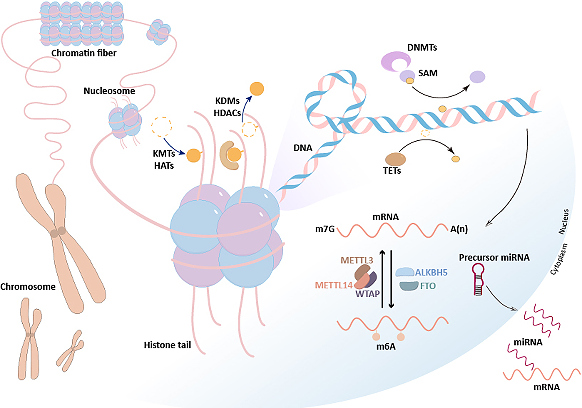
Histone used in this paper were all obtained by acid extraction. We used 12% SDS-PAGE to isolate 5ug of histones and detected the changes in the target protein. Protein was transferred to an immobilon-NC membrane (Merck Millipore, Ireland) by electroporation, and the membranes were incubated with the primary antibody, anti-Histone H3 (di methyl K79) antibody (1: 1000, abcam, Cambridge, UK), DOT1L (D1W4Z) Rabbit mAb (1: 1000, Cell Signaling Technology, USA), and Anti-Histone H3 antibody (1: 5000, Abcam, Cambridge, UK) and stored under four-degree refrigerator overnight. Membranes were washed with PBST for three times on the next day and incubated in secondary Peroxidase-conjugated goat anti-rabbit IgG antibody (1:5000, ZSGB-BIO, China) for 40 min.
ChIP-Seq and data analysis
107 mESC cells were used to obtain DNA with a concentration of 1 ng/ul and fragments were digested into100-500 bp for CHIP(chromatin immunoprecipitation) sequencing. SimpleChip KIT (Cell Signaling Technology, America). CHIP-Seq libraries were prepared according to the protocol and sequenced using Illumina NovaSeq 6000. After passing the quality check, all reads are mapped to mouse genome using Bowtie2 with default parameters. Two mismatches cannot appear in one cohort. The peak of the H3K79me2 on the genome was determined using MACS2 with a false detection rate of 0.05. Each peak-associated gene was identified by an internal program using Entrez gene annotation, where the Shh target gene was defined as a gene containing H3K79me2-peak in its transcription start site (TSS). The data set of H3K79me2 reads per kilobase per million (RPKM) values in the coding region of each gene was normalized.
Chromatin immunoprecipitation (ChIP) analysis
SimpleChip KIT (Cell Signaling Technology, America) was used. Chromatins were ultrasonicated for 100–500 bp DNA fragmentations, then immunoprecipitated with H3K79me2 XP Rabbit mouse antibody (CST, USA). The purified DNA was detected by QuantStudio 7 Flex with SYBR Green by RT-qPCR. The primers used for RT-qPCR are shown in Additional file 1: Table S1. Normal Rabbit IgG was used as a negative control.
RT-qPCR
Trizol was used to extract mRNA from mouse cranial neural tissues and cells. We used the RevertAid First Strand cDNA Synthesis Kit (Thermo, USA) to synthesize the single strand cDNA, followed by Maxima SYBR Green / ROX qPCR Master Mix (Thermo, USA) for subsequent qPCR experiments. The qPCR setup procedure is as follows: cDNA was initially denatured at 95 ºC for 3 min with 1 cycle, then held for 30 s, Annealing temperature was raised to 50 ºC for 30 s and elongated at 72 ºC, for 1 min, number of cycles from denaturation to extension was 40. Finial extension lasted for 5 min at 72 ºC. Primer sequences are shown in Additional file 1: Table S1.
Small interfering RNA (siRNA)
siRNA negative control, AUGUAUUGGCCUGUAUUAG.
DOT1L siRNA, GCUAUGGAGAAUUACGUUU.
Immunofluorescence
Cells were fixed in 4% paraformaldehyde and placed at room temperature for 10 min, then 1 ml of 0.1% Triton X-100 in PBS was added and shook on a shaker for 5 min. Cells were then blocked with 5% BSA blocking solution for 1 h at room temperature. Cells was incubated with primary antibodies DOT1L and H3K79me2 (1:50; CST) (1:50; Santa Cruz) and placed in a 4-degree refrigerator overnight. Followed by incubation with Alexa Fluor-conjugated secondary antibodies (1: 200; CST, USA) for 1 h at 37° C. DAPI staining was then added for 10 min. Images were observed under a Leica laser scanning confocal microscope. Colocalization values of DOT1L and H3K79me2 were calculated using FV10-ASW 3.0 Viewer.
Statistical analysis
All statistical analyses were performed with SPSS software, version 19.0. Student-t test was used to calculate means and standard deviation. Statistically significant is when p-value is under 0.05.


留言 (0)