
記住我


Long bone fractures are significant injuries with the potential to cause dramatic physical impairment.1 Despite improvements in surgical management, rates of nonunion, and malunion in long bone fractures remain high and are associated with significant morbidity including need for surgical revision, increased use of opioid medications, increased utilization of physical therapy, and increased overall cost for fracture related medical care.1–3 In response to these challenges, various surgical and biological approaches have been described to improve treatment options. Among biological options, bone autograft is widely considered the gold standard as it contains all the properties desired of graft material: osteoconduction, osteoinduction, and osteogenesis. These properties are the 3 key factors required for bone healing as described in the “diamond concept.”4 Bone autograft harvest is not without risk, primarily from donor site morbidity and limited amount/volume of bone available.2 Various attempts have been made to circumvent the limitations of traditional autograft harvesting, including the development of the Reamer Irrigator and Aspirator 2 (RIA 2) system (DePuy-Synthes). This RIA 2 system has been shown to have similar fusion rates and fewer complications compared with traditional iliac crest harvesting.3,5 Here we present a novel bone grafting technique in a patient with a complex nonhealing distal tibia pilon fracture utilizing the RIA 2 system to harvest ipsilateral tibial intramedullary bone graft and percutaneously deliver the graft to the distal fracture site through the intramedullary canal, avoiding the need for a separate surgical dissection and additional soft tissue disruption.
TECHNIQUEWhat follows is an illustrative example of the technique involving a nonhealing grade 43C1 distal tibia fracture with intra-articular extension 9 months after initial staged open reduction and internal fixation.
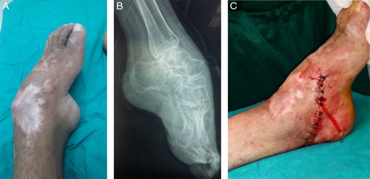
A 66-year-old otherwise healthy male presented 4 weeks after falling from a ladder with an open distal tibia pilon fracture (AO 43C1) which had previously undergone irrigation, debridement and external fixator placement at an outside hospital. He was subsequently treated with open reduction and internal fixation through a posterolateral approach and skin grafting to his anterior wound. Follow-up was concerning for poor fracture healing and delayed union of the metadiaphysis, but the patient elected to continue conservative treatment. The articular surface had successfully healed. At 8 months postoperatively, the patient complained of new onset pain with weight bearing. Radiographs in the office showed persistent metadiaphyseal fracture nonunion and hardware failure (Fig. 1). A computed tomography scan was obtained confirming the presence of a nonunion. Inflammatory labs at the time of this follow-up were all within normal limits. The patient was brought to the operating room for hardware removal, cultures of the nonunion, autologous bone grafting with Reamed Irrigator Aspirator 2 System (RIA 2) and placement of a circular external fixator.
 FIGURE 1:
 Anteroposterior and lateral radiograph of the right ankle showing nonunion of the tibia with hardware failure.
After removal of all prior hardware, the patient’s tibia was sized using the radiographic ruler and a 12 mm RIA head was selected. Using a standard suprapatellar approach to the tibia we placed our initial guidewires. Under fluoroscopic guidance the RIA 2 system was passed through the distal tibia and fracture site, thereby freshening the fracture ends. After thoroughly reaming the canal, we had a large amount of bone graft which we packed into the tubing of the RIA 2 system after removing the reamer head and cutting off the metal suction tip. This resulted in a compact tube of graft ideally sized for the reamed canal. We then inserted the tubing with graft into the reamed canal, and under fluoroscopic guidance introduced the graft at the fracture site utilizing the RIA 2 trocar to push the graft out of the tube into the fracture bed (Fig. 2). An osteotomy of the fibula was then performed and a circular external fixation was applied to compress the fracture site.
 FIGURE 2:
 Intraoperative images and fluoroscopy showing an anteroposterior view of the right ankle. Panel 1: Intraoperative photography showing harvested bone graft packed into the Reamer Irrigator and Aspirator (RIA)2 tubing. Panel 2: Removal of hardware. Panel 3: Bone graft in RIA tubing in the nonunion site. Panel 4: Placement of bone graft using RIA tubing and trochar. Panel 5: Presence of bone graft in tibia nonunion after removal of RIA tubing and trochar.
The patient was followed postoperatively. A computed tomography was performed at 12 weeks postopeartively showing bridging callus over nonunion. The external fixator was subsequently removed, and at last follow-up the patient was ambulating well. Images almost 9 months after grafting shows complete union of the tibia and fibula (Fig. 3).
 FIGURE 3:
 Anteroposterior and lateral radiographs 8 months after grafting procedure showing fracture union of the tibia and fibula.
EXPECTED OUTCOMESOur novel approach utilizing the RIA 2 system for enhanced intramedullary bone graft delivery can be applied to almost all long bone fractures, but in our trial patient the strength of our technique is particularly apparent. First, the patient has a complex open intra-articular distal tibia fracture. Despite years of evolving treatment approaches, this fracture remains a challenging case, and is frequently associated with complications. Second, our patient previously required a skin graft over the anterior tibia indicating poor healing potential and increasing the risk of complication following a secondary dissection for open graft placement. Through our technique, we were able to introduce a significant amount of bone graft without the morbidity associated with an incision in the area of grafted skin which would have been required through a traditional open graft placement.
Regarding long bone fractures in general, our technique reduces the technical challenge of introducing bone graft to distant fracture sites. Instead it relies on readily available systems to circumvent many of the problems, most notably donor site pain, with performing a traditional autograft with open graft placement and limited graft volume. Utilization of the RIA 2 tubing and trocar as a vehicle for graft delivery simplifies the process of graft placement without the need for additional equipment. Repurposing of equipment saves time and reduces cost in the operating room without increasing the technical difficulty of the procedure. In addition, this technique allows for the delivery of substantial quantities of bone graft as is radiologically evident on intraoperative x-ray.
COMPLICATIONSAs with any repair of nonunion there is always the potential for complications. This technique should be avoided with articular nonunions, to prevent inadvertent graft placement in the joint, as well as in the setting of active infection, to prevent propagating the infection throughout the tibia and knee joint. The bone graft can be challenging to visualize during delivery. Omnipaque could potentially be added to the graft to assist with visualization. As with any autografting procedure, the amount of bone graft obtained may be insufficient. Additional allograft bone can be added to augment the autograft bone if inadequate autograft bone is obtained. Although the use of the plastic sleeve is very effective for delivery of the graft, there is a potential of the plastic becoming entrapped in the medullary canal. This would require performing an open approach at the level of the nonunion to free or retrieve the plastic tubing. As with any treatment for nonunion, there is the potential of bone graft resorption and failure of fracture healing which would require repeat grafting with an open approach.
REFERENCES 1. Johnson L, Igoe E, Kleftouris G, et al. Physical health and psychological outcomes in adult patients with long-bone fracture non-unions: evidence today. J Clin Med. 2019;8:1998. 2. Marongiu G, Dolci A, Verona M, et al. The biology and treatment of acute long-bones diaphyseal fractures: overview of the current options for bone healing enhancement. Bone Rep. 2020;12:100249. 3. Antonova E, Le TK, Burge R, et al. Tibia shaft fractures: costly burden of nonunions. BMC Musculoskelet Disord. 2013;14:42. 4. Calori GM, Giannoudis PV. Enhancement of fracture healing with the diamond concept: the role of the biological chamber. Injury. 2011;42:1191–1193. 5. Nodzo SR, Kaplan NB, Hohman DW, et al. A radiographic and clinical comparison of reamer-irrigator-aspirator versus iliac crest bone graft in ankle arthrodesis. Int Orthop. 2014;38:1199–1203.
留言 (0)