Case 1
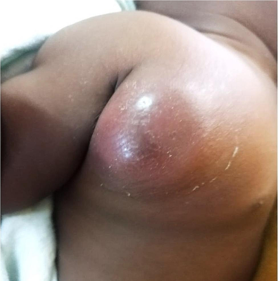
Thirty-year-old male without previous known medical history. He began on August 2022 with a single skin lesion in the glans penis. During the following days, similar lesions appeared throughout the trunk, neck, face, scalp, arms, and legs. After medical evaluation, he was started on valacyclovir, without clinical improvement. Positive skin PCR confirmed mpox. Two weeks later he developed facial edema, sialorrhea, poor oral intake, and worsening of the skin lesions, with phimosis and skin necrosis of the genital area.
On September 2022 he was referred to our Institution. On admission, he was afebrile, tachycardic (140 bpm), tachypneic (18 rpm), and hypoxemic (SpO2 89% on room air). Physical examination revealed a disseminated polymorphic dermatosis affecting all body segments (> 200 lesions) composed of giant ulcerative and crusted lesions with an edematous and indurated border surrounded by erythema; early lesions were pustules and umbilicated papules. HIV serology was positive (121 CD4+ cells/mm3 – 5% / viral load 312,077 copies/mL). HSV-1 coinfection was confirmed by PCR. Other sexually transmitted diseases were excluded.
Tracheal stricture and 2 pulmonary nodules were identified on admission computed tomography (CT) scan. Magnetic resonance imaging (MRI) of the pelvis showed signs of bilateral orchitis, urethritis, and superficial necrotic tissue around the penis body and glans. An emergency tracheostomy was performed to ensure airway patency. He was started on meropenem, vancomycin, acyclovir, and prophylactic trimethoprim-sulfamethoxazole. Six days later, after excluding opportunistic infections, antiretroviral therapy was initiated (bictegravir/tenofovir/emtricitabine).
Two weeks later, the patient´s clinical condition continued to deteriorate. Due to the unavailability of poxvirus antiviral agents, the local investigation board review (IRB) authorized the infusion of plasma from a previously healthy volunteer who had received two doses of the JYNNEOS vaccine > 28 days prior (the patient gave explicit consent for this extraordinary therapeutic measure). He received the infusion without complications but continued worsening thereafter (progression of skin lesions, preseptal cellulitis, increasing facial edema, acute kidney injury, neurologic deterioration, and respiratory failure). Subsequent chest CT revealed an increase in the size and number of pulmonary nodules. He was started on empiric liposomal amphotericin B, but he developed refractory shock and multiple organ failure (coagulopathy, elevated liver enzymes). He ultimately died four weeks after his admission. An extensive microbiological evaluation was negative for other pathogens. Serum PCR stayed positive for 28 days.
Case 2
Forty-four-year-old male with a past medical history of type 2 diabetes mellitus, with poor adherence to lifestyle changes and pharmacological treatment. He began on September 2022 with fever, malaise, myalgia, arthralgia, and disseminated rash characterized by vesicles, pustules, crusts, and eschar, as well as necrosis of the 5th left toe. Mpox was confirmed by PCR testing of skin lesions; he received symptomatic treatment.
On October 2022 he was referred to our Institution due to clinical worsening of the skin lesions. On admission, he was afebrile, tachycardic (110 bpm), and tachypneic (22 rpm), with normal oxygen saturation. Physical examination revealed a diffuse skin rash (> 100 lesions), with characteristic vesicular, pustular, umbilicated, and crusted lesions. The perianal region was notable for 2 ulcers with irregular borders. HIV serology was positive (106 CD4+ cells/mm3 – 6% / viral load 726,454 copies/mL). HSV-2 coinfection was confirmed by PCR. Other sexually transmitted diseases were excluded.
Multiple pulmonary nodules were identified by chest CT scan. An abdominopelvic CT scan revealed concentric thickening of the rectum with intramural collections, a perianal abscess, and a trans-sphincteric anal fistula. The patient was started on piperacillin-tazobactam, vancomycin, acyclovir, fluconazole, and prophylactic trimethoprim-sulfamethoxazole. Complete surgical drainage of the perianal abscess was performed, without complications. Six days after starting fluconazole, he presented with dysphagia. Upper endoscopy revealed elevated lesions with central umbilication in the distal esophagus, similar to those seen on the skin. No local treatment was applied, and he continued on fluconazole. Lung biopsy excluded infectious/malignant causes of the pulmonary nodules; mpox virus PCR was positive. After the exclusion of opportunistic infections, antiretroviral therapy was initiated (bictegravir/tenofovir/emtricitabine).
The patient´s skin lesions progressed to necrotic ulcerations in the following days. He developed acute kidney injury, respiratory failure, and distributive shock. Due to neurologic impairment, a lumbar puncture was performed. Cerebrospinal fluid (CSF) analysis was non-specific (WBC 0 mm3, RBC 465 cells/mL, protein 36.9 mg/dL, glucose 99 mg/dL); mpox virus PCR was positive. Despite orotracheal intubation, vasopressors, and renal replacement therapy, he developed multiple organ failure (coagulopathy, elevated liver enzymes, cardiac dysfunction) and died. An extensive microbiological evaluation was negative for other pathogens. Serum PCR stayed positive for 30 days.
Case 3
Thirty-seven-year-old male with recently diagnosed HIV infection (25 CD4+ cells/mm3 – 3% / viral load 31,385 copies/mL), and on < 1 month of antiretroviral treatment (bictegravir/tenofovir/emtricitabine). His past medical history was also positive for T. pallidum and C. trachomatis infections (previously treated). He began on September 2022, with localized dermatosis affecting the face, characterized by umbilicated papules.
On October 2022 he was referred to our Institution for dissemination of the dermatosis. On admission, he was afebrile, tachycardic (107 bpm), and without respiratory abnormalities. Physical examination revealed a disseminated polymorphic dermatosis (< 50 lesions) affecting all body segments, with central ulceration and crusting, indurated border, and surrounded by erythema. Oral candidiasis and necrotic proctitis were identified. The right hand and forearm were erythematous, with edema and disproportionate pain. CT imaging revealed cellulitis of the right distal forearm and hand. HSV-2 coinfection was confirmed by PCR. Other sexually transmitted diseases were excluded. The patient was started on piperacillin-tazobactam, vancomycin, fluconazole, doxycycline, acyclovir, and prophylactic trimethoprim-sulfamethoxazole.
He underwent anal canal surgical debridement and fasciotomy of the right forearm due to compartment syndrome. Notably, characteristic lesions were documented at surgical incision sites, which rapidly enlarged over the next days. Days later, he developed progressing respiratory distress. On head and neck CT, diffuse inflammation was observed in the buccopharyngeal space, with segmental obliteration of the glottis. He underwent emergency surgical tracheostomy, but complete occlusion of the airway conditioned severe hypoxemia and cardiac arrest. He died 21 days after admission. No opportunistic infections were identified. Serum PCR stayed positive for 21 days.
Case 4
Twenty-five-year-old male with HIV infection, on 6 months of antiretroviral treatment (68 CD4+ cells/mm3 – 7% / viral load 367 copies/mL). He was referred to our Institution on October 2022 for progressive mpox. Physical examination showed 2–3 cm round verrucous lesions with yellow crust, affecting the scalp, neck, torso, forearms, penis, and right foot. Soft tissue infection by methicillin-sensitive S. aureus was diagnosed. After a 2 week course of antibiotic therapy, he was discharged. Two months later he had no mpox lesions.
Case 5
Fifty-eight-year-old male with HIV infection, on > 6 months of antiretroviral treatment (177 CD4+ cells/mm3 – 8% / suppressed viral load). He was referred to our Institution on October 2022 to evaluate a large necrotic ulcer in the perianal region. Physical examination revealed a large ulcer (10 × 6 cm) in the perianal area with secondary bacterial infection, plus some disseminated crusted ulcers and umbilicated papules and vesicles, with necrotic center. Broad-spectrum antibiotics were started, and extensive surgical debridement was performed. Tissue cultures were positive for E. coli, E. faecium, and A. haemolyticum. After ten days of antibiotic therapy, he was discharged. Three months later, he still had mpox lesions (crusts) on the thighs, glutes, and perianal region. Virological assessment at that point was not made. Table 1 presents a detailed comparison of the clinical and molecular characteristics of the 5 cases described.
Table 1 Clinical and molecular characteristics of severely necrotic mpox in HIV-infected patients 

留言 (0)