
記住我


On March 4th, 2022, a 5-month-old male infant was admitted to the infant ward at Hospital Central de Maputo (HCM) with diagnoses of oral candidiasis, moderate anemia, and moderate acute malnutrition. He had been previously treated with nystatin at a local healthcare center without improvement. His immunizations were up to date per the Mozambique national schedule, including the BCG live-attenuated vaccine given in the right arm deltoid area at birth in South Africa. He had age-appropriate psychomotor development and maintained a diet of exclusive formula feeding since birth. His weight was 6 kg, with weight-for-age Z-score of -2, and weight-for-length Z-score of -2.2, indicating moderate acute malnutrition. The patient had no known TB exposures. His mother was a 29-year-old woman, diagnosed with HIV at her five-month antenatal visit and started ART at that time. Per the mother’s report, the patient received a DNA-PCR HIV test at one month of age in South Africa during November 2021. However, she returned to Mozambique before receiving the results. His initial treatment upon admission included therapeutic milk formula, fluconazole, and ceftriaxone.
Due to high suspicion of HIV and no record of previous test results from South Africa, a point-of-care nucleic acid test (PoC-NAT) was performed on March 7, 2022, which yielded a positive result, and was subsequently confirmed. He was given a WHO clinical HIV staging of III due to oral candida, anemia, and moderate acute malnutrition. Laboratory results of significance at this time included an absolute CD4 count of 955 cells/µL (15%) and a negative Xpert MTB/Rif® test performed on a gastric aspirate specimen. Admission full blood count had a total white blood cell count of 8,700/µL (neutrophils 21.3%; lymphocytes 57.0%), a hemoglobin of 9.7 g/dL, and a platelet count of 412,000/µL, He was started on the Mozambique standard pediatric first-line ART regimen (abacavir/lamivudine + dolutegravir dispersible tabs) on March 15, 2022 after inpatient extensive pre-ART counseling which included education on the signs and symptoms of BCG-IRIS. After 11 days of clinical improvement, he was discharged on ART, prophylactic cotrimoxazole, a multivitamin-anemia supplement, and follow-up scheduled at HCM.
On April 4th, three weeks after ART initiation, he presented to the emergency department at HCM with a one-week history of subjective intermittent fevers and progressive right axillary lymphadenopathy. Physical examination revealed a small ulcer over the right deltoid region and an enlarged right axillary lymph node without fluctuance or inflammatory signs. The child was then admitted for an inpatient work up. He was feeding well, had no cough or fever, and had no other complaints. The following day, he was discharged with a diagnosis of localized BCG-IRIS lymphadenitis without signs of disseminated BCG disease. The mother was advised to follow up as an outpatient until the resolution of the lesion while continuing ART, prophylactic cotrimoxazole, and the anemia-vitamin supplement.
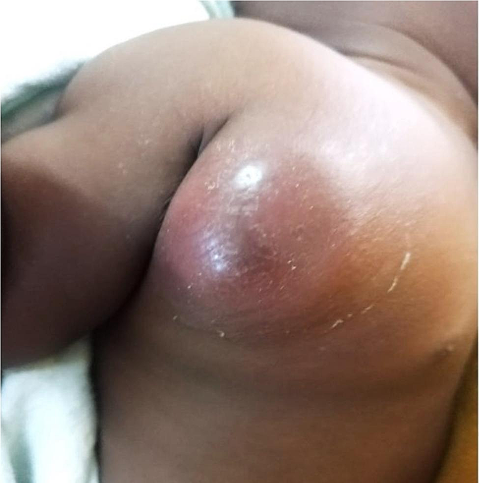
On April 14th, the patient returned with an enlarged, inflamed, and tender right axillary adenitis without fluctuance, spontaneous drainage, or signs of systemic BCG disease (Fig. 1). He was treated as an outpatient with paracetamol, ten days of oral prednisolone (2 mg/kg/day), and seven days of amoxicillin/clavulanate for possible bacterial superinfection.
Fig. 1
April 14th, 2022, ten days after initial presentation, and one month after ART initiation. Progression to localized inflammation and erythema with possible localized superinfection in the right axillary region.
On May 5th, at the following visit three weeks later, the child presented with five days of a completely eviscerated right axillary adenitis without fever or pain (Fig. 2). The mother confirmed good ART adherence since initiation. His laboratory results at this time were notable for an improved absolute CD4-count of 2,035 cells/µL (26%). The RNA-PCR viral load at this visit, which was less than two months since ART initiation, was 9,351 cp/mL with no baseline comparison since viral load testing is not routinely performed in Mozambique for new ART initiations. Other laboratory results were a white blood cell count of 18,100/µL (neutrophils 32.3%; lymphocytes 58.6%) and a hemoglobin of 9.4 g/dL. Pediatric surgery was consulted at this time and recommended against surgical intervention in favor of outpatient medical management due to concern about the increased risk of complications given the location and size of the mass. They advised treatment with topical silver nitrate with outpatient follow-up. After an additional consultation with a childhood TB expert in South Africa on May 17th, he was also treated topically with crushed INH tablets made into a paste while continuing silver nitrate. INH was initiated despite possible low-level INH resistance depending on the strain of M. bovis BCG he received with his birth BCG vaccine [11].
Fig. 2
Right axillary nodal evisceration and small right deltoid ulcer, seven weeks after ART initiation.
At a follow-up visit on June 3rd, approximately 2.5 months since ART initiation and two months after initial presentation with BCG-adenitis, there was a good clinical response to treatment with marked mass reduction, improvement of adenopathy and erythema, and correction of anemia (Fig. 3A). Despite improvement in the adenopathy, prednisolone 2 mg/kg/day was prescribed to further reduce the inflammation. On June 16th, he had further clinical improvement (Fig. 3B). At this visit, prednisolone treatment was continued. On July 4th, he weighed 8.2 kg (weight-for-length Z-score of 1.8), was asymptomatic, and had a largely healed lesion (Fig. 4A). A two-week taper of prednisolone was initiated, and the mother was advised to continue treatment with topical INH and silver nitrate. The patient’s wound was entirely closed on follow-up on July 18th at which time the topical treatments were discontinued, with complete recovery noted at a follow-up consultation on August 15th (Fig. 4B and C).
Fig. 3
Improved right axillary region suppuration and inflammation one month after treatment with topical isoniazid and oral prednisolone (A). Substantially improved inflammation with healing wound (B).
Fig. 4
Progression of wound healing during follow-up from July 4th (A), July 15th (B), and August 15th (C).
留言 (0)