
記住我
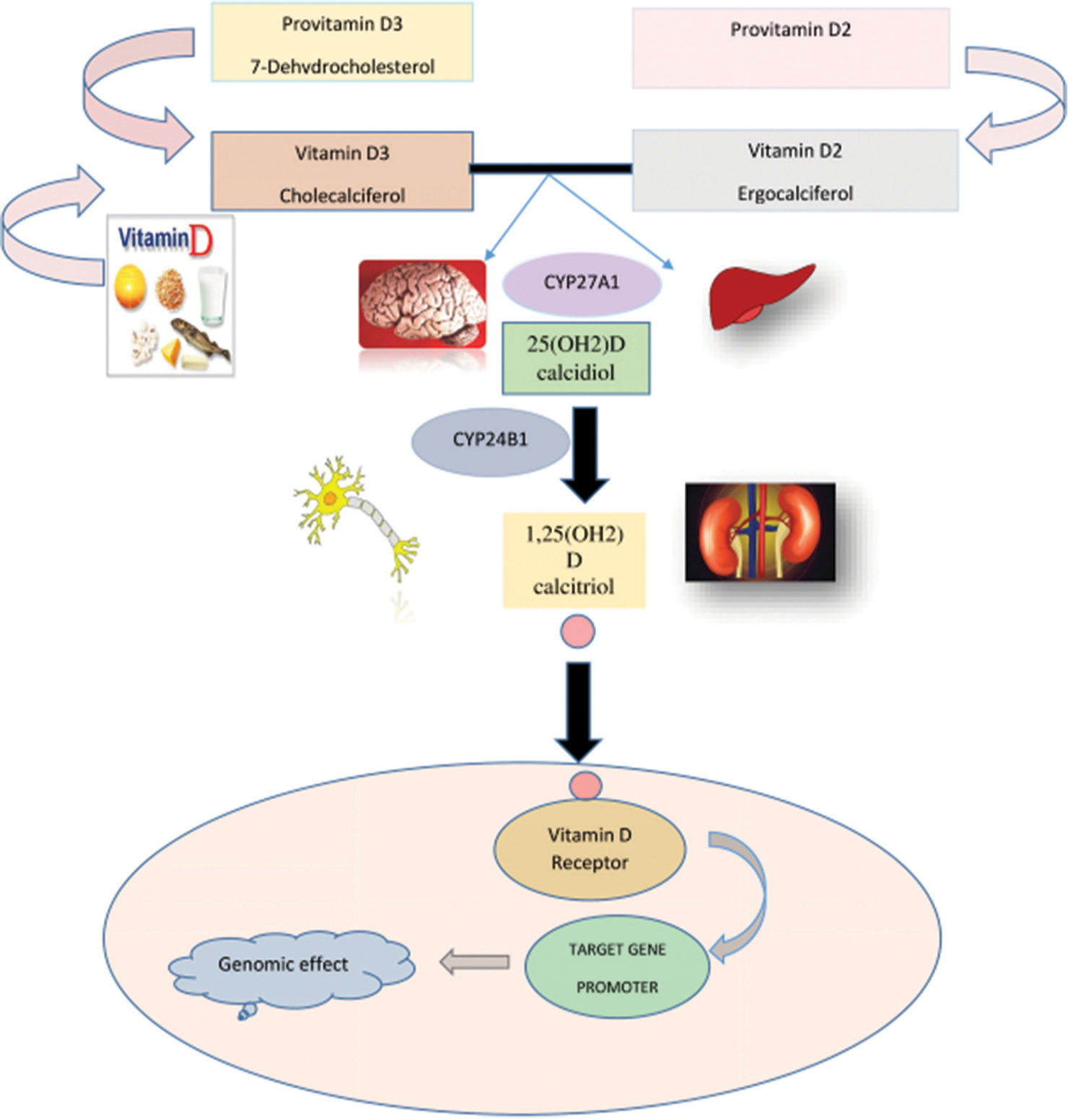
A lipid-soluble vitamin, Vitamin D (calciferol), belongs to the family of steroidal agents and is present in two different forms in plants and animals, namely, ergocalciferol (D2) and cholecalciferol (D3), respectively [Figure 1]. It acts as a hormone and affects different body organs with major actions on bones, intestines, and kidneys for the regulation of mineral homeostasis, specifically phosphorus, and calcium.[1] Calciferol is involved in numerous physiological functions and alterations in its concentrations and signaling pathways are associated with various pathological conditions.[2]
Export to PPT
Increasing evidence from recent research studies has pointed out the complex and crucial role of Vitamin D in the development and functioning of the nervous system including the brain, spinal cord, and related structures through modulation of many important processes including cellular proliferation, differentiation, immunoregulation, and neuroprotection.[3] The current review recaps the salient features of Vitamin D and revisits the evidence accrued thus far on its neurological functions and related clinical disorders [Table 1] An extensive literature exploration was carried out on this topic using databases, including Google Scholar, PubMed, Scopus, and Web of Science.
Table 1: Vitamin D in neurophysiology, neuropathology, and neurocognitive disorders.
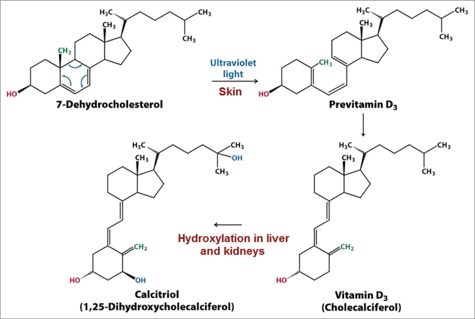
Physiological mechanism Pathological outcome in Vitamin D deficiency Associated clinical disease Neuroprotective function, regulation of gene expression, thrombolytic property, vasodilation, and antioxidant action Neurodisruption, vasoconstriction, and disintegration of blood-brain barrier Cerebrovascular accidents (Stroke) Clearance of amyloid plaque Accumulation of amyloid plaque Alzheimer’s disease Neuroprotection, anti-inflammatory effects, and anti-oxidant response Neuroinflammation, memory loss, and disorientation Cognitive dysfunction Restoration of dopamine levels Defective neurotransmission Parkinson’s disease Immunomodulation, modulation of interleukins and lymphocytes, and calcium homeostasis Immune dysregulation and calcium imbalance Multiple sclerosis Neurotrophic support and antioxidant activity Oxidative damage Amyotrophic lateral sclerosis VITAMIN D BIOCHEMISTRY Dietary sources and metabolic activationNaturally, Vitamin D is processed in the epidermis of the skin through the conversion of 7-dehydrocholesterol provided by the diet or produced endogenously in the presence of sunlight.
The dietary sources of Vitamin D include the yolk of eggs, different varieties of fish, margarine, fish oil, as well as Vitamin D-boosted products[2] [Figure 1]. The diet-obtained or cutaneous form of Vitamin D is inactive and requires two steps of hydroxylation. The steps primarily take place in the liver with the help of 25-hydroxylase, and in other organs including the prostate glands, kidneys, brain, placenta, and the immune system in the presence of 1-α-hydroxylase, forming active form of Vitamin D or 1,25-dihydroxycholecalciferol, also called calcitriol[2,4] [Figure 2]. In the serum, Vitamin D is predominantly present in the single hydroxylated form of 25-hydroxycholecalciferol. This form has less affinity for Vitamin D receptor (VDR) than the active 1,25-dihydroxycholecalciferol.[5] This hormone-like vitamin is associated with systemic disorders such as heart diseases, vascular problems, malignant tumors, brain hemorrhages, and diabetes mellitus to name a few. Vitamin D deficiency has also been associated with the impairment of cognition and dementia as well as psychiatric conditions such as psychosis and autism in addition to its well-established skeletal abnormalities including rickets and osteomalacia.[6] Hypervitaminosis D is not common but may cause severe health issues like hypercalcemia. The toxicity of Vitamin D occurs as a result of excessive ingestion of foods rich in Vitamin D or overdose with Vitamin D containing drugs either when administered repetitively or taken in a large dose.[7] The sign and symptoms resulting from Vitamin D toxicity are varied and typically include anxiety, confusion, gastric disturbances, cardiac impairment, and kidney dysfunction.[8]
Export to PPT
Receptor mechanismThe active form of Vitamin D exerts its function through genomic and non-genomic actions. In genomic actions, intracellular VDR that belongs to the nuclear receptors group is activated by Vitamin D to affect the target cell. VDR acts as a transcriptional factor and changes the expression of genes linked with different metabolic pathways. VDR has been found in almost all types of cells, which may elaborate its many functions in different tissues[9] [Figure 1]. 1,25-dihydroxycholecalciferol may act through another type of receptor present in the plasma membrane as membrane-associated rapid response steroid-binding (MARRS) receptors. These MARRS receptors help the hormonal form of Vitamin D in the regulation of calcium concentration in the cytosol.[2]
EFFECTS OF VITAMIN D ON NEUROLOGICAL HEALTHVitamin D is a neuroactive steroid that regulates various neurological functions in the body. Cholecalciferol and its metabolites are penetrable through the blood–brain barrier and can be metabolized locally due to the presence of VDR and enzyme CYP27B1 for the formation and CYP24A1 for the degradation of Vitamin D and its metabolites in neural cells.[10-12]
Location of receptorsThe receptors for Vitamin D, VDR, are present almost in every tissue of the human body and their distribution is similar to that seen in rats and hamsters.[3] These receptors are located in different parts of the brain and spinal cord including the hippocampus, hypothalamus, thalamus, ventricles, cortex, cerebellum, and substantia nigra. The localization of VDR in various brain areas has helped in the identification of specific roles of Vitamin D in various neurological functions.[13]
Neurological developmentVitamin D imparts great benefits to neurological development and growth.[14] In the human brain, production of the active form of Vitamin D occurs in the neonatal and adult brains due to the presence of the gene CYP27B1, which is responsible for the synthesis of the enzyme that converts the inactive form to the active form.[15,16]
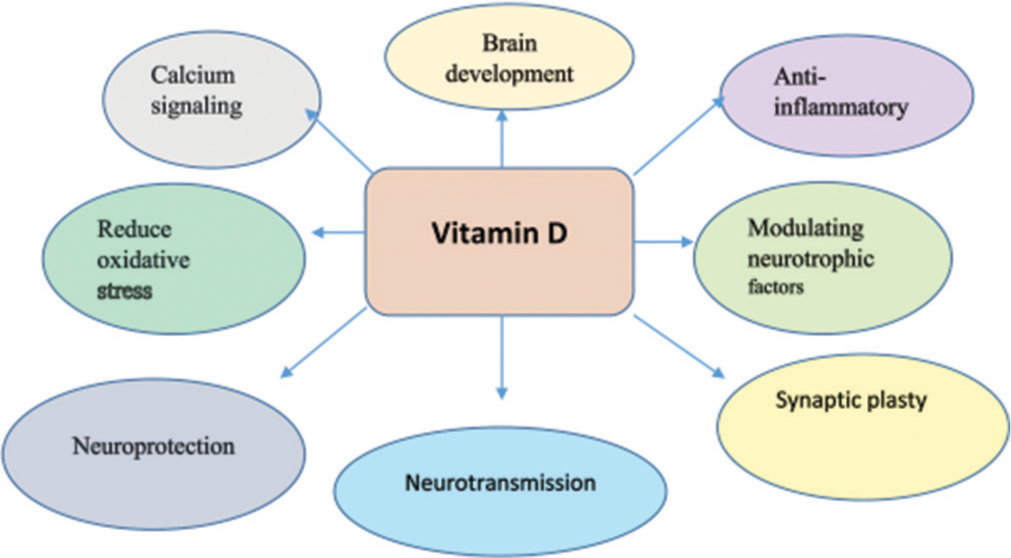
Vitamin D has been reported to affect the elementary processes in fetal brain development, involving changes in synapses and the support of the cytoskeleton.[17] In addition, new mechanisms have revealed how Vitamin D earmarks gene expression and alteration of Vitamin D signaling could affect critical events such as the elongation of exons, neurotransmitter synthesis, and production of neurotrophin in the development and functioning of the brain[16] [Figure 3].
Export to PPT
Calciferol contributes to the development of dopaminergic neurons. At first, VDR was found in the substantia nigra[18,19] During the development phase, Vitamin D deficiency has been shown to decrease certain factors that are mandatory for the growth of neurons involved in dopamine synthesis, change the turnover of dopamine, and interfere with the formation of substantia nigra.[20-22] These abnormalities due to maternal deficiency of Vitamin D can be avoided with cautious supplementation of Vitamin D during pregnancy under medical advice.[23] Vitamin D is postulated to interact with nervous tissue extracellular matrix aggregates and perineuronal nets to regulate brain plasticity and Vitamin D deficiency-induced dysregulation of these interactions may manifest as neurologic deficits which can have implications in neuropsychiatric disorders.[24,25] Vitamin D supplementation in age-related cognitive decline in rats has been shown to modulate synaptic plasticity and improve cognitive function.[26]
Actions on the central nervous system (CNS)Vitamin D promotes genomic expression in the brain through the linkage of the VDR/retinoic acid receptor complex with the Vitamin D-responsive elements in the CNS.[27] It influences the release of neurotransmitters in CNS and affects multiple neuronal functions including differentiation of cells, neurotrophin expression, movement of calcium ions, reduction in oxidative stress and inflammation, and normal physiology of neurons through the manipulation of genetic expression[14] [Figure 3].
Actions on the peripheral nervous system (PNS)The PNS connects the CNS with the peripheral organs. Cells of PNS are involved more in nerve regeneration than CNS.[28] Both VDR and 1-α-hydroxylase enzymes are present in the Schwan cells and peripheral glial cells that affect the excitatory conduction velocity of motor neurons, modulate the production of growth factors, and provide aid in injury and restoration of nerve.[29]
Actions on the autonomic nervous system (ANS)The sympathetic and parasympathetic systems constituting the ANS are considered a fundamental component of the peripheral efferent nervous system, which modulates the functions of visceral organs, endocrine glands, and the smooth muscle of blood vessels.[30] This system innervates nearly all organs of the body.[31-33] Vitamin D contributes to the stability of the autonomic system and potentially behaves as the chief neuroactive substance. It affects the autonomic activities of the brain, heart, and digestive system by acting as an antioxidant and releasing serotonin.[34,35]
Cognitive physiology and psychologyVDRs in the different neuronal cells are responsible for cognitive function and demonstrating these roles through mechanisms such as neuroprotective induction, oxidative stress modification, inflammatory suppression, and calcium balance[36] [Figure 3]. In elderly patients, there is an association between cholecalciferol and white matter disease of the brain.[37] A positive correlation has been reported between Vitamin D levels and the concentration of neurotrophic growth factor, neurogelin-1 (NRG1) has been shown along with an association between the concentration of NRG1 and symptoms of schizophrenia. While a direct link between Vitamin D concentrations and schizophrenia symptoms was not observed, there is a possibility that the relationship may be mediated through NRG1.[38]
ALTERED VITAMIN D STATUS AND NEUROLOGICAL DYSFUNCTION Cerebrovascular accidents (CVA)CVAs are the major reason for significant long-term disability worldwide and are an immense source of the global disease burden. Low Vitamin D levels are linked to a heightened risk of vascular diseases including CVA. Deficits of Vitamin D have been shown to be one of the factors responsible for endothelial dysfunction.[39] Vitamin D deficiency has been suggested as an independent risk factor for the occurrence of acute ischemic stroke.[40] The exact mechanism of altered levels of Vitamin D leading to stroke has not been elucidated yet. The neuroprotective action of Vitamin D takes effect by its regulation of the gene expression of insulin-like growth factor that has anti-thrombolytic properties and helps in dendritic and axonal degeneration. 1,25-dihydroxycholecalciferol helps in vasodilation and activation of nitric oxide synthase, thereby facilitating the reduction of blood pressure directly and indirectly by inhibiting the renin-angiotensin system that is a potent vasoconstrictor.[41] Low levels of Vitamin D are linked to vascular stiffness and reduction in the diameter of blood vessels which may lead to their occlusion.[42] Research on rat models has revealed its function as an antioxidant that helps in the prevention of the destruction of blood–brain barrier integrity after stroke.[43]
Alzheimer’s diseaseAlzheimer’s disease is the most common cause of dementia, characterized by memory loss and deteriorating cognition. Pathologically, extracellular amyloid-beta (Aβ) plaques and intracellular neurofibrillary tangles primarily describe Alzheimer’s disease.[44] In the processing pathway of Aβ in neuronal cultures, cholecalciferol modulates gene expression and helps in the clearance of amyloid plaque. Hence, Vitamin D deficiency may result in Alzheimer’s pathology through disturbed gene expression.[45] Supplementation with Vitamin D has been shown to improve the brain’s memory function through a potential reversal of amyloidopathy.[46]
Cognitive dysfunctionVitamin D deficiency is associated with different cognitive dysfunctions including schizophrenia, sadness, and loss of attention.[24] A number of observational studies have reported the association between a decline in cognitive function and a decreased level of Vitamin D in serum.[47,48] The exact mechanisms of Vitamin D-related cognitive decline are not known, though the neuroprotective properties, anti-inflammatory actions, and antioxidant effects of Vitamin D may possibly serve its potential as a modifiable risk factor for cognitive disease.[36]
Parkinson’s diseaseParkinson’s disease is a neurodegenerative disorder characterized by the loss of neurons of the substantia nigra that produces dopamine. The degeneration of dopaminergic pathways induces motor and non-motor dysfunction in the body. Multiple studies have shown the relationship between decreased serum Vitamin D levels and an increased risk of Parkinson’s disease.[49,50] VDR and 1-α-hydroxylase are present in substantia nigra. The neuroprotective action of Vitamin D helps maintain dopamine levels by upregulating the nerve growth factor.[51,52]
Multiple sclerosis and amyotrophic lateral sclerosisA neurological autoimmune disorder, multiple sclerosis affects the brain and spinal cord through the demyelination of nerve fibers.[53,54] Scientific studies have suggested that hypovitaminosis D contributes as a negative element in the development of multiple sclerosis. The exact pathophysiology of this is not known yet, but Vitamin D’s role as a neuroimmunomodulator and its actions on interleukins and lymphocytes may be involved.[55,56] Vitamin D and aerobic training have recently been shown to positively modulate neurotrophic and inflammatory profiles in female patients with multiple sclerosis.[57] Amyotrophic lateral sclerosis is a neurological illness characterized by the deterioration of motor neurons. The disruption of the neuroprotective effects of Vitamin D through the reduction of oxidative stress is thought to play a role in the pathogenesis of amyotrophic lateral sclerosis.[58]
CONCLUSIONVitamin D has been highlighted as a crucial micronutrient in neural development, growth, and maturation. Recent work has also pointed out the link between Vitamin D and diseases that cause neurocognitive dysfunction such as Parkinson’s and Alzheimer’s. However, most of the work has been limited by a lack of resources, inadequate study designs, inappropriate statistical analyses, and insufficient scientific documentation. It is imperative to undertake properly designed laboratory studies exploring the biochemical signaling pathways of Vitamin D to potentially determine the preventive and therapeutic molecular targets of neurologic pathologies. Vitamin D may then be incorporated as a synergistic agent in the treatment regimens of implicated neurological deficits based on evidence gathered from future interventional studies and randomized control trials.
留言 (0)