This is a study to investigate the clinical characteristics of patients with COPD having OSA, determined using type-3 portable monitors. We believe that the results of the study are useful even in primary care settings, as PSG is often not available in primary care centers. In this study, type-3 portable monitors objectively revealed an OSA prevalence of 77.2% in Japanese patients with COPD. The observed prevalence is slightly higher than those reported previously2. The reason for this discrepancy is not clear, but it may reflect the different populations under investigation and the different methods or criteria for the diagnosis of overlap syndrome. As coexisting OSA worsens the prognosis of COPD patients3 and the survival of patients with overlap syndrome that is not treated with nocturnal positive airway pressure is significantly inferior to that of patients with overlap syndrome that is appropriately treated3,18, the diagnosis of OSA in COPD patients is important. However, overlap syndrome is frequently undiagnosed4. This study showed that OSA is a common comorbidity in Japanese patients with COPD. Two novel observations of potential relevance were made. First, the severity of airflow limitation, hyperinflation, and upper airway collapsibility were found to affect the REI in COPD patients through detailed spirometric assessment. Second the nutritional status, represented by BMI, is one of the most influential factors on the REI in COPD patients, even within the normal range.
Many factors related to coexisting COPD have been reported to modify the formation of apnea and hypopneas in overlap syndrome. Among them, decreased tethering of airways through the loss of lung recoil19, hyperinflation-augmented vagal-mediated reflex20, neural reflex-induced nasal obstruction21, and blunted responses of the respiratory center to various stimuli, particularly during REM sleep22, have been considered as supporting factors for apneas and hypopneas. On the other hand, hyperinflation-augmented stiffness of the upper airway walls23 is thought to be an important inhibitory factor against apnea and hypopnea formation. In fact, data from clinical trials revealed inverse relationships between AHI and FEV1 or radiological emphysema in patients with overlap syndrome5,6. The significant role of high lung volume in the inhibition of extrathoracic upper airway obstruction in subjects with no COPD was originally demonstrated by Spann and Hyatt in 197123. Caudal traction through hyperinflation may unfold the airway or stiffen the airway walls to create a net ventral force that opens the extrathoracic upper airway, which leads to the inhibition of obstructive apnea and hypopnea formation (tracheal tug theory)23,24,25. Herein, we showed an important role of airflow limitation or TLC- and RV-related hyperinflation in restraining apnea and hypopnea formation. Our findings are consistent with the view that the hyperinflation-related inhibitory force against apnea and hypopnea formation is practically canceled out by the forces that promote apnea and hypopnea formation, including the decreased tethering of airways caused by the loss of elastic recoil in the destroyed lung parenchyma in patients with COPD.
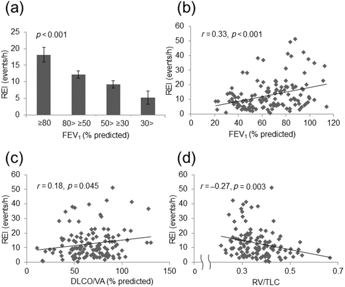
The upper airway collapsibility provided by the PIF/PEF was associated with airflow limitation and hyperinflation, but the correlation between PIF/PEF and the REI was not significant (p = 0.09). Taken together, these findings suggest that although the PIF/PEF detects upper airway collapsibility under a specific condition, the PIF/PEF-represented upper airway opening force may be insufficient to alleviate the airflow limitation-related apnea and hypopnea formation, or the PIF/PEF does not reflect a dynamic change in upper airway collapsibility during sleep. No reports on the association between the PIF/PEF and other spirometric parameters in COPD patients are available. Therefore, to draw a definite conclusion regarding the effect of airflow limitation on upper airway collapsibility in association with apnea and hypopnea formation, it may be necessary to introduce a more precise method that allows the detection of dynamic changes in the upper airway cross-sectional area during sleep, such as CT26, MRI27, fluoroscopy28, or video-assisted nasoendoscopy29. However, these methods present difficulty in preserving natural sleep when applied to a sleeping subject.
The BMI and FFMI were significantly correlated with the REI. These results were consistent with those of a previous study, which showed that obesity is the most common risk factor for OSA in the general population9. Even if the BMI was within the normal range, a BMI > 21.96 kg/m2 was a predictor of moderate to severe OSA in our COPD population, especially in the preserved lung function groups (FEV1 ≥ 50% of predicted value). The relationship between obesity and the REI of overlap syndrome patients remains controversial3. While the reason for this discrepancy is not known, ethnic differences may be partly responsible. Asians including Japanese are generally less obese than Westerners. Thus, the threshold of BMI to predict the occurrence of OSA may differ among countries. Further, several studies, including our previous study, have demonstrated that the cachexia and emphysema phenotypes are more common in Japanese patients with COPD than in Western patients with COPD30. Therefore, whether this examination applies to Westerners should be determined henceforth. According to the results of this study, high BMI was associated with high AHI in patients with mild obstructive disorders, but not in those with severe obstructive disorders. Asians who are generally less obese than Westerners have been reported to be at a risk for OSA even in mild obesity31. It can be deduced that even a mild increase in BMI in this study was associated with high AHI in the mild COPD group. On the other hand, nutritional modulation has no effect on apnea and hypopnea development among patients with advanced COPD having an FEV1 below 50% of the predicted value. There are two possible reasons for the lack of such an association in severe COPD. One is that the effects of obstructive dysfunction and hyperinflation were more strongly associated with AHI than with high BMI. Therefore, the association with BMI may not have been apparent. The other reason is that few cases with a high BMI were included in the severe COPD group, making it difficult to appreciate the differences. In previous studies, severe COPD has been associated with a lower BMI and FFMI32,33. In fact, in our study, only 10.3% (N = 14/136) of patients with severe COPD had a higher BMI > 21.96 kg/m2. To examine the exact group differences, it may be necessary to study an even larger number of GOLD 1–4 patients.
In addition to airflow obstruction, we also investigated the impact of CAT score and history of exacerbations. There were no differences in REI between the three groups of A, B, and E based on the GOLD classification 2023. These results indicate that it may be appropriate to first focus on the degree of airflow obstruction and the risk of OSA complications rather than on subjective symptoms and exacerbations in primary care settings. In this study, there was only a small number of patients in Group E. This result was consistent with previous studies that reported that Japanese patients with COPD have a lower frequency of exacerbations34,35; however, more extensive studies are needed. There are several limitations to the present study. First, this is a cross-sectional study with only a single time point evaluation. As such, no conclusion can be drawn regarding the causal relationship between the abnormality of spirometric parameters or nutritional status and OSA severity. Second, the proportion of women was relatively small (8.8%), and the average age of the participants was higher than in the studies conducted in Western countries. Third, the study population included outpatients of a relatively large hospital. Therefore, caution should be exercised while applying the results of our study to patients visiting a clinic. Further, the number of cases is relatively small, and further investigations, such as a multicenter study in a clinic or a population-based study, are needed to determine whether our findings are applicable to the Japanese population as a whole as well as to the world general population. Fourth, the mechanism of upper airway obstruction has not yet been determined, as it has not been possible to directly observe the pharyngeal area in otolaryngology; some inferences about the mechanism have been made based on indirect data. Finally, lung function tests were not performed under the post-bronchodilator condition, and tests to exclude asthmatic elements, such as FeNO and peripheral blood eosinophils, were also not performed. Therefore, COPD may have been overdiagnosed, and cases with a combined asthmatic component were not completely removed. Many studies have shown that asthma has a negative impact on sleep apnea19,36,37,38, and it is possible that asthma complications have some impact on the pathogenesis of sleep apnea. The present study also did not measure eosinophils or FeNO, nor did it confirm the degree of reversibility, as mentioned above. Therefore, the effect of asthma complications on the results needs to be further investigated.
In conclusion, In Japanese COPD patients, airflow limitation and lung hyperinflation had a significant impact on the severity of OSA, with higher severity leading to lower REI. Further, BMI and FFMI were correlated with REI, especially in the preserved lung function groups. These findings may be useful in identifying high-risk groups for OSAS and the pathogenesis of overlap syndromes.


留言 (0)