
記住我

A 65-year-old Caucasian male came to our attention with a 4-month history of purpuric lesions of the lower limbs (Fig. 1A), arthralgias, low-grade fever, weight loss, abdominal pain, and nausea. The patient denied any ear-nose-throat, lung, neurological or genitourinary symptom. Petechiae extended up to the buttocks, they were palpable, slightly pruritic, sub-centimetric in diameter and did not blanch when pressure was applied. His health records were unremarkable apart from systemic hypertension and Epstein-Barr virus (EBV) infection two years earlier. Routine laboratory tests showed normochromic normocytic anaemia, white blood cell count 6250/µL (neutrophils 3390/µL, lymphocytes 1540/µL, eosinophils 540/µL), erythrocyte sedimentation rate 40 mm/h, C-reactive protein 1.6 mg/dL, normal kidney function and urinalysis, and pronounced polyclonal hypergammaglobulinemia with raised IgA (660 mg/dL, ULN 500 mg/dL) and IgG (2790 mg/dL, ULN 2530 mg/dL). The autoimmune panel detected ANA 1:160 cytoplasmic fibrillar pattern (AC-15,16,17), with negative anti-ENA, anti-dsDNA, rheumatoid factor, anti-neutrophil cytoplasmic antibodies and cryoglobulins; complement was not consumed. The patient was negative for hepatitis B and C infection. Serum and urine immunofixation were negative, and the haematology consultant ruled out an ongoing lymphoproliferative disease.
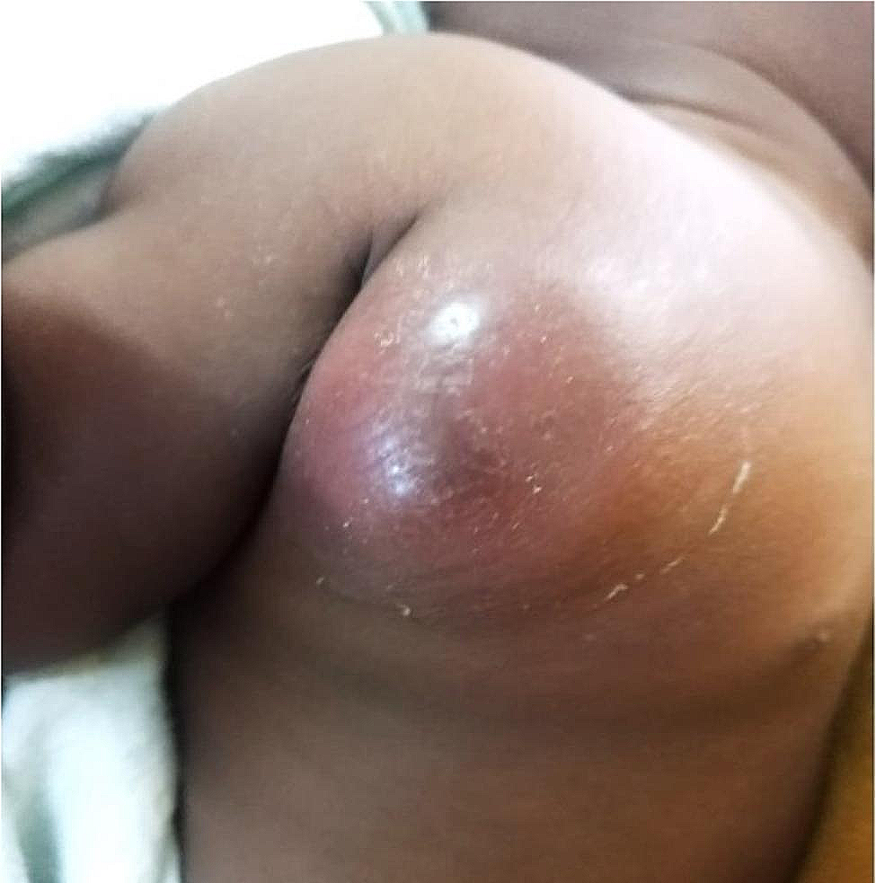
Fig. 1
A Purpuric lesions of the lower limbs at disease presentation. B Histology of caecal mucosa (Haematoxylin and Eosin): granulation tissue with endothelial hyperplasia, widespread infiltration of eosinophils, histiocytes and macrophages, intranuclear Owl’s eye inclusion bodies consistent with CMV infection (arrows). C CMV immunohistochemistry on caecal mucosa: positive reaction within the nuclei of CMV-infected cells. D Purpuric lesions of the lower limbs after treatment
IgAV was suspected, thus further studies aimed at investigating vasculitis were performed. Punch skin biopsy showed leukocytoclastic vasculitis and direct immunofluorescence detected IgA deposits within small vessels of the dermis. Urine sediment analysis did not reveal any sign of active glomerulopathy. Colonoscopy found a caecum ulcer of 25 mm in diameter. A diagnosis of IgAV with cutaneous and suspected gastrointestinal involvement was confirmed, hence the patient started prednisone 50 mg/day with disappearance of arthralgias and abdominal pain, with partial resolution of petechiae. Upon glucocorticoid tapering, he experienced a recrudescence of cutaneous pruritic rash involving the trunk and lower limbs. Prednisone was increased to 37.5 mg/day and azathioprine 100 mg/day was added with moderate improvement of skin lesions.
Meanwhile, he developed perianal vesicular lesions positive for herpes simplex virus 1 and 2, and the histological examination of the intestinal ulcer revealed intranuclear CMV inclusion bodies (Fig. 1B, C). Following these findings, blood tests for EBV-DNA and CMV-DNA were performed, resulting in 102,420 copies/mL and 2997 copies/mL respectively. Considering the multiple infections and the clinical picture not completely responding to high-dose glucocorticoids, HIV infection was suspected. HIV antibody and p24 antigen resulted positive, with a viral load of 122,791 copies/mL and 12 CD4+ T lymphocytes/µL. The patient was diagnosed with acquired immunodeficiency syndrome (AIDS), disseminated CMV infection with CMV colitis, EBV reactivation, and cutaneous IgAV. He started dolutegravir/lamivudine and ganciclovir together with prophylaxis for opportunistic infections, whereas azathioprine was interrupted, and glucocorticoids slowly tapered. After antiviral treatment initiation, viral loads progressively decreased and the CD4+ T cell count rose slightly. Nonetheless, the patient experienced a flare of petechial lesions on the lower limbs one month after glucocorticoid discontinuation. Colchicine was started with prompt improvement of the skin rash (Fig. 1D).
留言 (0)