2.1 Isolation and expansion of HUMSCs and HUVECs
This study was approved by the Institutional Review Board of the Second Affiliated Hospital of Shantou University Medical College. After written informed consent, umbilical cords from term (gestational age (GA) ≥ 37 weeks, n = 2, term) and extremely preterm (GA < 28 weeks or birth weight < 1000 g, n = 5) babies without intrauterine infection and congenital malformations were collected in a sterile environment. HUMSCs were isolated from umbilical cord as previously described [9]. The cells were cultured in fresh growth medium (containing the DMEM/F-12 with 10% fetal bovine serum) in a 37 °C humidified incubator with 5% CO2, when the media was replaced every 2 days. HUVECs were obtained from umbilical cord samples of healthy term infants using enzymatic digestion protocols described as Edyta [24]. The cells were cultured in M199 medium(Gibco, Grand Island, NY, USA) (containing 20% FBS and VEGF[Sciencell Research Laboratories, Carlsbad, CA, USA]) at 37 °C in 5% CO2 and used in passage 3–4.
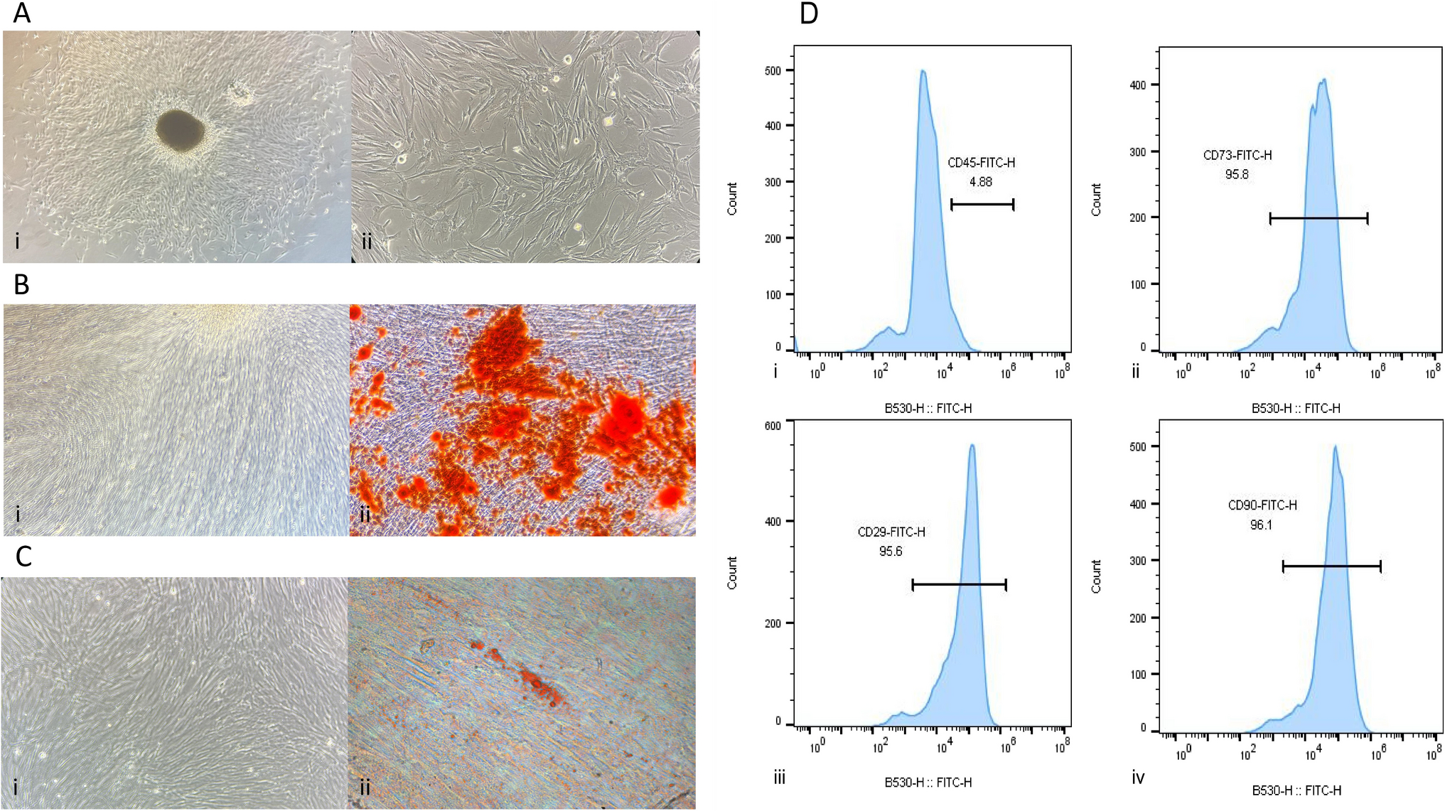
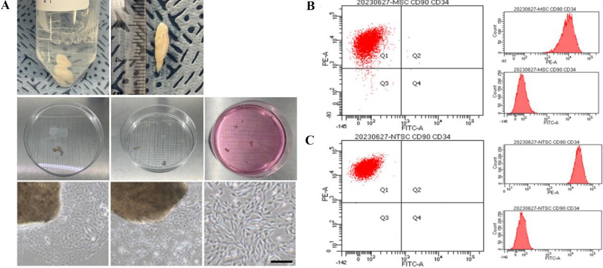
2.2 Flow cytometry phenotyping of HUMSCs
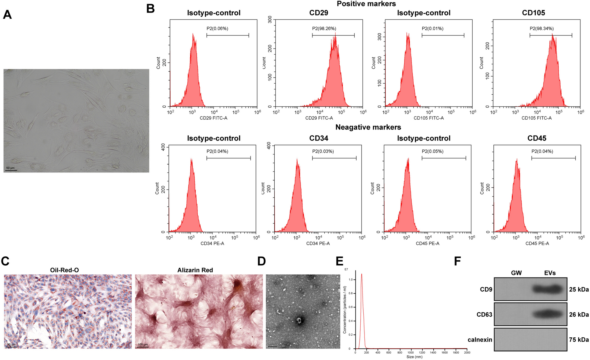
HUMSCs isolated from different gestation ages were characterized by flow cytometry in passage 3 or 4. Cells were washed in PBS for twice, then incubated in the dark with coupled mouse anti-human antibodies:FITC-CD90 (BioLegend, San Diego, CA, USA), PerCPCyTM5.5-CD105 (BD Biosciences, Heidelberg, Germany), APC-CD73 (BD Biosciences),PE-HLA-DR (BD Biosciences),PE-CD11b (BioLegend), PE-CD19 (BioLegend),PE-CD45 (BioLegend) and mouse anti-human PE-CD34 (BioLegend). After staining, the cells were washed twice with PBS and analyzed in a FACSCanto II cytometer (BD Biosciences).At least 20,000 cells were recorded, cells without adding any antibody as a negative control.
2.3 Reverse transcription polymerase chain reaction and quantitative RT-PCR analysis
Total RNA was isolated from cells using the Eastep® super total RNA extraction kit (Promega, Madison, WI, USA) and 1-mg total RNA was used for reverse transcription using the GoScript™ reverse transcriptase (Promega). qRT-PCR was performed on a LightCycler 480 high flux Real-Time Fluorescence Quantitative PCR System (Roche, Indianapolis, IN, USA) using GoTaq ® qPCR Master Mix (Promega). PCR primer sequences are listed in Table 1 (Invitrogen, Shanghai, China).All the data were analyzed using GAPDH as an internal control. Relative copy numbers of target genes were determined using the 2-ΔΔCt method.
Table 1 List of primers used in RT-PCR2.4 Cell proliferation assay
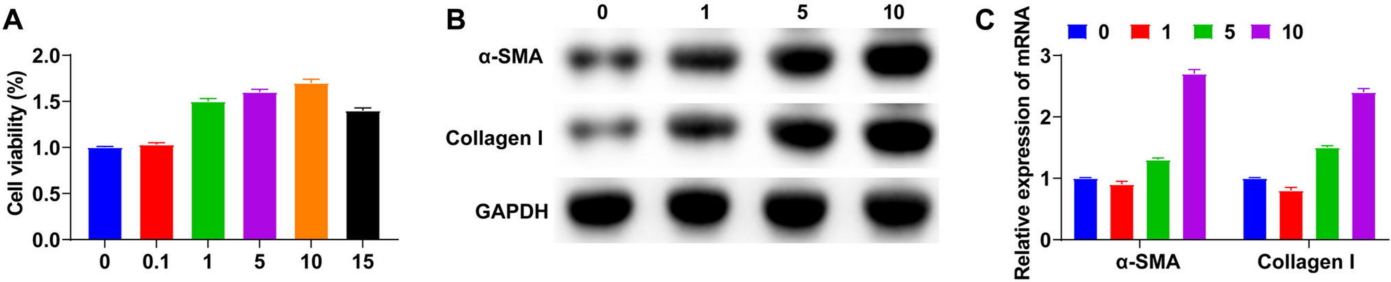
Proliferation of HUMSCs was analyzed using cell counting kit-8 (CCK–8, Promega) according to the manufacturer’s protocols. HUMSCs were plated at a density of 2 × 10³ cells with 100ul medium into each well of a 96-well flat-bottom tissue culture plate (BD Falcon, San Jose, CA, USA),cultured for 4 h,24 h,48 h,72 h. CCK8 solution was added to each well and incubated in standard culture conditions for 1 h. The absorbance was analysed at 450 nm using a microplate reader (BioTek, Winooski, VT, USA), wells without cells as blanks. The results were expressed as mean absorbance in nanometers. All experiments were performed in triplicate.
2.5 Cell injury model and co-culture system
HUVECs were seeded in 6-well plates and incubated for 12 h. The inflammatory model was treated with LPS(10 μg/ml). HUMSCs were pretreated with mitomycin for 24 h and then digested, 106 cells were seeded on the upper layer chambers of Transwell plates (0.4 μm pore size; Corning Life Sciences, Tewksbury, MA, USA) at a ratio of HUVECs:HUMSCs of 2: 1. The supernatant was collected at 4 h, 24 h, and 48 h after co-culture, and the expression of inflammatory factor TGFβ1 was detected by Elisa. After 24 h’ co-culture, HUMSCs in the upper chambers were discarded. HUVEC in lower chambers were washed by PBS, and cultured in DMEM/FBS for Wound healing assay, or collected for the vasculogenesis assay.
2.5.1 ELISA
In co-cultured system, the supernatant was collected at 4 h, 24 h, 48 h and centrifuged (1000 × g, 5 min) to remove the cells. The levels of secreted TGF-β1and IL6 in the supernatant were measured according to the manual of the enzyme-linked immunosorbent assay (ELISA) kit(Human TGF-β1 elisa kit: Neobiosience, Shenzhen, China, EHC107b.96. AuthentiKine Human IL-6 ELISA Kit, Proteintech, USA. Human IL-1 beta ELISA Kit, Proteintech).The absorbance value at a wavelength of 450 nm was measured with a TriStar2 LB 942 Multimode Microplate Reader (Berthold Technologies, Bad Wildbad, Germany).
2.6 Wound healing assay
LPS induced HUVECs were cultured with HUMSCs in transwell chambers(Tissue culture plate inserts, BIOFIL, Taipei, Taiwan) for 24 h, the upper chambers of HUMSCs were discarded, washed by PBS for twice,and cultured in DMEM/FBS for 24 h. scratch was made using a 200 μl pipette tip across the plates. After plates were washed with culture medium, HUVECs were grown for 24 h and photographs were taken.
2.7 Tube formation
HUVECs were harvested after 24 h cocultured, and cells were (3 × 10⁴ cells per well) seeded onto matrigel plates (Matrigel356234; BD Biosciences) and cultured at 37 °C in 5% CO2. And observed by an inverted microscopy over the course of 24 h for tube formation. Networks formed by the HUVECs were quantified with Image J software (National Institutes of Health, Bethesda, MD, USA).
2.8 Statistics analysis
All data were represented by mean ± standard deviations, and were analyzed by one-way ANOVA and Bonferroni’ multiple comparison test. Each result was completed by at least three independent experiments. The difference was statistically significant when p value < 0.05. Charts are drawn using GraphPad Prism(version 8.0 for Windows, GraphPad Software Inc., San Diego, CA, Bethesda, MD, USA).


留言 (0)